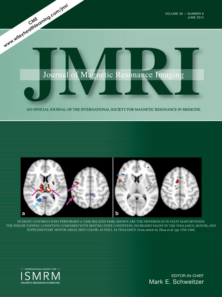
Characterization of thalamo-cortical association using amplitude and connectivity of functional MRI in mild traumatic brain injury
Yongxia Zhou PhD
Department of Radiology / Center for Biomedical Imaging, NYU Langone Medical Center, New York, New York, USA
Search for more papers by this authorYvonne W. Lui MD
Department of Radiology / Center for Biomedical Imaging, NYU Langone Medical Center, New York, New York, USA
Search for more papers by this authorXi-Nian Zuo PhD
Laboratory for Functional Connectome and Development, Key Laboratory of Behavioral Science, Magnetic Resonance Imaging Research Center, Institute of Psychology, Chinese Academy of Sciences, Beijing, China
Search for more papers by this authorMichael P. Milham MD, PhD
Child Mind Institute, New York, New York, USA
Search for more papers by this authorJoseph Reaume BS
Department of Radiology / Center for Biomedical Imaging, NYU Langone Medical Center, New York, New York, USA
Search for more papers by this authorRobert I. Grossman MD
Department of Radiology / Center for Biomedical Imaging, NYU Langone Medical Center, New York, New York, USA
Search for more papers by this authorCorresponding Author
Yulin Ge MD
Department of Radiology / Center for Biomedical Imaging, NYU Langone Medical Center, New York, New York, USA
Address reprint requests to: Y.G., Center for Biomedical Imaging, Department of Radiology, New York University School of Medicine, 660 First Avenue, 4th floor, New York, NY 10016 USA. E-mail: [email protected]Search for more papers by this authorYongxia Zhou PhD
Department of Radiology / Center for Biomedical Imaging, NYU Langone Medical Center, New York, New York, USA
Search for more papers by this authorYvonne W. Lui MD
Department of Radiology / Center for Biomedical Imaging, NYU Langone Medical Center, New York, New York, USA
Search for more papers by this authorXi-Nian Zuo PhD
Laboratory for Functional Connectome and Development, Key Laboratory of Behavioral Science, Magnetic Resonance Imaging Research Center, Institute of Psychology, Chinese Academy of Sciences, Beijing, China
Search for more papers by this authorMichael P. Milham MD, PhD
Child Mind Institute, New York, New York, USA
Search for more papers by this authorJoseph Reaume BS
Department of Radiology / Center for Biomedical Imaging, NYU Langone Medical Center, New York, New York, USA
Search for more papers by this authorRobert I. Grossman MD
Department of Radiology / Center for Biomedical Imaging, NYU Langone Medical Center, New York, New York, USA
Search for more papers by this authorCorresponding Author
Yulin Ge MD
Department of Radiology / Center for Biomedical Imaging, NYU Langone Medical Center, New York, New York, USA
Address reprint requests to: Y.G., Center for Biomedical Imaging, Department of Radiology, New York University School of Medicine, 660 First Avenue, 4th floor, New York, NY 10016 USA. E-mail: [email protected]Search for more papers by this authorAbstract
Purpose
To examine thalamic and cortical injuries using fractional amplitude of low-frequency fluctuations (fALFFs) and functional connectivity MRI (fcMRI) based on resting state (RS) and task-related fMRI in patients with mild traumatic brain injury (MTBI).
Materials and Methods
Twenty-seven patients and 27 age-matched controls were recruited. The 3 Tesla fMRI at RS and finger tapping task were used to assess fALFF and fcMRI patterns. fALFFs were computed with filtering (0.01–0.08 Hz) and scaling after preprocessing. fcMRI was performed using a standard seed-based correlation method, and delayed fcMRI (coherence) in frequency domain were also performed between thalamus and cortex.
Results
In comparison with controls, MTBI patients exhibited significantly decreased fALFFs in the thalamus (and frontal/temporal subsegments) and cortical frontal and temporal lobes; as well as decreased thalamo-thalamo and thalamo-frontal/ thalamo-temporal fcMRI at rest based on RS-fMRI (corrected P < 0.05). This thalamic and cortical disruption also existed at task-related condition in patients.
Conclusion
The decreased fALFFs (i.e., lower neuronal activity) in the thalamus and its segments provide additional evidence of thalamic injury in patients with MTBI. Our findings of fALFFs and fcMRI changes during motor task and resting state may offer insights into the underlying cause and primary location of disrupted thalamo-cortical networks after MTBI. J. Magn. Reson. Imaging 2014;39:1558–1568. © 2013 Wiley Periodicals, Inc.
REFERENCES
- 1
Silver JM,
McAllister TW,
Yudofsky SC. Textbook of traumatic brain injury, 2nd edition. Washington, DC: American Psychiatric Publishing, Inc; 2011.
10.1176/appi.books.9781585624201 Google Scholar
- 2 Ge Y, Patel MB, Chen Q, et al. Assessment of thalamic perfusion in patients with mild traumatic brain injury by true FISP arterial spin labelling MR imaging at 3T. Brain Inj 2009; 23: 666–674.
- 3 Kirov I, Fleysher L, Babb JS, Silver JM, Grossman RI, Gonen O. Characterizing ‘mild’ in traumatic brain injury with proton MR spectroscopy in the thalamus: initial findings. Brain Inj 2007; 21: 1147–1154.
- 4 Zhou Y, Milham MP, Lui YW, et al. Default-mode network disruption in mild traumatic brain injury. Radiology 2012; 265: 882–892.
- 5 Maxwell WL, Pennington K, MacKinnon MA, et al. Differential responses in three thalamic nuclei in moderately disabled, severely disabled and vegetative patients after blunt head injury. Brain 2004; 127(Pt 11): 2470–2478.
- 6 Jones EJ. The thalamus, 2nd edition. Cambridge University Press; 2007.
- 7 Chatila M, Milleret C, Rougeul A, Buser P. Alpha rhythm in the cat thalamus. C R Acad Sci III 1993; 316: 51–58.
- 8 Lopes da Silva FH, Hoeks A, Smits H, Zetterberg LH. Model of brain rhythmic activity. The alpha-rhythm of the thalamus. Kybernetik 1974; 15: 27–37.
- 9 Schreckenberger M, Lange-Asschenfeldt C, Lochmann M, et al. The thalamus as the generator and modulator of EEG alpha rhythm: a combined PET/EEG study with lorazepam challenge in humans. Neuroimage 2004; 22: 637–644.
- 10 Buzsaki G, Wang XJ. Mechanisms of gamma oscillations. Annu Rev Neurosci 2012; 35: 203–225.
- 11 Engel AK, Fries P, Konig P, Brecht M, Singer W. Temporal binding, binocular rivalry, and consciousness. Conscious Cogn 1999; 8: 128–151.
- 12 Pollack R. The missing moment: how the unconscious shapes modern science. Boston: Houghton Mifflin; 1999.
- 13 Kempf F, Brucke C, Salih F, et al. Gamma activity and reactivity in human thalamic local field potentials. Eur J Neurosci 2009; 29: 943–953.
- 14 Sun Y, Farzan F, Barr MS, et al. gamma oscillations in schizophrenia: mechanisms and clinical significance. Brain Res 2011; 1413: 98–114.
- 15 Ben-Simon E, Podlipsky I, Arieli A, Zhdanov A, Hendler T. Never resting brain: simultaneous representation of two alpha related processes in humans. PLoS One 2008; 3: e3984.
- 16 Zou Q, Long X, Zuo X, et al. Functional connectivity between the thalamus and visual cortex under eyes closed and eyes open conditions: a resting-state fMRI study. Hum Brain Mapp 2009; 30: 3066–3078.
- 17 Muthukumaraswamy SD, Edden RA, Jones DK, Swettenham JB, Singh KD. Resting GABA concentration predicts peak gamma frequency and fMRI amplitude in response to visual stimulation in humans. Proc Natl Acad Sci U S A 2009; 106: 8356–8361.
- 18 Haacke EM, Duhaime AC, Gean AD, et al. Common data elements in radiologic imaging of traumatic brain injury. J Magn Reson Imaging 2010; 32: 516–543.
- 19 Tang L, Ge Y, Sodickson DK, et al. Thalamic resting-state functional networks: disruption in patients with mild traumatic brain injury. Radiology 2011; 260: 831–840.
- 20 Sun FT, Miller LM, D'Esposito M. Measuring interregional functional connectivity using coherence and partial coherence analyses of fMRI data. Neuroimage 2004; 21: 647–658.
- 21 Zang YF, He Y, Zhu CZ, et al. Altered baseline brain activity in children with ADHD revealed by resting-state functional MRI. Brain Dev 2007; 29: 83–91.
- 22 Zou QH, Zhu CZ, Yang Y, et al. An improved approach to detection of amplitude of low-frequency fluctuation (ALFF) for resting-state fMRI: fractional ALFF. J Neurosci Methods 2008; 172: 137–141.
- 23 Hoptman MJ, Zuo XN, Butler PD, et al. Amplitude of low-frequency oscillations in schizophrenia: a resting state fMRI study. Schizophr Res 2010; 117: 13–20.
- 24 Zhang G, Yin H, Zhou YL, et al. Capturing amplitude changes of low-frequency fluctuations in functional magnetic resonance imaging signal: a pilot acupuncture study on NeiGuan (PC6). J Altern Complement Med 2012; 18: 387–393.
- 25 Zuo XN, Di Martino A, Kelly C, et al. The oscillating brain: complex and reliable. Neuroimage 2010; 49: 1432–1445.
- 26 Bajo R, Maestu F, Nevado A, et al. Functional connectivity in mild cognitive impairment during a memory task: implications for the disconnection hypothesis. J Alzheimers Dis 2010; 22: 183–193.
- 27 Duff EP, Johnston LA, Xiong J, Fox PT, Mareels I, Egan GF. The power of spectral density analysis for mapping endogenous BOLD signal fluctuations. Hum Brain Mapp 2008; 29: 778–790.
- 28 Sparing R, Meister IG, Wienemann M, Buelte D, Staedtgen M, Boroojerdi B. Task-dependent modulation of functional connectivity between hand motor cortices and neuronal networks underlying language and music: a transcranial magnetic stimulation study in humans. Eur J Neurosci 2007; 25: 319–323.
- 29 Biswal BB. Resting state fMRI: a personal history. Neuroimage 2012; 62: 938–944.
- 30 Power JD, Barnes KA, Snyder AZ, Schlaggar BL, Petersen SE. Spurious but systematic correlations in functional connectivity MRI networks arise from subject motion. Neuroimage 2012; 59: 2142–2154.
- 31 Bennett CM, Wolford GL, Miller MB. The principled control of false positives in neuroimaging. Soc Cogn Affect Neurosci 2009; 4: 417–422.
- 32 Garcia-Panach J, Lull N, Lull JJ, et al. A voxe-based analysis of FDG-PET in traumatic brain injury: regional metabolism and relationship between the thalamus and cortical areas. J Neurotrauma 2011; 28: 1707–1717.
- 33 Shim WH, Baek K, Kim JK, et al. Frequency distribution of causal connectivity in rat sensorimotor network: resting-state fMRI analyses. J Neurophysiol 2013; 109: 238–248.
- 34 Aron AR, Schlaghecken F, Fletcher PC, et al. Inhibition of subliminally primed responses is mediated by the caudate and thalamus: evidence from functional MRI and Huntington's disease. Brain 2003; 126(Pt 3): 713–723.
- 35 Steriade M. Sleep, epilepsy and thalamic reticular inhibitory neurons. Trends Neurosci 2005; 28: 317–324.
- 36 Saad ZS, Ropella KM, Cox RW, DeYoe EA. Analysis and use of FMRI response delays. Hum Brain Mapp 2001; 13: 74–93.
- 37 Muller K, Lohmann G, Neumann J, Grigutsch M, Mildner T, von Cramon DY. Investigating the wavelet coherence phase of the BOLD signal. J Magn Reson Imaging 2004; 20: 145–152.
- 38 Salomon RM, Karageorgiou J, Dietrich MS, et al. MDMA (Ecstasy) association with impaired fMRI BOLD thalamic coherence and functional connectivity. Drug Alcohol Depend 2012; 120: 41–47.
- 39 Kalcher K, Boubela RN, Huf W, et al. RESCALE: voxel-specific task-fMRI scaling using resting state fluctuation amplitude. Neuroimage 2013; 70: 80–88.
- 40 Schiff ND, Giacino JT, Kalmar K, et al. Behavioural improvements with thalamic stimulation after severe traumatic brain injury. Nature 2007; 448: 600–603.


