Volume 30, Issue 10 pp. 1201-1203
CASE VIDEO

Usefulness of texture and color enhancement imaging in peroral pancreatoscopy

Yuki Tanisaka

Corresponding Author

Yuki Tanisaka

Department of Gastroenterology, Saitama Medical University International Medical Center, Saitama, Japan

Correspondence

Yuki Tanisaka, Department of Gastroenterology, Saitama Medical University International Medical Center, 1397-1, Yamane, Hidaka, Saitama 350-1298, Japan.

Email: [email protected]

Search for more papers by this author
Masafumi Mizuide

Masafumi Mizuide

Department of Gastroenterology, Saitama Medical University International Medical Center, Saitama, Japan

Search for more papers by this author
Shomei Ryozawa

Shomei Ryozawa

Department of Gastroenterology, Saitama Medical University International Medical Center, Saitama, Japan

Search for more papers by this author
First published: 03 February 2023

Graphical Abstract

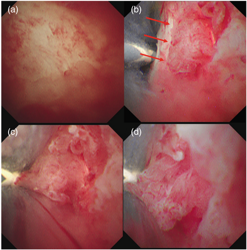
Tanisaka and colleagues report the usefulness of texture and color enhancement imaging provided by a new-generation image-enhanced endoscopy system in a patient with intraductal papillary mucinous neoplasm who had undergone peroral pancreatoscopy. Texture and color enhancement imaging clearly showed structural changes of the lesion and improved the diagnostic quality of peroral pancreatoscopy.

CONFLICT OF INTEREST STATEMENT

All authors declare no conflicts of interest for this article.

The full text of this article hosted at iucr.org is unavailable due to technical difficulties.