A time-saving–modified Giemsa stain is a better diagnostic method of Helicobacter pylori infection compared with the rapid urease test
Abstract
Background
Despite having chronic gastritis, most people infected by Helicobacter pylori (H. pylori) are asymptomatic and have no specific clinical signs and symptoms. H. pylori infection can be diagnosed by several detection methods. Giemsa stain and rapid urease test (CLO test) are the most performed tests of H. pylori infection at first-line clinical examination because of their simplicity and reliability. However, the sensitivity of CLO test is significantly reduced in patients with atrophic gastritis and intestinal metaplasia, and the weaknesses of Giemsa stain are higher cost and time-consuming.
Methods
The Giemsa stain was modified in several staining solutions and procedures based on the simplified Giemsa technique described by Gray, Wyatt, & Rathbone (1986). The modified Giemsa stain is examined its efficacy and compared with the CLO test using 233 H. pylori-infected patients with gastric disease.
Results
The modified Giemsa stain is comparable to the traditional one. Statistical analysis indicated that the modified Giemsa stain obtains greater accuracy in H. pylori-infected patients with gastritis and ulcer than the CLO test (48.1% vs. 43.7%). Moreover, considering the prognosis of different symptoms of gastric diseases, the modified Giemsa stain has a more accurate prognosis than combination symptoms (P = 1.8E-05 vs. P = 5.49E-05). The modified Giemsa stain is confirmed to be better than CLO test using 233 H. pylori-infected patients with gastric disease.
Conclusions
The modified Giemsa stain is more simplified and time-saving than traditional Giemsa stain, which is comparable to the traditional one and is confirmed to be better than CLO test using 233 H. pylori-infected patients with gastric disease. In clinical examination, this modified Giemsa stain can be applied to routine examination and provides quick and accurate diagnosis and prognosis to H. pylori-infected patients with gastric diseases.
1 INTRODUCTION
More than 50% of the population worldwide were infected by Helicobacter pylori (H. pylori) which harbored in their upper gastrointestinal tract.1 H. pylori2 belongs to gram-negative bacterium, which is usually found in microaerophilic circumstance of the gastric epithelium.3, 4 H. pylori utilizes the strong activity of urease as a protective buffering enzyme to hydrolyze urea into ammonia and carbon dioxide against gastric acid and for survival in low pH environment of human stomac H. Currently, the first-line treatment for H. pylori is triple combination therapy, including proton-pump inhibitor (PPI), clarithromycin, and amoxicillin. However, as increasing drug-resistance strains of H. pylori were found, different therapies were applied to overcome the drug-resistance, such as sequential therapy, high-dose dual therapy, and concomitant therapy.5, 6 Despite having chronic gastritis, most people infected by H. pylori are asymptomatic and have no specific clinical signs and symptoms. For many gastric diseases, such as chronic gastritis, gastroduodenal ulcers, and even gastric carcinogenesis, are majorly caused by H. pylori infection.7
H. pylori infection can be diagnosed by several detection methods. These tests include non-invasive and invasive methods. The non-invasive method includes urea breath test, stool antigen test, and serology. The invasive methods include culture, histological examination, and rapid urease test, which requires the use of endoscopy to collect biopsy specimens.8-10 It is generally believed that invasive test is more accurate than non-invasive test for H. pylori infection.11-13 Regarding the histological examination, hematoxylin and eosin (H&E) staining, Genta stain, immunohistochemical (IHC) stain, and Giemsa stain were developed. In general, H&E stain is the first and routine examination performed at pathologist desk before other specialized methods. IHC stain has major advantages of higher sensitivity and reliability compared with H&E and Giemsa stain even in patients treated for gastritis.11, 14, 15 Unfortunately, the expense and time-consuming nature of IHC stain make it disadvantageous for using a routine method in many laboratories. Therefore, H&E and Giemsa stains are better histological methods due to simplicity of use and consistency. However, the disadvantage of H&E stain is low specificity, and Giemsa stain has several drawbacks, such as higher cost, time-consuming, and interobserver variability.11 In addition, the rapid urease test, Campylobacter-like organism test (CLO test), is another routine examination with speed, easy diagnosis, and high accuracy,10 especially with patients who are not treated with proton-pump inhibitor (PPI). However, the sensitivity of CLO test was significantly reduced in patients with atrophic gastritis (AG) and intestinal metaplasia (IM).11
In most hospitals, the fastest and convenient diagnostic methods for H. pylori detection are the invasive tests including the CLO test and Giemsa stain that are most commonly used in first-line routine clinical practice.10 In this study, we improved the traditional Giemsa stain and provide an easier and less time-consuming method with the same accuracy. Furthermore, we compared the diagnosis results from the modified Giemsa stain with results from the CLO test. The modified Giemsa stain we developed gives greater accuracy in diagnosis of H. pylori-infected patients with gastric diseases than the CLO test and has a comparable accuracy with the traditional Giemsa stain.
2 PATIENT AND METHODS
Tissue specimens of 233 patients with gastritis or/and gastric ulcer were collected and retrieved from diagnostic resection specimens in the Departments of Pathology at Mackay Memorial Hospital, Taipei, Taiwan. The informed consent was obtained from each patient in advance before the specimen was collected into the archive. The experiment was performed with approval from the Institutional Review Board (No. 18MMHIS032). All experiments were performed in accordance with relevant guidelines and regulations.
2.1 Modified Giemsa stain and CLO test
All samples from peptic ulcer patients were obtained by biopsy forceps of endoscope at least 5 mm in diameter. Tissue specimens of gastric mucosa were separated into three parts for the traditional Giemsa stain, modified Giemsa stain, and CLO test. The modified Giemsa stain was amended from the simplified Giemsa technique described by Gray et al.16 The 2-3 mm tissue sample was stored in 10% formalin and then fixed by paraffin. The paraffin sections of the tissue blocks were transferred to an adhesive-coated slide system. The sample slide was deparaffinized and hydrated in distilled water. The slide was incubated in fresh-made HP (H. pylori) solution for 30 min. HP solution was made by Giemsa stain solution (Merck) in distilled water (1:20) with 12.5% methanol (Nihon Shiyaku). Followed by rinsing in distilled water for a while, the slide was dehydrated with 95% alcohol instead of air dry. Finally, the slide was mounted and sealed by coverslip to prevent from drying and evaluated the stain under microscope. For CLO test (Kimberly-Clark, Medex Supply, USA), 2-3 mm specimen was placed into medium containing urea and was examined by the test following manufacturer's instructions. The CLO test results were evaluated 2 to 12 h after by endoscopy. The evaluation of the modified Giemsa stain and CLO test was performed at the Department of Pathology in Mackay Memorial Hospital.
2.2 Statistical analysis
Fisher's exact test was used to examine the significance of the association between two kinds of classification. The log-rank test was used to examine the difference in survival distributions between two groups. All data were analyzed using the R statistical software (version 3.4.3). In all analysis, a p-value of < 0.05 was considered as statistically significant.
3 RESULTS
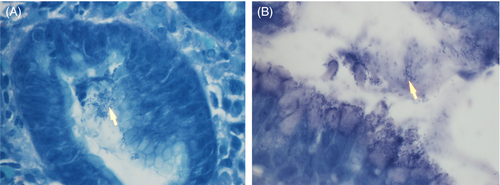
To confirm and compare the accuracy of the modified Giemsa stain and CLO test, two hundred and thirty-three biopsies from gastric disease patients were used to perform two tests to examine the H. pylori infection. The detailed procedures of the modified Giemsa stain and CLO test were described in the Patients and Methods section. The examination results from two tests were double confirmed by the traditional Giemsa staining used as a positive control. The blue stain of H. pylori was presented adjacent to the gastric mucosa by the traditional Giemsa stain (Figure 1). All test samples were examined simultaneously by the modified Giemsa stain and CLO test and compared together to the positive control from traditional Giemsa stain. The test samples with the same pattern as the positive control were assigned to positive results of H. pylori infection.

The enrolled 233 patients were diagnosed and displayed gastric diseases, including gastritis, ulcer, or polyps. Our results found that the modified Giemsa stain we developed has the same accuracy as the traditional Giemsa stain. Seventy-seven of the 173 gastritis patients (44.5%) were diagnosed as positive for H. pylori infection and 96 (55.5%) as negative by the modified Giemsa stain (Table 1). The same cohort in CLO test revealed that 72 (41.6%) are positive for H. pylori infection and 101 (58.4%) are negative. As for the patients with ulcer, twenty-three of 35 patients (65.7%) are positive and 12 patients (34.3%) are negative by the modified Giemsa stain. The same groups of ulcer patients examined by the CLO test revealed 19 (54.3%) positive and 16 (45.7%) negative. These results showed that the modified Giemsa stain has more accurate diagnosis results of H. pylori infection than the CLO test (44.5% vs. 41.6%; 65.7% vs. 54.3%; Table 1). All the results from the CLO test were believed with sensitivity of 91% (94/103) and specificity of 100% (130/130) for H. pylori infection compared with the modified Giemsa stain. Moreover, considering the prognosis of different symptoms of gastric diseases, statistics analysis significantly indicated that the modified Giemsa stain has a more accurate prognosis of gastric diseases caused by H. pylori infection than the CLO test (P = 1.80E-05 vs. P = .00041; Table 1). Therefore, the analysis indicates that the modified Giemsa stain is better than the CLO test regarding the diagnostic accuracy and the prognosis of gastric diseases caused by the H. pylori infection.
| Modified Giemsa stain | Gastritis | Ulcer | Polyp | None | Total | P-value |
|---|---|---|---|---|---|---|
| Positive | 77 (44.5%) | 23 (65.7%) | 0 | 3 | 103 | 1.80E-05 |
| Negative | 96 | 12 | 17 | 5 | 130 |
| CLO test | ||||||
|---|---|---|---|---|---|---|
| Positive | 72 (41.6%) | 19 (54.3%) | 0 | 3 | 94 | 0.000411 |
| Negative | 101 | 16 | 17 | 5 | 139 | |
Numerous studies have shown that H. pylori is a causative agent of gastric cancer, and WHO classified it as class 1 human carcinogen.17 Therefore, patients with different types of gastric malignancy were examined for H. pylori infection by the modified Giemsa stain and CLO test. The 233 patients were separated into different five groups according to different pathologic diagnosis, including hyperplasia/low-grade dysplasia, ulcer, hemorrhage, and focal glandular adenocarcinoma atypia. By the modified Giemsa staining, 22 of 233 patients were positive, including 20 hyperplasia/low-grade dysplasia, and 1 focal glandular adenocarcinoma atypia, and 91 test samples were negative and without any malignancy. However, there was no significant difference in the diagnosis of gastric malignancy by the modified Giemsa stain (P = .0945; Table 2). Similarly, there was no significant difference in the diagnosis of gastric malignancy by the CLO test (P = .2588, Table 2). These results indicated that the diagnosis accuracy of H. pylori infection examined by the modified Giemsa stain and the CLO test is not able to prognose gastric cancer in the current cohort, although the modified Giemsa stain still has a trend that it is better than the CLO test (P = .0945 vs. P = .2588; Table 2).
| Modified Giemsa stain | Hyperplasia/Low-grade dysplasia | Ulcer | Hemorrage | Focal glandular adenocarcinoma atypia | None | p-value |
|---|---|---|---|---|---|---|
| Positive | 21 | 0 | 0 | 1 | 81 | 0.0945 |
| Negative | 34 | 3 | 2 | 0 | 91 |
| CLO test | ||||||
|---|---|---|---|---|---|---|
| Positive | 20 | 0 | 0 | 0 | 74 | 0.2588 |
| Negative | 35 | 3 | 2 | 1 | 98 | |
4 DISCUSSION
In this study, to efficiently diagnose H. pylori infection, we improved the traditional Giemsa stain and provided an easier and time-saving stain with the same accuracy. Comparison of diagnostic values of the modified Giemsa stain against the CLO test in 233 patients with gastric diseases was investigated because both tests are routinely used in clinical practice.10 Statistical results indicated that the modified Giemsa stain is more accurate than the CLO test in the diagnosis of H. pylori-infected patients with gastric diseases.
H. pylori utilizes the strong activity of urease as a protective buffering enzyme to hydrolyze urea against gastric acid for survival in low pH environment of human stomach.5, 6 The rapid urease test, Campylobacter-like organism test (CLO test), is a routine examination based on the strong urease activity under H. pylori infection. The CLO test is a examination with speed and easy diagnosis,10 which indeed obtains the first read in 2 hours after collecting samples by endoscopy. However, the CLO test is prone to produce false-negative results because of several factors, including a quick reaction of CLO test,18 the treatment of bismuth, proton-pump inhibitor (PPI), and antibiotics,19-23 peptic ulcer bleeding,13, 24 and ulcer healing medication.11, 18, 25 Usually, accurate diagnosis with the CLO test takes more than 24 hours to obtain. In addition, the sensitivity of urease is reduced once the acidic condition is changed to less acidic or neutral, such as the endoscopy samples from the patients with atrophic gastritis (AG) and intestinal metaplasia (IM).11, 26, 27
Comparing to the CLO test, results showed that the modified Giemsa stain has higher sensitivity and the same specificity. Giemsa stain increases the sensitivity versus the CLO test because it produces a lower false-negative result than the CLO test.28, 29 The reason is that Giemsa stain depends on the morphology of H. pylori regardless of activity, which avoids the problem of a lower urease activity of H. pylori in less acidic environment.30-32 Thus, no matter what region of gastrointestinal samples obtained by endoscopy, the Giemsa stain is able to overcome the pH value issue and provides more accurate results. Lee and Kim compared different diagnostic tests for the detection of H. pylori infection, and they concluded that histology method provides more correct information about H. pylori infection.11 Routinely, Giemsa stain is a better histological method because of advantages of simplicity and consistency. However, the histological method, like Giemsa stain, has several drawbacks, such as higher cost, time-consuming, and interobserver variability.11 In this article, the modified Giemsa stain we developed improves the time-consuming issue of the traditional Giemsa stain, which only takes less a hour instead of 12-15 hours of the traditional stain. In addition, fewer organic chemicals are needed in the modified Giemsa stain, such as methanol and acetic acid used in the washing step. Most importantly, the modified Giemsa stain has the same accuracy as the traditional stain and has a better performance than CLO test. However, as mentioned before, although it solves the drawbacks of higher cost and time-consuming of histological methods and avoids the problem of low detection efficacy in less acidic environment of CLO test, the modified Giemsa stain still depends on the morphology of H. pylori under microscope and has the drawback of interobserver variability.
In summary, we developed a time-saving, cheaper, and easier modified Giemsa stain with the same accuracy diagnosis as the traditional Giemsa stain. In addition, the modified Giemsa stain obtains more accurate diagnosis results of H. pylori infection than the CLO test. In clinical examination, this modified Giemsa stain can be applied to the first routine examination and provides quick and accurate diagnosis for H. pylori-infected patients with gastric diseases.
ACKNOWLEDGMENTS
The authors thank Department of Pathology, Mackay Memorial Hospital, Taipei, Taiwan, for tissue specimens.
CONFLICT OF INTEREST
The author(s) declared no potential conflicts of interest with respect to the research, authorship, and/or publication of this article.
AUTHOR CONTRIBUTIONS
C-C.F, C-LK, and AY-LL conceived the project. C-C.F performed most of the experiments. C-C.F collected tissue blocks of clinical patients and performed the Giemsa stain. C.-HC performed Fisher's extract test for the statistical analysis. T.-Y. K and ANC provided discussion and suggestions to the experiment or the article. AY-LL and C.-LK wrote the article with input from all authors.
Open Research
DATA AVAILABILITY STATEMENT
No additional data available. Patients were not involved in the design and the recruitment to and conduct of the study.


