Sex-Specific Muscular Mediation of the Relationship Between Physical Activity and Cortical Bone in Young Adults
ABSTRACT
Muscle mass is a commonly cited mediator of the relationship between physical activity (PA) and bone, representing the mechanical forces generated during PA. However, neuromuscular properties (eg, peak force) also account for unique portions of variance in skeletal outcomes. We used serial multiple mediation to explore the intermediary role of muscle mass and force in the relationships between cortical bone and moderate-to-vigorous intensity PA (MVPA). In a cross-sectional sample of young adults (n = 147, 19.7 ± 0.7 years old, 52.4% female) cortical diaphyseal bone was assessed via peripheral quantitative computed tomography at the mid-tibia. Peak isokinetic torque in knee extension was assessed via Biodex dynamometer. Thigh fat-free soft tissue (FFST) mass, assessed via dual-energy X-ray absorptiometry, represented the muscular aspect of tibial mechanical forces. Habitual MVPA was assessed objectively over 7 days using Actigraph GT3X+ accelerometers. Participants exceeded MVPA guidelines (89.14 ± 27.29 min/day), with males performing 44.5% more vigorous-intensity activity relative to females (p < 0.05). Males had greater knee extension torque and thigh FFST mass compared to females (55.3%, and 34.2%, respectively, all p < 0.05). In combined-sex models, controlling for tibia length and age, MVPA was associated with strength strain index (pSSI) through two indirect pathways: (i) thigh FFST mass (b = 1.11 ± 0.37; 95% CI, 0.47 to 1.93), and (i) thigh FFST mass and knee extensor torque in sequence (b = 0.30 ± 0.16; 95% CI, 0.09 to 0.73). However, in sex-specific models MVPA was associated with pSSI indirectly through its relationship with knee extensor torque in males (b = 0.78 ± 0.48; 95% CI, 0.04 to 2.02) and thigh FFST mass in females (b = 1.12 ± 0.50; 95% CI, 0.37 to 2.46). Bootstrapped CIs confirmed these mediation pathways. The relationship between MVPA and cortical structure appears to be mediated by muscle in young adults, with potential sex-differences in the muscular pathway. If confirmed, these findings may highlight novel avenues for the promotion of bone strength in young adults. © 2019 American Society for Bone and Mineral Research.
Introduction
Approximately 20% to 40% of bone mineral accrual is attributable to lifestyle behaviors.1 Specifically, there is strong evidence that activity generating high-impact loading, typically represented by time spent in moderate-to-vigorous–intensity physical activity (MVPA), is highly osteogenic.2 One hypothesized mechanism behind these osteogenic effects, grounded in Frost's mechanostat theory,3 is that bone remodeling is modulated through the mechanical forces applied to the skeleton by muscles,4, 5 which are among the largest forces experienced by bones.6 In fact, muscular forces transfer more than 70% of the bending moments onto the femur.7 On a cellular level, this generates liquid-flow–induced shear forces in the osteocyte lacunocanalicular network and stimulates the release of anabolic factors to promote bone formation.8, 9 Indeed, muscle has emerged as a potential mediator of the relationship between physical activity (PA) and bone,2 though few have formally tested its mediating role,10 typically inferring based upon changes in regression coefficients in multivariate models.
When examining the relationship between PA and skeletal outcomes, muscle mass or size-based proxies such as cross-sectional area or fat-free soft tissue mass are often used to account for the potential mechanical role of muscle. This is justified by the strong relationship between muscle size and force/power output11; however, such an assumption fails to take into account the many other neurophysiological aspects of muscle that also dictate force capacity,11, 12 especially in resistance-trained individuals where force may be increased without noticeable change in muscle size. Importantly, data in youth and young adults highlight independent roles of muscle size and neuromuscular function (peak muscular forces) as they relate to bone status.13-16 Young adult (18 to 21 years) muscle size (2.1% to 7.5%), peak muscular torque (1.0% to 4.5% of variance), and peak power (0.8% to 5.5% of variance) all accounted for unique portions of variance in cortical structural outcomes, independent of time spent in MVPA. Similarly, peak isokinetic torque accounted for 2% to 5% of the variance in bone mineral content, area, and estimated strength, after accounting for leg lean mass and length in children (6.5 to 8.9 year).11 The independent roles of muscle size and function highlight a gap in our understanding of the specific pathways through which muscle might act as an intermediary for the effect of PA on bone and the contribution of mechanisms aside from the simple application of mechanical forces.
When considering the mechanisms responsible for the relationship between PA and bone status, potential sex-differences in this relationship must also be addressed.17 Sex-specific hormonal environments drive bone accrual at different rates in girls and boys during childhood and adolescence, leading to differences in both bone mass and structure in adulthood.18 For example, women tend to have a larger bone area than men for a given muscle size, specifically at habitually loaded sites.19, 20 Women may also have a lower relative muscle force (force output per unit of cross-sectional area) than their male counterparts,14 suggesting a difference in the mechanical relationship between muscle and bone status. However, the relative importance of these sex-specific mechanical and physiological environments in controlling adaptation to habitual loading remains incompletely characterized. Defining the pathways through which PA may be associated with bone status is therefore crucial to optimizing interventions and recommendations to optimize bone accrual.
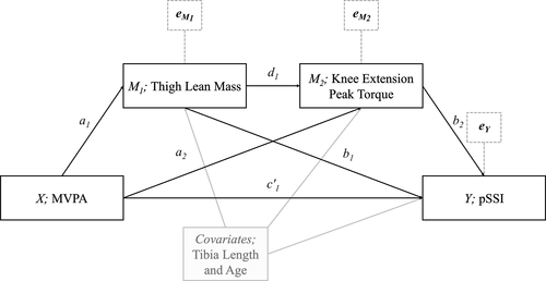
Accordingly, there is a need to characterize the specific muscular pathway(s) through which PA may be indirectly associated with bone status, and to examine whether this differs in men compared to women. Techniques such as path analysis, an aspect of structural equation modeling, extend the capabilities of standard multiple regression to offer a means by which we might explore these potential mechanisms through the quantification of indirect effects; the effect of one variable on another through a prespecified network of intermediary variables.21, 22 Specifically, a “serial mediation” approach to path analysis would build on prior attempts to quantify such relationships via simple mediation,10 whereby only one muscular mediator (mass) could be assessed, allowing for the assessment of a series of potential mediators. A key strength of path analysis is the ability to utilize non-experimental data in mechanistic models; however, results should be interpreted with caution, because inference of causation from such data is inappropriate. In the context of muscular characteristics, this would provide an opportunity to test a priori hypotheses regarding how independent predictors of skeletal outcomes (ie, muscle mass and force) may influence each other in the context of PA. Our hypothesized serial mediation model is depicted in Fig. 1. Consequently, the aims of this study were twofold: (i) to examine via path analysis the indirect association between PA and cortical bone through potential muscular mediators in a predefined, theoretically based series (MVPA → muscle mass → muscle force → bone), and (ii) to explore potential sex differences in these pathways. Combined and sex-specific serial mediation models included measures of thigh fat-free soft tissue (FFST) mass and knee extensor peak torque as aspects of muscular mechanical force.
Subjects and Methods
Recruitment and participant characteristics
Participants included within this cross-sectional analysis were recruited via email, posted flyer, and word-of-mouth advertising from fall 2016 to spring 2017, as described.16 Briefly, participants (n = 147; n = 77 female) were aged 18 to 21 years to ensure that most had achieved the majority of their longitudinal growth and skeletal maturation, minimizing the effect of maturational differences on cortical outcomes.23, 24 Inclusion criteria also dictated that participants were (i) free of orthopaedic limitations that precluded participation in exercise and PA; (ii) not current smokers (past 6 months); (iii) not pregnant or planning to become pregnant for the duration of their participation, and had not given birth in the last 12 months; (iv) not taking medications known to affect bone metabolism (ie, glucocorticoids), habitual dietary intake, or PA; (v) free of any medical conditions know to affect bone metabolism (ie, Crohn's disease); (vi) not currently diagnosed with an eating disorder; and (vii) had not undergone recent weight-loss surgery (bariatric or gastric bypass). All participants provided written informed consent prior to participation. The Institutional Review Board of the University of Georgia approved all aspects of the protocol.
Anthropometric measures
Standing height was measured by a stadiometer (Novel Products Inc., Rockton, IL, USA) to the nearest 0.1 cm. Body mass was measured with a digital scale (Seca Bella 840; Seca, Columbia, MD, USA) to the nearest 0.1 kg. Tibia length was measured with a sliding anthropometer to the nearest 0.1 cm as the distance from the distal edge of the medial malleolus to the tibial plateau.
Physical activity
Objectively measured PA was assessed using a tri-axial ActiGraph GT3X+ accelerometer (Firmware v3.2.1; Actigraph, LLC, Pensacola, FL, USA), which collected data at 30 Hz. Each participant was asked to wear the accelerometer on the mid axillary line of his/her right hip during all waking hours for a ≥7-day period, via an elastic band around the waist, except during water-based activities (eg, showering and swimming). Data were included only if the participant had accumulated a minimum of 10 hours/day of wear determined using the macro developed by the National Cancer Institute (CDC/National Center for Health Statistics, Bethesda, MD, USA) from the Troiano algorithm,25 for at least 3 days including 1 weekend day. Periods with consecutive raw activity values of zero (with a 2-min spike tolerance of ≤100 counts) for 60 min or longer were interpreted as “non-wear” and excluded from this analysis.26 Wear-time analysis and data scoring was performed using Actilife software (v6.10.1; Actigraph, LLC, Pensacola, FL, USA). The Freedson Adult VM3 cut points were used to classify PA intensity across 15-s epochs, specifically, 0 to 2690 for light, 2691 to 6166 for moderate, ≥6167 for vigorous, and ≥2691 for MVPA.27 A weighted average PA time [(weekday average × 5) + (weekend average × 2) /7] was used to represent mean weekly activity variables. MVPA was adjusted for wear time by regressing MVPA on wear time and using the resulting residuals within analyses.28
Bone assessments
Bone strength was assessed via peripheral quantitative computed tomography (pQCT; Stratec XCT-3000; Stratec Medizintechnic GmbH, Pforzheim, Germany). A scan of the nondominant tibia was performed with a 0.4-mm voxel at a slice thickness of 2.4 mm at the 38% site relative to the total leg length from the distal metaphysis. An automatic scout view positioned the cross-sectional measurement using the participant's medial endplate as an anatomic marker. Image processing and calculation of the various bone indices was performed using the Stratec software (version 6.20). The following parameters were assessed at the tibia 38% site: cortical volumetric bone mineral density (Ct.vBMD; mg/mm3), cortical area (Ct.Ar; mm2), cortical thickness (Ct.Th; mm), periosteal circumference (PC; mm), endosteal circumference (EC; mm), and polar strength-strain index (pSSI, mm3). Cortical bone parameters were assessed using Cortmode 2 and the default threshold of 710 mg/cm3, except for pSSI, which utilized a threshold of 480 mg/cm3. All pQCT measures were performed and analyzed by one operator who was trained for acquisition and analysis following guidelines provided by Bone Diagnostic (Spring Branch, TX, USA). The manufacturer-supplied phantom was scanned daily to maintain quality assurance (Stratec Medizintechnik GmbH, Pforzheim, Germany).
Muscular assessment
Thigh FFST mass (g) was assessed via a dual-energy X-ray absorptiometry (DXA; Discovery A; Hologic Inc., Marlborough, MA, USA) total body scan with a region of interest that passed proximally through the femoral neck and distally across the mid-line of the knee. One trained operator acquired and analyzed all scans and the manufacturer-supplied phantom was scanned daily to maintain quality assurance (Hologic Inc.). The short-term reliability of musculoskeletal and anthropometric measures in our laboratory has been reported,29 based on repeat measures on 10 volunteers on the same day.
Absolute peak isokinetic torque (N∙m) during knee extension was assessed using a Biodex System Pro 4 isokinetic dynamometer (Biodex Medical Systems, Inc., Shirley, NY, USA) with the participant positioned per manufacturer guidelines. Using the nondominant leg, five maximal effort voluntary knee extensions were performed at 60 degrees/s. Relative peak torque (N∙m/g) was then calculated by dividing absolute peak torque by thigh FFST mass. Peak torque during knee extension assessed via isokinetic dynamometry has been found to be highly reliable in young adults,30 and was chosen as the major force outcome because it reflects a muscle-bone unit with direct force transfer onto the tibia.
Statistical analysis
Statistical analyses were performed using SPSS for Windows (SPSS 22.0; IBM Corp., Armonk, NY, USA) with significance set at an α level of p <0.05. The normal distribution of residuals, linearity, and homogeneity of variance were examined across all combinations of outcome variables. Multicollinearity between independent variables was also assessed via variance inflation factor (VIF), with a VIF of <10 indicating the absence of collinearity31; no variables met this criteria. Data were also screened for influential multivariate outliers based on Mahalonobis distance critical values for a chi square distribution with k degrees of freedom (k = number of predictors in the model); no outliers were present. Sex-specific means and standard deviations (mean ± SD) were calculated for all participant characteristics and primary outcome variables, and independent t tests then identified any differences between sexes; see Table 1. Partial correlations were calculated to assess the strength of relationships between variables of interest while holding constant the confounding variables, tibia length and age; see Table 2. Tibia length and age were chosen based on prior research describing the importance of bone length as a covariate in models assessing the functional muscle-bone unit14 and to control for developmental differences among participants in the context of bone mineral accrual, respectively. Moreover, both variables exhibited significant bivariate correlations with various cortical and muscular outcomes (data not shown).
| Males (n = 70) | Females (n = 77) | p | |
|---|---|---|---|
| Age (years) | 19.7 ± 0.7 | 19.7 ± 0.7 | 0.734 |
| Body mass index (kg/m2) | 23.6 ± 2.9 | 22.8 ± 3.7 | 0.162 |
| Tibia length (cm) | 39.5 ± 2.3 | 36.8 ± 2.1 | <0.001 |
| Thigh FFST (g) | 3218.0 ± 454.0 | 2398.0 ± 327.8 | <0.001 |
| Knee extension peak torque (N∙m) | 161.5 ± 34.1 | 104.0 ± 21.6 | <0.001 |
| Relative knee extension peak torque (N-m/g) | 0.050 ± 0.008 | 0.044 ± 0.008 | <0.001 |
| Accelerometer wear time (hours/day) | 14.4 ± 1.0 | 14.1 ± 1.1 | 0.112 |
| MVPA (min/day) | 92.8 ± 26.7 | 85.8 ± 27.5 | 0.121 |
| Moderate (% of MVPA) | 84.0 ± 9.9 | 88.3 ± 6.9 | 0.003 |
| Vigorous (% of MVPA) | 16.0 ± 9.9 | 11.7 ± 6.9 | 0.001 |
| pQCT - 38% tibia bone measures | |||
| Ct.vBMD (mg/cm3) | 1160.1 ± 21.4 | 1186.0 ± 20.8 | <0.001 |
| Ct.Ar (mm2) | 345.6 ± 52.3 | 270.8 ± 35.7 | <0.001 |
| Ct.Th (mm) | 6.3 ± 0.8 | 5.5 ± 0.6 | <0.001 |
| PC (mm) | 74.7 ± 4.9 | 66.5 ± 4.0 | <0.001 |
| EC (mm) | 35.2 ± 4.3 | 32.0 ± 4.1 | <0.001 |
| pSSI (mm3) | 2035.8 ± 384.4 | 1471.3 ± 247.7 | <0.001 |
- Values are mean ± SD. Bold values are significant at p < 0.05.
- FFST = fat-free soft tissue; MVPA = moderate-to-vigorous intensity physical activity; Ct.vBMD = cortical volumetric bone mineral density; Ct.Ar = cortical area; Ct.Th = cortical thickness; PC = periosteal circumference; EC = endosteal circumference; pSSI = polar stress strain index.
| Variable | 1 | 2 | 3 | 4 | 5 | 6 | 7 | 8 | 9 | 10 | 11 | |
|---|---|---|---|---|---|---|---|---|---|---|---|---|
| 1 | BMI (kg/m2) | – | 0.38b | 0.49a | 0.25c | −0.01 | −0.21 | 0.49a | 0.35b | 0.51a | 0.13 | 0.51a |
| 2 | Thigh FFST (g) | 0.29c | – | 0.50a | −0.20 | 0.14 | −0.34b | 0.69a | 0.59a | 0.63a | −0.01 | 0.63a |
| 3 | Knee extension peak torque (N∙m) | 0.33b | 0.29c | – | 0.74a | 0.21 | −0.25c | 0.56a | 0.44a | 0.56a | 0.08 | 0.55a |
| 4 | Relative knee extension peak torque (N∙m/g) | 0.14 | −0.39b | 0.76a | – | 0.12 | −0.03 | 0.11 | 0.05 | 0.16 | 0.10 | 0.15 |
| 5 | MVPA (min/day) | 0.14 | 0.36b | 0.25c | 0.01 | – | 0.03 | 0.04 | 0.06 | 0.01 | −0.05 | 0.01 |
| 6 | Ct.vBMD (mg/cm3) | −0.11 | −0.02 | −0.11 | −0.08 | −0.22 | – | −0.35b | −0.15 | −0.50a | −0.33b | −0.37b |
| 7 | Ct.Ar (mm2) | 0.58a | 0.54a | 0.42a | 0.06 | 0.32b | −0.13 | – | 0.88a | 0.87a | −0.09 | 0.91a |
| 8 | Ct.Th (mm) | 0.45a | 0.53a | 0.43a | 0.09 | 0.36b | 0.05 | 0.89a | – | 0.54a | −0.55a | 0.61a |
| 9 | PC (mm) | 0.56a | 0.40a | 0.32b | 0.04 | 0.22 | −0.31b | 0.86a | 0.53a | – | 0.41b | 0.97a |
| 10 | EC (mm) | 0.06 | −0.18 | −0.16 | −0.05 | −0.18 | −0.36b | −0.12 | −0.56a | 0.41a | – | 0.30c |
| 11 | pSSI (mm3) | 0.58a | 0.45a | 0.31b | 0.00 | 0.17 | −0.17 | 0.91a | 0.63a | 0.97a | 0.26c | – |
- Values are r. Above the diagonal = male; below the diagonal = female. Bold values are significant at p < 0.05 or stronger. Tibia length (cm) and age (years) were used as covariates in all partial correlations.
- BMI = body mass index; FFST = fat-free soft tissue; MVPA = moderate-to-vigorous intensity physical activity; Ct.vBMD = cortical volumetric bone mineral density; Ct.Ar = cortical area; Ct.Th = cortical thickness; PC = periosteal circumference; EC = endosteal circumference; pSSI = polar stress strain index.
- a p < 0.001.
- b p < 0.01.
- c p < 0.05.
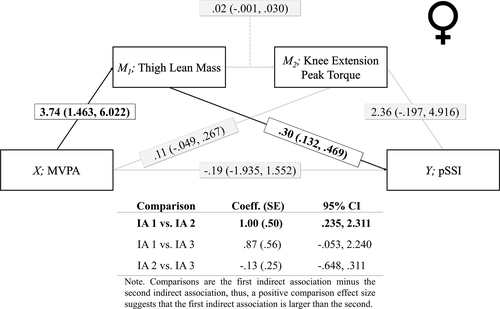
Serial mediation analysis was performed via model 6 of the PROCESS macro for SPSS, provided by Hayes32 (Figs. 1-4). This model determines whether the relationship between an antecedent variable (X) and a consequent variable (Y), is mediated by multiple intermediary variables (M 1, M 2, …, M k) in a predefined series. Assumptions of mediation are similar to that of multiple regression, including linearity, homoscedasticity, normality of estimation error, and independence of observations.33 The results of a mediation analysis are typically termed direct and indirect effects, with the term “effect” and the a priori sequencing of variables having causal inference. However, it is important to note that results from path analysis using non-experimental data should be interpreted with caution, because causal inference is not appropriate for observational data. Thus, throughout the remaining sections of this work we use the terms “association” and “relationship” in place of “effect” to avoid inadvertent implication of causation. Using bootstrapping, 10,000 random samples were taken with replacement to construct 95% bias-corrected bootstrap confidence intervals (CIs), which were reported in place of p values, with a CI entirely above or below zero highlighting a significant relationship. All models controlled for tibia length and age; however, sensitivity analyses consisting of less parsimonious, combined-sex models including body mass index and total percent fat as additional covariates were also performed (results not presented), yielding similar results to our primary analyses. Moreover, though dietary factors such as calcium and vitamin D3 are known correlates of bone mineral density,34 they were not included in the current analysis because of a lack of relationship with cortical outcomes in this sample, as reported.16 As is depicted in Fig. 1, the serial mediation model tested the hypothesis that the sex-specific relationship between MVPA and cortical bone status is mediated by thigh FFST mass and absolute knee extension peak torque in series; ie, the relationship between MVPA and bone status is indirectly carried first through thigh FFST mass and then through absolute knee extension peak torque. Although relationships between muscle size and force capacity are complex, likely influenced by sex-related and training status-related factors,35 in absolute terms, an increase or decrease in muscle cross-sectional area (CSA) typically leads to an increase or decrease in peak force production, respectively.36 Thus, our choice of a serial mediation model assumes that muscle mass is a contributory causal factor in muscle force gains.35, 36 The model assessed three indirect associations: indirect association 1, the association between X (MVPA) and Y (cortical outcomes), indirectly via M1 (thigh FFST mass); indirect association 2, X and Y, indirectly via M1 and M2 (knee extension peak torque) in series; and indirect association 3, X and Y, indirectly via M2. The model also assessed the direct association between X and Y while controlling for all mediators and covariates included in the model.
Results
Recruitment and sample characteristics
A total of (n = 452) individuals completed the online screening survey between September 2016 and February 2017, of which (n = 108) were deemed ineligible based on inclusion/exclusion criteria, as described.16 Potential participants (n = 344) were contacted via email, of which (n = 184) were scheduled for testing. Prior to testing or between sessions (n = 24) participants withdrew from the study without reason, with a further (n = 6) participants data excluded due to missing data in major outcomes, leaving a total useable sample of (n = 154). Due to the known moderating effect of race on bone outcomes, only white, non-Hispanic, or Latino participants were included, producing a final analyzed sample of (n = 147).
Preliminary analysis – descriptives
Because the main analysis examined sex-specific serial mediation, participant characteristics are presented separately for males and females in Table 1. Measures of body size, including tibia length and thigh FFST mass, were significantly greater in males than females (7.3% and 34.2%, respectively; both p < 0.001). Muscle torque was also 55.3% greater in males than females (p < 0.001). When expressed relative to thigh FFST mass, males had 13.6% greater force output per gram than females (p < 0.001). MVPA did not differ between males and females (p > 0.05); however, females tended to accrue more of their MVPA as moderate-intensity activity than males (p = 0.010), whereas males tended to accrue more as vigorous-intensity activity (p = 0.002). Bone geometric properties and estimated strength including cortical area, cortical thickness, periosteal circumference, and pSSI were greater in males than females (27.6%, 14.5%, 12.3%, and 38.4%, respectively; all p < 0.001). In contrast, females had smaller, more dense bones with a smaller medullary cavity, as reflected by a 2.2% greater Ct.vBMD, and 10.0% smaller endosteal circumferences, coupled with the already identified smaller periosteal circumference (all p < 0.001).
Preliminary analysis – partial correlations
Partial correlations controlling for tibia length and age are shown in Table 2. MVPA was positively related to thigh FFST mass (r = 0.36, p < 0.01) and absolute knee extension peak torque (r = 0.25, p < 0.05), but not relative knee extension peak torque in females (p > 0.05). However, in males MVPA was not related to any measure of muscle force or mass (all p > 0.05). MVPA was positively related to Ct.Ar and Ct.Th in females (r = 0.32 and r = 0.36, respectively; both p < 0.01), but was not related to any other cortical outcome. Conversely, MVPA was not related to any cortical outcomes in males (all p < 0.05). Both thigh FFST mass and absolute knee extension peak torque were positively related to cortical outcomes of Ct.vBMD, Ct.Ar, Ct.Th, PC, and pSSI in males and Ct.Ar, Ct.Th, PC, and pSSI in females, with relationships in males appearing stronger for all cortical outcomes (male versus female: r range, 0.25 to 0.69 versus r range, 0.28 to 0.49; all p < 0.01).
Serial mediation analysis
Traditional mediation analysis such as Barron and Kenney's37 causal step approach requires all individual paths within a mediation analysis to be significant. However, Hayes32 and Mackinnon and Fairchild38 suggest that this reasoning should be abandoned because the indirect associations assessed in mediation and serial mediation analyses are a product of multiple paths and an inferential test for an indirect association should be predicated on the product rather than on the testing of individual paths. Moreover, Kenny and Judd39 argue that the standard test of the association between X on Y prior to running a mediation analysis is typically done with less power than are tests of indirect associations of equal size, making them less trustworthy as a deciding factor for future analyses. Thus, despite the apparent lack of relationship between some outcomes, the primary analysis continued as planned because of reasons listed above and the theoretical basis of muscle's role in transferring force to bone during skeletal loading.
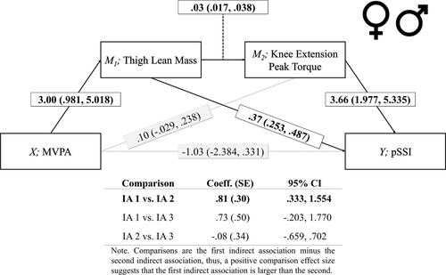
Results of the serial mediational analysis can be found in Tables 3 to 5, with individual pathways and comparisons between indirect associations depicted in Figs. 2 to 4. In combined-sex models (Table 3; Fig. 2), when controlling for age and tibia length, MVPA was positively associated with cortical outcomes through two indirect pathways: (i) thigh FFST mass (pSSI; b = 1.11 ± 0.37; 95% CI, 0.47 to 1.93), and (ii) thigh FFST mass and knee extension peak torque in series (pSSI; b = 0.30 ± 0.16; 95% CI, 0.09 to 0.73), in both cases significant relationships were seen with cortical structural outcomes Ct.Ar, Ct.Th, PC, and pSSI, but not with Ct.vBMD. The indirect association through thigh FFST mass solely was significantly stronger than the indirect association through both thigh FFST mass and knee extension peak torque in series (pSSI; Comparison coefficient = 0.81 ± 0.30; 95% CI, 0.33 to 1.55). Tests for direct associations were nonsignificant, suggesting that there was no relationship between MVPA and cortical bone when controlling for thigh FFST mass and knee extension peak torque.
| Combined sample | ||||||
|---|---|---|---|---|---|---|
| Variable | Ct.vBMD (mg/cm3) | Ct.Ar (mm2) | Ct.Th (mm) | PC (mm) | EC (mm) | pSSI (mm3) |
| Indirect association 1 | −0.023 ± 0.018 | 0.180 ± 0.059a | 0.003 ± 0.001a | 0.014 ± 0.005a | −0.003 ± 0.004 | 1.109 ± 0.373a |
| (X→M1→Y) | (−0.068 to 0.006) | (0.072–0.306) | (0.001–0.005) | (0.006–0.025) | (−0.012 to 0.004) | (0.466–1.930) |
| Indirect association 2 | −0.008 ± 0.009 | 0.045 ± 0.021a | 0.001 ± 0.000a | 0.004 ± 0.002a | 0.000 ± 0.002 | 0.300 ± 0.157a |
| (X→M1→M2→Y) | (−0.033 to 0.005) | (0.014–0.101) | (0.000–0.001) | (0.001–0.010) | (−0.003 to 0.004) | (0.091–0.730) |
| Indirect association 3 | −0.010 ± 0.012 | 0.057 ± 0.039 | 0.001 ± 0.001 | 0.005 ± 0.003 | 0.000 ± 0.002 | 0.381 ± 0.257 |
| (X→M2→Y) | (−0.047 to 0.005) | (−0.017 to 0.140) | (−0.000 to 0.002) | (−0.001 to 0.012) | (−0.003 to 0.006) | (−0.083 to 0.944) |
| Direct association | −0.030 ± 0.062 | −0.036 ± 0.094 | 0.001 ± 0.002 | −0.007 ± 0.010 | −0.014 ± 0.012 | −1.026 ± 0.687 |
| (−0.153 to 0.093) | (−0.222 to 0.151) | (−0.002 to 0.005) | (−0.027 to 0.012) | (−0.039 to 0.010) | (−2.384 to 0.331) | |
- Values are unstandardized regression coefficients ± bootstrap standard error (95% CI). Tibia length (cm), age (years), and sex were used as covariates. Bold values are significant associations based on 95% CI.
- Indirect association 1 = MVPA, thigh FFST, and diaphyseal cortical bone; Indirect association 2 = MVPA, thigh FFST, knee extension peak torque, and diaphyseal cortical bone; Indirect association 3 = MVPA, knee extension peak torque, and diaphyseal cortical bone; FFST = fat-free soft tissue; Ct.vBMD = cortical volumetric bone mineral density; Ct.Ar = cortical area; Ct.Th = cortical thickness; PC = periosteal circumference; EC = endosteal circumference; pSSI = polar stress strain index.
- a Significant difference between indirect associations 1 and 2, p < 0.05.
| Males | ||||||
|---|---|---|---|---|---|---|
| Variable | Ct.vBMD (mg.cm3) | Ct.Ar (mm2) | Ct.Th (mm) | PC (mm) | EC (mm) | pSSI (mm3) |
| Indirect association 1 | −0.022 ± 0.024 | 0.119 ± 0.113 | 0.002 ± 0.002 | 0.009 ± 0.009 | −0.001 ± 0.004 | 0.740 ± 0.705 |
| (X→M1→Y) | (−0.093 to 0.013) | (−0.102 to 0.347) | (−0.001 to 0.005) | (−0.008 to 0.028) | (−0.013 to 0.004) | (−0.631 to 2.227) |
| Indirect association 2 | −0.003 ± 0.010 | 0.026 ± 0.031 | 0.000 ± 0.000 | 0.003 ± 0.003 | 0.001 ± 0.002 | 0.203 ± 0.247 |
| (X→M1→M2→Y) | (−0.044 to 0.006) | (−0.015 to 0.115) | (−0.000 to 0.002) | (−0.002 to 0.012) | (−0.001 to 0.008) | (−0.133 to 0.892) |
| Indirect association 3 | −0.013 ± 0.026 | 0.100 ± 0.064 | 0.001 ± 0.001 | 0.010 ± 0.006 | 0.003 ± 0.004 | 0.775 ± 0.481 |
| (X→M2→Y) | (−0.088 to 0.025) | (0.001–0.261) | (0.000–0.004) | (0.001–0.025) | (−0.003 to 0.015) | (0.040–2.024) |
| Direct association | 0.076 ± 0.087 | −0.190 ± 0.146 | −0.002 ± 0.003 | −0.019 ± 0.014 | −0.008 ± 0.019 | −1.638 ± 1.100 |
| (−0.112 to 0.237) | (−0.481 to 0.100) | (−0.007 to 0.004) | (−0.047 to 0.009) | (−0.046 to 0.031) | (−3.835 to 0.559) | |
- Values are unstandardized regression coefficients ± bootstrap standard error (95% CI). Tibia length (cm) and age (years) were used as covariates. Bold values are significant associations based on 95%CI.
- Indirect association 1 = MVPA, thigh FFST, and diaphyseal cortical bone; Indirect association 2 = MVPA, thigh FFST, knee extension peak torque, and diaphyseal cortical bone; Indirect association 3 = MVPA, knee extension peak torque, and diaphyseal cortical bone; FFST = fat-free soft tissue; Ct.vBMD = cortical volumetric bone mineral density; Ct.Ar = cortical area; Ct.Th = cortical thickness; PC = periosteal circumference; EC = endosteal circumference; pSSI = polar stress strain index.
| Females | ||||||
|---|---|---|---|---|---|---|
| Variable | Ct.vBMD (mg.cm3) | Ct.Ar (mm2) | Ct.Th (mm) | PC (mm) | EC (mm) | pSSI (mm3) |
| Indirect association 1 | 0.022 ± .041 | 0.185 ± 0.076a | 0.005 ± 0.001a | 0.014 ± 0.007a | −0.005 ± 0.006 | 1.123 ± 0.497a |
| (X→M1→Y) | (−0.046 to 0.119) | (0.069–0.389) | (0.001–0.006) | (0.003–0.033) | (−0.020 to 0.006) | (0.371–2.457) |
| Indirect association 2 | −0.004 ± 0.010 | 0.027 ± 0.019a | 0.003 ± 0.000a | 0.002 ± 0.002a | −0.001 ± 0.002 | 0.128 ± 0.107a |
| (X→M1→M2→Y) | (−0.036 to 0.008) | (0.005–0.090) | (0.000–0.002) | (0.000–0.009) | (−0.007 to 0.002) | (0.016–0.511) |
| Indirect association 3 | −0.009 ± 0.019 | 0.055 ± 0.043 | 0.001 ± 0.001 | 0.004 ± 0.003 | −0.002 ± 0.005 | 0.258 ± 0.208 |
| (X→M2→Y) | (−0.069 to 0.015) | (−0.014 to 0.164) | (−0.000 to 0.004) | (−0.001 to 0.014) | (−0.017 to 0.003) | (−0.049 to 0.790) |
| Direct association | −0.156 ± 0.086 | 0.120 ± 0.124 | 0.003 ± 0.002 | 0.006 ± 0.014 | −0.014 ± 0.016 | −0.191 ± 0.874 |
| (−0.326 to 0.015) | (−0.128 to 0.368) | (−0.001 to 0.008) | (−0.022 to 0.035) | (−0.046 to 0.018) | (−1.935 to 1.552) | |
- Values are unstandardized regression coefficients ± bootstrap standard error (95% CI). Tibia length (cm) and age (years) were used as covariates. Bold values are significant associations based on 95% CI.
- Indirect association 1 = MVPA, thigh FFST, and diaphyseal cortical bone; Indirect association 2 = MVPA, thigh FFST, knee extension peak torque, and diaphyseal cortical bone; Indirect association 3 = MVPA, knee extension peak torque, and diaphyseal cortical bone; FFST = fat-free soft tissue; Ct.vBMD = cortical volumetric bone mineral density; Ct.Ar = cortical area; Ct.Th = cortical thickness; PC = periosteal circumference; EC = endosteal circumference; pSSI = polar stress strain index.
- a Significant difference between indirect associations 1 and 2, p < 0.05.
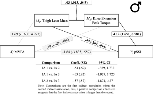
When mediation pathways were assessed in males only (Table 4; Fig. 3), MVPA was indirectly associated with Ct.Ar, Ct.Th, PC, and pSSI, through knee extension peak torque (pSSI; b = 0.78 ± 0.48; 95% CI, 0.04 to 2.02). However, relationships were not mediated through any other pathway. In contrast, the indirect association between MVPA and Ct.Ar, Ct.Th, PC, and pSSI in females (Table 5; Fig. 4) existed through thigh FFST mass (pSSI; b = 1.12 ± 0.50; 95% CI, 0.37 to 2.46) with no mediation through knee extension peak torque. The serial mediation pathway through both muscular outcomes was also significant for Ct.Ar, Ct.Th, PC, and pSSI (pSSI; b = 0.13 ± 0.11; 95% CI, 0.02 to 0.51); however, when these relationships were compared, the serial indirect association through both mediators was significantly smaller than the indirect association through thigh FFST mass alone (pSSI; Comparison coefficient = 1.00 ± 0.50; 95% CI, 0.24 to 2.31). Similar to combined-sex models, no statistically significant direct associations were seen between MVPA and any skeletal outcome in either males or females.
Discussion
The aims of this study were twofold: (i) to examine via path analysis the indirect association between PA and cortical bone through potential muscular mediators in a predefined, theoretically based series (MVPA → muscle mass → muscle force → bone), and (ii) to explore potential sex differences in these pathways. We found that in combined-sex models, MVPA was positively associated with cortical strength and structural outcomes through two indirect pathways: (i) thigh FFST mass solely, and (ii) thigh FFST mass and knee extensor peak torque in series, with the former being significantly stronger than the latter. When sex-specific models were applied, the pathway through which the indirect association between MVPA and cortical strength and structure was transmitted differed between males and females. Specifically, a significant indirect association through solely knee extensor peak torque was seen in males, whereas indirect association through solely thigh FFST mass was seen in females. In both combined and sex-specific instances, there was no direct association between MVPA and skeletal outcomes when controlling for these muscular pathways. Contextually, this suggests that in our sample, without the relationship between MVPA and muscle (males: torque versus females: mass) and the subsequent relationship between muscle and bone, there would be no relationship between MVPA and skeletal outcomes. Moreover, that our path analysis exhibits potential sex-specific relationships suggest that the muscle-bone unit may function differently between males and females in the context of habitual PA during young adulthood. Whether this difference could be attributed to the characteristics of the activities performed or the specific biochemical and/or mechanical environment is unclear and confirmation in non-observational datasets is warranted prior to inference of any causal mechanism.
In a review of the effects of PA on bone strength in children and adolescents, muscle emerged as a potential mediator2; when muscular variables such as FFST mass or muscular power were added to regression models, the association between PA and bone was often reduced. However, the mediating role of muscle had not been formally tested until a recent analysis of longitudinal data from the Iowa Bone Development Study cohort.10 Zymbal and colleagues10 assessed whether the relationship between PA trajectories throughout youth and proximal femur areal bone mineral density (aBMD) and geometry was mediated through leg FFST mass. Similar to the findings of the current study, leg FFST mass appeared to mediate the relationship between MVPA and aBMD at the proximal femur in both males and females, despite an absence of direct associations between MVPA and aBMD or geometric outcomes in females. Moreover, and importantly, the mediating role of muscle differed by sex with leg FFST mass explaining 43% to 49% of the relationship between MVPA and aBMD in females but only 27% to 32% in males. Similar findings with regard to proximal femur geometry were seen in the indirect association between MVPA and hip axis length via muscle in both males (b = 0.08 ± 0.04; 95% CI, 0.02 to 0.17) and females (b = 0.10 ± 0.04; 95% CI, 0.05 to 0.18), and on the narrow neck width in males (b = 0.02 ± 0.01; 95% CI, 0.00 to 0.05). Our data agree with these longitudinal findings, suggesting a sex-specific mediating role of thigh FFST mass in the relationship between MVPA and skeletal outcomes in females. Although the lack of a direct association seen in the female sample of Zymbal and colleagues10 suggest that though thigh FFST may mediate the relationship between MVPA and skeletal outcomes, other pathways likely also account for some of the variance in this relationship. It should be noted, however, that Zymbal and colleagues10 utilized DXA-derived proximal femur aBMD and hip geometry, so our lower-extremity volumetric measures cannot be directly compared to their areal measures. Thus, future research should aim to confirm the potential mediating roles of muscle force, in males, and muscle mass, in females, on the relationship between PA and bone, exploring the mechanisms through which these independent muscular pathways may exist.
Potential sexual dimorphisms in the muscle-bone relationship, in the context of PA have been suggested40; however, mechanisms beyond quantity of muscle mass have not been explored. Our serial mediation model attempted to address this gap by including a measure of muscular function as well as mass and by assessing sex-specific pathways. These mediators begin to highlight the potential combined contribution of the mechanical and the hormonal muscular environment, which may be key to understanding sex-specific adaptation to PA.19, 20 For example, traditional discourse has highlighted muscle's role to be the structure that applies mechanical forces to bone, primarily during dynamic loading, but also to maintain postural stability. Specifically, the deformation of bone tissue due to muscle forces at attachment sites generates hydrostatic pressure gradients within the lacuna-canalicular network,8, 9 stimulating periosteal and trabecular apposition via osteocyte perturbation. The degree to which bone formation occurs as a result of dynamic loading is directly related to the customary strain stimulus (ie, load characteristics such as peak strain, loading frequency, and number of loading cycles).41 Thus, PA characteristics are key to determining the degree of bone formation modeling through direct stimulation of osteogenic cells. Furthermore, global and localized hormonal environments may also play a key role in modulating sex-specific mechanosensitivity through factors such as the concentration of estrogen receptor-α, which is dependent on estrogen concentrations,42, 43 or concentrations of anabolic hormones such as growth hormone (GH) and insulin-like growth factor-1 (IGF-1).18, 44 Though our serial mediation model begins to address these potential mechanisms, we did not measure hormones or specific loading rates so are only able to speculate on the relative importance of mechanical versus biochemical mechanisms by which muscular factors may mediate the relationship between PA and bone status, an important area for future research.
With regard to the mechanical stimulation of bone by muscle during PA, it is plausible that differences in PA characteristics may account for the sex-specific indirect associations seen in our data, specifically, the types of activity performed by men and women. The type of activity is not captured by accelerometry, though a comparison of the proportions of time spent in moderate or vigorous PA between sexes in our data and those of others45 may hint toward such activity choices. For example, males performed a greater proportion of their MVPA as vigorous activity, which would induce greater mechanical strains using a larger percentage of their peak muscular torque. Moreover, vigorous muscular activities such as jumping have been shown to apply greater strain to the mid-femoral neck than lower intensity activities, through greater activation of the gluteal muscles.46 These data support our findings of the importance of peak muscle force in males and those of Zymbal and colleagues,10 who reported that leg FFST mediated the relationship between PA and proximal femur narrow neck width in males but not females. Seemingly, vigorous PA requiring a high degree of muscular force may play a key role in the promotion of bone accrual in young adults, especially in males; however, we do not have the data to fully test this hypothesis.
Although our data are novel and of interest, several limitations must be discussed. Primarily, our use of cross-sectional data limit the inference of causality from our hypothesized path model. Our models highlight potential relationships inherent to the functional-muscle bone unit in young adults that should be interpreted cautiously until confirmed through experimental research that removes the potential influence of confounding. Though our data are tightly controlled with regard to chronological age, we did not measure maturational status or history in our sample. Because the timing of pubertal onset is inversely related with peak bone mass and may also affect bone geometric properties during young adulthood,47, 48 future research should aim to clarify whether interactions exist with regard to PA and the muscle-bone unit, especially in females where menarche-related increases in estrogen may alter patterns of mineralization following PA (ie, endosteal versus periosteal).18, 49 Excess adiposity may also affect the muscle-bone unit in the context of adaptation to habitual PA.50, 51 Results of our sensitivity analyses suggested that further controlling for relative adiposity did not alter the outcomes of our models; however, participants in the current study were healthy and not obese. Moreover, although the narrow age-range of this healthy white non-Hispanic or Latino sample is a strength of this analysis by reducing variance and potential for confounding, this also means that our data are not generalizable to other populations. In addition, it is possible that such a highly active sample of college students meant that we were unable to distinguish the relationship between lower levels of MVPA and bone status, or the mediation of this relationship through muscle characteristics, giving an incomplete picture of the role of PA in this age group. With regard to the measurement of skeletal loading, waist-worn accelerometers do not distinguish between activities of a high and low osteogenic value, nor do they measure all activities that might be beneficial to bone status (ie, resistance training), which may have led to some PA being misclassified. Similarly, objectively measured MVPA during this transitional college period may not be indicative of typical activity accrued during childhood and adolescence that predominantly drives PA-related bone accrual, as evidenced by average MVPA being substantially higher than expected across sexes. Future research should therefore strive to implement more specific measures of osteogenic PA that assess impact-based activities over a longer period of time, giving a more accurate view of the habitual loading likely to be associated with greater bone accrual.
Conclusion
Skeletal loading during habitual MVPA is indirectly associated with improvements in cortical structure in male and female young adults; importantly, this relationship may be mediated by muscle. Specifically, peak muscle force appears to mediate the relationship between PA and cortical structure and strength in males, whereas muscle mass appears to mediate the same relationship in females. Characteristic differences in physical activity (mechanical strain) and/or muscle mass–related factors may contribute; however, the mechanism responsible for these differing pathways is unclear. To our knowledge, this study is the first to assess the relationships between MVPA and the muscle-bone unit using a serial mediation model of both muscle mass and a measure of neuromuscular performance, in a sample of young adults. Future studies should look to confirm the sex-specific mediation of the PA and bone relationship and to do so in a more diverse sample at key periods of bone accrual, such as during childhood and early adolescence, to clarify whether sex-specific testing and training guidelines are warranted.
Disclosures
The authors declare no conflict of interest.
Acknowledgments
Authors' roles: Study design: SH, MDS, EME, and RDL. Study conduct: SH, CMS, and MV. Data collection: SH, CMS, and MV. Data analysis: SH. Data interpretation: SH. Drafting manuscript: SH. Revising manuscript content: SH, MDS, EME, and RDL. Approving final version of manuscript: SH, CMS, MV, MDS, EME, and RDL. SH takes responsibility for the integrity of the data analysis.


