Estradiol and Follicle-Stimulating Hormone as Predictors of Onset of Menopause Transition-Related Bone Loss in Pre- and Perimenopausal Women
ABSTRACT
The menopause transition (MT) may be an opportunity for early intervention to prevent rapid bone loss. To intervene early, we need to be able to prospectively identify pre- and perimenopausal women who are beginning to lose bone. This study examined whether estradiol (E2), or follicle-stimulating hormone (FSH), measured in pre- and perimenopausal women, can predict significant bone loss by the next year. Bone loss was considered significant if bone mineral density (BMD) decline at the lumbar spine (LS) or femoral neck (FN) from a pre- or early perimenopausal baseline to 1 year after the E2 or FSH measurement was greater than the least detectable change. We used data from 1559 participants in the Study of Women's Health Across the Nation and tested E2 and FSH as separate predictors using repeated measures modified Poisson regression. Adjusted for MT stage, age, race/ethnicity, and body mass index, women with lower E2 (and higher FSH) were more likely to lose BMD: At the LS, each halving of E2 and each doubling of FSH were associated with 10% and 39% greater risk of significant bone loss, respectively (p < 0.0001 for each). At the FN, each halving of E2 and each doubling of FSH were associated with 12% (p = 0.01) and 27% (p < 0.001) greater risk of significant bone loss. FSH was more informative than E2 (assessed by the area under the receiver-operator curve) at identifying women who were more versus less likely to begin losing bone, especially at the LS. Prediction was better when hormones were measured in pre- or early perimenopause than in late perimenopause. Tracking within-individual change in either hormone did not predict onset of bone loss better than a single measure. We conclude that measuring FSH in the MT can help prospectively identify women with imminent or ongoing bone loss at the LS. © 2019 American Society for Bone and Mineral Research.
Introduction
The menopause transition (MT) is a period of rapid bone loss that contributes to a woman's risk of osteoporosis and fracture in later life. Bone mineral density (BMD) decline, at rates commonly observed during the MT,1 can be associated with irreversible deterioration in bone microarchitecture2-4 and with increased fracture risk.5-7 Indeed, in some studies, fast BMD decline in midlife is associated with appendicular and vertebral fractures within the first postmenopausal decade.8-10 This suggests that the MT may be an opportune time for early, short-term intervention to prevent rapid BMD decline and reduce the risk of future fracture.11
To intervene before substantial bone loss has occurred, we first need to be able to predict whether a pre- or perimenopausal woman is about to begin losing bone. MT-related BMD decline accelerates approximately 1 year before the final menstrual period (FMP).1 Currently, however, this time point can only be identified retrospectively, ie, after ≥12 months of amenorrhea when the FMP date can be assigned.12 By the time the FMP date can be defined, many women will have already been losing bone for the preceding 2 years. Becasue the rate of BMD decline at the lumbar spine during the MT averages 2.5% per year,1 even a relatively short period of bone loss can be significant. The objective of this study was, therefore, to determine whether markers of ovarian function—estradiol (E2) or follicle-stimulating hormone (FSH)—measured in pre- and perimenopause can help prospectively identify the onset of significant bone loss in advance of substantial BMD decline.
E2 is the major sex steroid hormone in women, and the likely effector estrogen at the estrogen receptor.13, 14 FSH is produced by the anterior pituitary under negative feedback inhibition by estrogen. We considered E2 and FSH as potential predictors of imminent BMD decline because an increase in FSH and decrease in E2 temporally precedes the MT-related acceleration in bone loss.1, 15-17 We thus designed this study to address two questions: 1) Can measuring E2 or FSH during pre- (regular menstrual bleeding), early peri- (less predictable bleeding at least once every 3 months), or late perimenopause (less predictable bleeding at least once every 3 to 12 months) help determine if a woman will have significant decline in BMD (from an earlier baseline) by the next 12 months; and 2) Does tracking within-individual change in E2 or FSH improve this determination?
This study was conducted in the Study of Women's Health Across the Nation (SWAN), a longitudinal cohort study of the MT in a multi-ethnic, community-based cohort of women with annual measurements of E2, FSH, and BMD.
Materials and Methods
SWAN is a multicenter, longitudinal study of the MT in a multiracial/ethnic cohort of ambulatory, community-dwelling women.18 SWAN was initiated in 1996, when participants were aged 42 to 52, and in pre- (no change in menstrual bleeding in the past year) or early perimenopause (less predictable menstrual bleeding at least once every 1 to 3 months in the past year). A total of 3302 SWAN participants were recruited at seven clinical sites: Boston, MA; Chicago, IL; Detroit, MI; Pittsburgh, PA; Los Angeles, CA; Newark, NJ; and Oakland CA. The SWAN Bone Cohort includes 2417 participants from five sites (excluding the Chicago and Newark sites). Among these women, E2, FSH, and BMD were measured at baseline and at each follow-up visit thereafter. Each clinical site obtained IRB approval, and all participants provided written informed consent.
Study sample
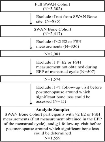
Of 2417 SWAN bone cohort participants, 336 women were excluded because they did not have at least two measurements (at baseline visit and at least one follow-up visit) of E2 or FSH. The most common reason for exclusion was starting a bone-modifying medication (including sex steroid hormones, oral glucocorticoids, aromatase inhibitors, chemotherapy for breast cancer, and osteoporosis medications [bisphosphonates, selective estrogen receptor modulators, calcitonin, parathyroid hormone]) before the second E2 or FSH measurement. Of the remaining participants, another 507 women were excluded because their first E2 or FSH measurement was not obtained during the early follicular phase (days 2 to 5) of the menstrual cycle. We lastly excluded 15 women who did not have at least one follow-up visit before postmenopause (defined as ≥1 year after the FMP), around which we could determine whether significant bone loss occurred. We could not assess for BMD loss if there was missing baseline or follow-up BMD data, or if a bone-modifying medication was initiated before the second dual-energy X-ray absorptiometry (DXA) scan. Our analytic sample was thus 1559 women (Fig. 1). Among these participants, a total of 3618 follow-up visits starting from the first follow-up visit to the last visit before the clinical diagnosis of postmenopause could be made were included in our analyses.

Predictors
Every effort was made to perform phlebotomy before 10:00 a.m. during the early follicular phase (between days 2 and 5) of a spontaneous menstrual cycle. If a follicular phase sample could not be obtained after two attempts, a random fasting sample was taken within a 90-day window of the anniversary of the baseline visit. Collected specimens were initially stored between −20°C and −80°C at individual study sites for up to 30 days and then shipped to the Central Lab at the University of Michigan (Ann Arbor, MI, USA), and stored at −80°C. Assays were then performed in batch mode. Serum E2 was measured in duplicate with a modified, offline ACS:180 (E2–6) immunoassay using an ACS:180 automated analyzer (Bayer Diagnostics Corp., Tarrytown, NY, USA). The average between duplicates was recorded in the data set and used in the analyses in this study. The lower limit of detection was 1.0 pg/mL, and inter- and intraassay coefficients of variation (CV) were 10.6% and 6.4%, respectively. Serum FSH was measured in singlicate with a 2-site chemiluminometric assay (Bayer Diagnostics Corp.). The lower limit of detection was 1.05 mIU/mL, and inter- and intraassay CV were 12.0% and 6.0%, respectively.
Outcomes
BMD at the lumbar spine (LS) and femoral neck (FN) BMD was measured by DXA. At study inception, the Pittsburgh and Oakland sites used the Hologic (Waltham, MA, USA) QDR 2000 machine, and the Boston, Los Angeles, and Michigan sites used the Hologic QDR 4500A model. At follow-up visit 8, Pittsburgh and Oakland upgraded to the 4500A models. To develop cross-calibration regression equations, each site obtained duplicate scans using the old and new hardware in 40 volunteers within a maximum of 90 days. Of the 3618 observations included in our analyses, only 56 occurred after the machine changes. To determine the short-term in vivo precision error, each study site measured LS and FN BMD twice in 5 women with complete subject repositioning between duplicate scans. Using the root mean square SD approach, the precision error in SWAN was 1.4% at the LS and 2.2% at the FN. An anthropomorphic spine phantom was circulated between sites for cross-site calibration. Standard quality-control phantom scans were conducted before each BMD measurement session. If necessary, these were used to adjust for longitudinal machine drift.
For each follow-up visit N, we calculated the percentage decline in LS and FN BMD from SWAN baseline to follow-up visit N + 1. Significant BMD decline was defined as loss of BMD that exceeded the site-specific least significant change (LSC). LSC is the amount of change that is considered statistically significant using a two-sided type I error (alpha) of 5%, given the measure's precision error (coefficient of variation [CV]). The LSC (which is 2.8 times the measurement's CV) is thus 3.9% for LS BMD and 6.2% for FN BMD.
Covariates
Body mass index (BMI) was calculated from weight and height measurements (BMI = weight in kilograms/[height in meters]2). Clinical MT stage was determined using menstrual bleeding patterns. Premenopause was defined as no change in menstrual regularity in the past year. Early perimenopause was defined as less predictable menstrual bleeding at least once every 3 months. Late perimenopause was defined as less predictable menstrual bleeding at least once every 3 to 12 months.
Statistical analysis
We generated descriptive statistics for all variables and assessed the distributions of continuous variables. E2 and FSH had skewed distributions and were thus log transformed to base 2 for all analyses.
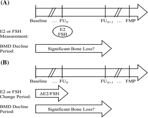
In our first set of analyses, we assessed whether a one-time measurement of E2 or FSH could predict imminent bone loss by the next year. We used repeated measures, modified Poisson regression with E2 or FSH measured at each follow-up visit N as primary predictor, and significant bone loss (yes versus no) at the LS or FN from SWAN baseline to follow-up visit N + 1 (yes/no) as the dependent variable (Fig. 2 A). E2 and FSH were tested in separate models. Models were adjusted for MT stage (pre- versus early peri- versus late perimenopause) and relevant clinical covariates (age, race/ethnicity, BMI, SWAN study site, and whether follow-up E2 or FSH more measured during the early follicular phase of the menstrual cycle).

In our second set of analyses, we examined the ability of within-individual change in E2 or FSH to predict imminent bone loss by the next year of the second hormone measurement. We again used repeated measures, modified Poisson regression, this time with change in log-transformed E2 or FSH from SWAN baseline to each follow-up visit N as primary predictor, and significant bone loss at the LS or FN from SWAN baseline to follow-up visit N + 1 (yes/no) as dependent variable (Fig. 2 B). Models were adjusted for MT stage, and relevant clinical covariates as above. Time-varying covariates were obtained at the time of the second hormone measurement.
In both sets of analyses, we compared the abilities of E2 and FSH to discriminate women who were more likely from those less likely to be losing bone, using the area under the receiver operating characteristic curve (AUC) metric (estimated using logistic regression).19 We also tested for interactions of each hormone with race/ethnicity, MT stage, and whether the hormone was measured during the early follicular phase (EFP, days 2 to 5) of the menstrual cycle to see if the strength of each hormone's association (effect size) differed by those factors.
Lastly we conducted three sets of sensitivity analyses. First, we examined whether excess weight loss or weight gain affected the associations of E2 and FSH with significant bone loss. Specifically, we excluded observations for which change in weight from the baseline visit to the exposure visit was in the bottom 5% or top 5% of the population distribution (ie, weight loss >5.8 kg or weight gain >9.2 kg). Second, the SWAN protocol for cross-calibration after a DXA hardware change did not meet the International Society for Clinical Densitometry's (ISCD's) current recommendation to obtain duplicate scans on old and new machines within 60 days.20 To determine if this affected our findings, we excluded the 56 of 3618 (approximately 1.5%) observations that occurred after the Pittsburgh and Oakland machine upgrades. Third, SWAN's DXA precision estimates did not meet the ISCD's current recommendation to obtain triplicate or duplicate scans in 15 or 30 subjects.20 We thus conducted sensitivity analyses using the ISCD's limit of acceptable LSC thresholds (5.3% at the LS and 6.9% at the FN) as alternative definitions for significant bone loss.
Results
Participant characteristics: study baseline
This study included 1559 SWAN participants. Half were white, 25% black, 11% Chinese, and 14% Japanese. At study baseline, 58% were premenopausal, and 42% were in early perimenopause. Mean BMD values at the LS and FN were 1.071 and 0.837 g/cm2, respectively. E2 and FSH had skewed distributions, with median E2 being 52.5 pg/mL (interquartile range [IQR] 32.8–82.1) and median FSH being 15.1 mIU/mL (IQR 11.1–23.3) (Table 1).
| Descriptive statistic, N = 15591 | |
|---|---|
| Age (years)2 | 46.1 (2.6) |
| Race/ethnicity3 | |
| Black | 383 (25%) |
| Chinese | 171 (11%) |
| Japanese | 222 (14%) |
| White | 783 (50%) |
| Body mass index (kg/m2)2 | 27.2 (7.8) |
| Menopause transition stage3 | |
| Premenopause | 907 (58%) |
| Early perimenopause | 652 (42%) |
| Hormone predictors4 | |
| Estradiol (pg/mL) | 52.5 (32.8, 82.1) |
| Follicle-stimulating hormone (mIU/mL) | 15.1 (11.1, 23.3) |
| Bone mineral density2 | |
| Lumbar spine (g/cm2) | 1.071 (0.1) |
| Femoral neck (g/cm2) | 0.837 (0.1) |
- 1 All participants were pre- or early perimenopausal at SWAN baseline.
- 2 Continuous variables with normal distributions expressed as mean (standard deviation).
- 3 Categorical variables expressed as count (proportion).
- 4 Continuous variables with skewed distributions expressed as median (interquartile range).
Participant characteristics: repeated measures
Among the 1559 participants, a total of 3618 follow-up visits after SWAN baseline and before the first postmenopausal visit (defined as ≥1 year after the FMP) were included in our analyses. Eleven percent of these follow-up visits occurred during premenopause, 75% in early perimenopause, and 14% in late perimenopause. Median E2 was similar during pre- (40.1 pg/mL) and early perimenopause (44.9 pg/mL) but was significantly lower in late perimenopause (21.7 pg/mL) (p < 0.001 for comparison of late perimenopause versus pre- or early perimenopause). Analogously, median FSH was similar in pre- (15.5 mIU/mL) and early perimenopause (18.9 mIU/mL) but was significantly higher in late perimenopause (83.6 mIU/mL) (p < 0.001 for comparison of late perimenopause versus pre- or early perimenopause) (Table 2).
| No. of observations2 | Premenopause n = 399 | Early perimenopause n = 2715 | Late perimenopause n = 504 |
|---|---|---|---|
| Age (years)3 | 48.3 (2.4) | 48.6 (2.9) | 51.8 (2.4) |
| Body mass index (kg/m2)3 | 26.7 (6.6) | 27.2 (6.6) | 28.1 (7.1) |
| Absolute level of hormone level at follow-up visit N4 | |||
| Estradiol (pg/mL) | 40.1 (25.6, 66.5) | 44.9 (26.6, 88.3) | 21.7 (13.7, 55.3) |
| Follicle-stimulating hormone (mIU/mL) | 15.5 (12.1, 24.0) | 18.9 (12.0, 36.0) | 83.6 (50.6, 114.0) |
| Change in hormone level from SWAN baseline to follow-up visit N4 | |||
| Estradiol (pg/mL) | −7.9 (−28.4, +7.8) | −4.9 (−29.5, +27.1) | −18.9 (−61.3, +0.4) |
| Follicle-stimulating hormone (mIU/mL) | +2.8 (−1.2, +9.0) | +4.3 (−1.8, +19.5) | +58.5 (+23.8, +90.5) |
| Annualized change in bone mineral density from SWAN baseline to follow-up visit N+13 | |||
| Lumbar spine (g/cm2*year) | −0.4 (1.4) | −0.9 (1.4) | −1.5 (1.5) |
| Femoral neck (g/cm2*year) | −0.4 (1.3) | −0.7 (1.4) | −1.3 (1.4) |
| Significant bone loss (yes versus no) from SWAN baseline to follow-up visit N+15 | |||
| Lumbar spine | 15 (3.8%) | 312 (11.6%) | 190 (38.4%) |
| Femoral neck | 8 (2.0%) | 121 (4.5%) | 50 (10.1%) |
- 1 All follow-up visits after SWAN baseline for each participant transitioned to postmenopause.
- 2 Number of visits across all participants in each menopause transition stage.
- 3 Continuous variables with normal distributions expressed as mean (standard deviation).
- 4 Continuous variables with skewed distributions expressed as median (interquartile range).
- 5 Categorical variables expressed as count (percentage).
BMD decreased at a higher rate in early perimenopause (0.9% per year [LS]; 0.7% per year [FN]) compared with premenopause (0.4% per year [LS]; 0.4% per year [FN]) (p < 0.001), and in late perimenopause (1.5% per year [LS]; 1.3% per year [FN]) compared with early perimenopause (p < 0.001). As a consequence, the proportion of observations that were associated with significant bone loss was lowest in premenopause and greatest in late perimenopause (Table 2). The risk of imminent bone loss at the LS was 2.1-fold greater in early peri- versus premenopausal women (risk ratio [RR] 2.1, p = 0.008), after accounting for clinical covariates (age, BMI, race/ethnicity, and SWAN study site). Similarly, risk of imminent bone loss at the LS and FN was 2.1-fold greater in late peri- versus early perimenopausal women (risk ratio [RR] 2.1, p < 0.0001).
Single measure of E2 or FSH as predictor of imminent bone loss
In repeated measures modified Poisson regression, after adjusting for MT stage (pre- versus early peri- versus late perimenopause) and clinical covariates (age, BMI [at the time of E2 measurement], race/ethnicity, SWAN study site, and whether E2 was measured during the EFP of the menstrual cycle), lower E2 was associated with greater risk of imminent bone loss at both the LS and FN. With each 50% decrement in E2, risk of significant bone loss was 10% and 12% greater at the LS (p < 0.0001) and FN (p = 0.01), respectively. The ability of E2 (combined with MT stage and clinical covariates) to identify women with imminent bone loss, as assessed by the model AUC, was 0.756 for the LS (compared with 0.752 for MT stage and clinical covariates alone, p = 0.07) and 0.740 for the FN (compared with 0.735 for MT stage and clinical covariates alone, p = 0.01) (Table 3).
| Relative risk (RR) of significant bone loss by the next year (per 50% decrement [halving] of E2, per 100% increment [doubling] of FSH) | ||||||||
|---|---|---|---|---|---|---|---|---|
| Lumbar spine | Femoral neck | |||||||
| RR (95% CI) | p Value2 | AUC | p Value3 | RR (95% CI) | p Value2 | AUC | p Value3 | |
| Single measures | ||||||||
| E2 | 1.10 (1.06, 1.15) | <0.0001 | 0.756 | 0.07 | 1.12 (1.02, 1.23) | 0.01 | 0.740 | 0.1 |
| FSH | 1.39 (1.30, 1.49) | <0.0001 | 0.782 | <0.0001 | 1.27 (1.11, 1.44) | <0.001 | 0.751 | 0.02 |
| Within-individual change | ||||||||
| E2 | 1.09 (1.04, 1.12) | <0.0001 | 0.759 | 0.04 | 1.09 (1.00, 1.19) | 0.05 | 0.745 | 0.1 |
| FSH | 1.17 (1.10, 1.24) | <0.0001 | 0.757 | 0.04 | 1.06 (0.94, 1.19) | 0.3 | 0.739 | 0.8 |
| Covariates only model | N/A | N/A | 0.752 | N/A | N/A | N/A | 0.735 | N/A |
- 1 Associations estimated using modified Poisson regression on repeated measures from all follow-up visits up to the last visit before postmenopause (1 year after the FMP). Separate models were run for each hormone predictor level and within-woman change. Bone loss considered significant if decrease in bone mineral density (from SWAN baseline to the follow-up visit around 1 year after the hormone measurement) was greater than the site-specific least significant change (3.9% for the lumbar spine and 6.2% for the femoral neck). All models included the following covariates: menopause transition stage, age (years), race/ethnicity, clinical site, body mass index (kg/cm2), and whether samples were collected during the early follicular phase of the menstrual cycle (yes/no). The area under the receiver operator curves (AUC) for each model was estimated using logistic regression to assess the model's ability to discriminate between women who were more versus less likely to have significant bone loss in the next year.
- 2 For hormone predictor.
- 3 For AUC of model containing hormone predictor with covariates compared with model with covariates only.
Higher FSH was also associated with greater risk of imminent bone loss at both the LS and FN, adjusted for the same covariates. For each twofold increment in FSH, risk of significant bone loss at the LS and FN was 39% and 27% greater (p < 0.0001 for both sites), respectively. When combined with MT stage and clinical covariates, the ability of FSH to identify women with imminent bone loss (as assessed by AUC) was 0.782 (p < 0.0001 compared with MT stage and clinical covariates alone) at the LS and 0.751 (p = 0.02 compared with MT stage and clinical covariates alone) at the FN (Table 3).
Within-woman change in E2 or FSH as predictor of imminent bone loss
Greater within-individual declines in E2 and greater increases in FSH were associated with greater risk of imminent bone loss at the LS, but not the FN, after adjusting for MT stage (pre- versus early peri- versus late perimenopause) and clinical covariates. Similarly, the AUCs for the hormone-plus-covariates models were significantly higher than the AUCs for the covariates-only models for the LS but not the FN (Table 3).
Single measure of FSH as predictors of imminent bone loss, stratified analyses
Because identification of women with significant bone loss was greatest for single measures of FSH (ie, the model AUC was greatest) and obtaining single measures of FSH is more practical than checking within-individual change E2 or FSH, our remaining analyses focused on one-time measures of FSH. We further characterized the association of FSH with imminent bone loss by examining whether the association was modified by race/ethnicity, MT stage, or timing of hormone measurements within the menstrual cycle. Formal interaction testing confirmed that the ability of FSH to predict significant bone loss was similar during pre- and early perimenopause (interaction p = 0.8 [LS]; interaction p = 0.4 [FN]) but was different between early perimenopause versus late perimenopause (interaction p = 0.03 [LS]; interaction p = 0.04 [FN]). FSH prediction was not modified by race/ethnicity or whether the hormone level was measured during the early follicular phase of the menstrual cycle.
In analyses stratified by MT stage (pre- and early perimenopause in one stratum, late postmenopause in a second stratum), predictions were better earlier in the MT. During pre- and early perimenopause (stratum 1), each twofold increment in FSH was associated with 45% and 22% greater risk of significant bone loss at the LS (p < 0.001) and FN (p = 0.01), respectively, after accounting for MT stage (pre- versus early perimenopause) and clinical covariates. The AUC for FSH plus MT stage and clinical covariates to predict bone loss at the LS was 0.777 (compared with 0.732 for MT stage and clinical covariates alone, p < 0.0001) and 0.732 at the FN (compared with 0.732 for MT stage and clinical covariates alone, p = 0.8) (Table 4). During late perimenopause (stratum 2), each twofold increment in FSH was associated with 21% and 71% greater risk of significant bone loss at the LS (p = 0.001) and FN (p = 0.001), respectively. As in pre- and early perimenopause, discrimination for imminent bone loss was greater with FSH plus MT stage and covariates compared with MT stage and covariates alone at the LS (AUC 0.725 versus 0.642, p < 0.0001) but not the FN (AUC 0.621 versus 0.603, p = 0.4) (Table 4). Table 5 reports the sensitivity and specificity of various FSH thresholds for imminent bone loss.
| Relative risk (RR) of significant bone loss by the next year (per twofold increment of FSH) | ||||||||
|---|---|---|---|---|---|---|---|---|
| Lumbar spine | Femoral neck | |||||||
| MT stage | RR (95% CI) | p Value2 | AUC | p Value3 | RR (95% CI) | p Value2 | AUC | p Value3 |
| Pre- and early perimenopause | ||||||||
| FSH | 1.46 (1.34, 1.59) | <0.0001 | 0.777 | <0.0001 | 1.22 (1.04, 1.43) | 0.01 | 0.732 | 0.8 |
| Covariates only model | N/A | N/A | 0.732 | N/A | N/A | N/A | 0.732 | N/A |
| Late perimenopause | ||||||||
| FSH | 1.21 (1.09, 1.36) | 0.001 | 0.725 | <0.0001 | 1.71 (1.23, 2.37) | 0.001 | 0.621 | 0.4 |
| Covariates only model | N/A | N/A | 0.642 | N/A | N/A | N/A | 0.603 | N/A |
- 1 Associations estimated using modified Poisson regression on repeated measures from all follow-up visits up to the last visit before postmenopause (1 year after the FMP). Bone loss considered significant if decrease in bone mineral density (from SWAN baseline to the follow-up visit around 1 year after FSH measurement) was greater than the site-specific least significant change (3.9% for the lumbar spine and 6.2% for the femoral neck). All models included the following covariates: age (years), race/ethnicity, clinical site, body mass index (kg/cm2). In the pre- and early perimenopause stratum, models also included a flag for pre- versus early perimenopause and a flag for whether samples were collected during the early follicular phase of the menstrual cycle (yes/no). The area under the receiver operator curves (AUC) for each model was estimated using logistic regression to assess the model's ability to discriminate between women who were more versus less likely to have significant bone loss in the next year.
- 2 For hormone predictor.
- 3 For AUC of model containing hormone predictor with covariates compared to model with covariates only (all comparisons made within each MT stage stratum).
| Sensitivity and specificity1 of various FSH thresholds for significant bone loss by the next year | ||||
|---|---|---|---|---|
| Pre- and early perimenopause | Lumbar spine | Femoral neck | ||
| Sensitivity (95% CI) | Specificity (95% CI) | Sensitivity (95% CI) | Specificity (95% CI) | |
| FSH ≥8 mIU/mL | 97.8 (95.7, 99.0) | 3.6 (3.0, 4.3) | 96.8 (92.6, 98.9) | 3.5 (2.9, 4.2) |
| FSH ≥16 mIU/mL | 77.0 (72.3, 81.2) | 46.9 (45.1, 48.6) | 66.9 (58.9, 74.2) | 45.1 (43.3, 46.8) |
| FSH ≥32 mIU/mL | 45.2 (40.0, 50.5) | 83.4 (82.0, 84.7) | 31.2 (24.0, 39.1) | 81.0 (79.6, 82.3) |
| FSH ≥64 mIU/mL | 16.7 (13.0, 20.9) | 96.8 (96.2, 97.4) | 11.0 (6.6, 17.1) | 95.7 (94.9, 96.4) |
| Late perimenopause | Lumbar spine | Femoral neck | ||
|---|---|---|---|---|
| Sensitivity (95% CI) | Specificity (95% CI) | Sensitivity (95% CI) | Specificity (95% CI) | |
| FSH ≥8 mIU/mL | 100.0 (98.1, 100.0) | 2.3 (0.9, 4.7) | 100.0 (92.9, 100.0) | 1.6 (0.6, 3.2) |
| FSH ≥16 mIU/mL | 95.8 (91.9, 98.2) | 11.5 (8.1, 15.6) | 98.0 (89.4, 99.9) | 9.5 (6.9, 12.6) |
| FSH ≥32 mIU/mL | 88.9 (83.6, 93.0) | 30.5 (25.4, 36.0) | 86.0 (73.3, 94.2) | 24.1 (20.2, 28.4) |
| FSH ≥64 mIU/mL | 41.6 (34.5, 48.9) | 72.1 (66.7, 77.1) | 52.0 (37.4, 66.3) | 69.1 (64.6, 73.4) |
- 1 Sensitivity and specificity reported as %.
Sensitivity analyses
We performed three sets of sensitivity analyses. First, we excluded observations for which change in weight from SWAN baseline to the exposure follow-up visit was in the bottom 5% or top 5% of the population distribution. Second, we excluded observations that occurred after the DXA machine upgrade at the Pittsburgh and Oakland sites. Third, we used the ISCD's limit of acceptable LSC thresholds at the LS or FN as alternative definitions for significant bone loss. For each set of sensitivity analyses, the associations of E2 and FSH with significant bone loss were similar to the primary analyses in both unstratified and stratified models (data not shown).
Discussion
This study had two objectives. The first was to determine if E2 or FSH, measured once early in the MT, could predict if there will be significant MT-related bone loss by the next year. The second was to determine if within-individual change in E2 or FSH was superior at this prediction compared with one-time measures of these hormones. We report that single measures of both E2 and FSH predict imminent bone loss by the next year at the LS and FN, independent of MT stage and clinical covariates. When combined with these covariates, FSH was better than E2 at identifying women who were more versus less likely to begin losing significant bone, based on the AUC metric. Tracking within-individual change in E2 and FSH did not afford superior prediction of significant bone loss by the following year.
Plausibly, FSH may offer superior prediction of significant bone loss because it is a better marker of average estrogen-mediated bioactivity than is E2.21 Although osteoclasts and osteoblasts are target cells of E2,22-26 circulating E2 levels may not accurately reflect the amount of E2 that enters these cells to carry out its biological function. In contrast, FSH is produced by the anterior pituitary gland under feedback inhibition by E2. The amount of circulating FSH is thus a direct reflection of E2-mediated bioactivity at the level of the target cell (ie, the pituitary). This rationale is analogous to why thyroid stimulating hormone (TSH) is considered a better marker of thyroid hormone status than either thyroxine (T4) or triiodothyronine (T3).27 Adding FSH to clinical covariates increases the AUC for predicting bone loss by the next year at the LS (from 0.732 to 0.777 in pre- and early perimenopause and from 0.642 to 0.725 in late perimenopause).
Our second main finding was that tracking within-individual change in E2 or FSH was not better than using single measures of these hormones for identifying women who were more likely to lose significant BMD by the next year. We hypothesize that this is attributable to unavoidable measurement error. Because E2 and FSH values fluctuate markedly throughout the menstrual cycle, serial measures of these hormones should be obtained at the same point in the menstrual cycle.15, 28 This becomes less feasible as menstrual cycles became increasingly irregular in perimenopause. In fact, whereas 100% of SWAN visits in premenopause occurred during the EFP (dates 2 to 5 of the menstrual cycle), only 57% and 6% of visits in early peri- and late perimenopause, respectively, occurred during the EFP.
Our third key finding is that one-time measures of both E2 and FSH were stronger predictors of significant bone loss by the next year at the LS than at the FN. We suspect that this is attributable to the lesser BMD decline at the FN site during the MT, compounded by the larger CV of FN BMD measures. In our study sample, the mean annual rate of decline in FN BMD during late perimenopause (when MT-related decline is fastest) was lower than the SWAN CV.
Strengths of this study include its multiracial/ethnic composition; longitudinal study design with repeated measures of BMD, E2, and FSH; and careful documentation of the FMP. However, our study has several limitations that warrant mention. First, although we tried to collect serum samples during the EFP of the menstrual cycle, this was not always possible, especially in the late perimenopausal visits. Because E2 and FSH values vary markedly during the menstrual cycle, tracking within-individual change in measurements obtained at different time points introduces measurement error. Second, SWAN protocols (initiated ~20 years ago) for computing cross-calibrations after a DXA hardware change and for calculating the in vivo precision error of DXA scans do not meet the current ISCD recommendations.20 To address this, we conducted sensitivity analyses that: 1) excluded the 56 of 3618 observations that occurred after the machine changes at the Pittsburgh and Oakland sites; and 2) used the ISCD's limit of acceptable LSC thresholds at the LS or FN as alternative thresholds for significant bone loss. Results from sensitivity analyses were essentially unchanged from primary analyses. Third, although many studies suggest that BMD decline, at rates commonly observed during the MT,1 is a risk factor for fracture5-9, 29-31 and fast BMD decline in midlife is associated with appendicular and vertebral fractures,8, 9, 31 the relative contributions of peak bone mass versus BMD loss to fracture risk have not been established. For example, the risk associated with BMD loss may depend on starting BMD30 and fracture site (eg, vertebral versus hip),32 but at least one study reported that peak bone mass and BMD loss are equally important.31
In conclusion, both lower E2 and greater FSH values, measured once during pre- or perimenopause, were associated with greater risk of imminent bone loss, independent of relevant clinical risk factors, especially at the LS. However, FSH was better than E2 at identifying women who were more likely to lose significant BMD by next year, and tracking within-individual change in E2 or FSH was not better than using one-time measures. Future studies will test FSH in combination with clinical covariates and other biomarkers (eg, anti-Mullerian hormone or bone turnover markers) to develop models that can prospectively identify women who are about to begin losing bone. This, in turn, will enable us to test whether early, time-limited interventions can prevent MT-related bone loss and ultimately whether this reduces the risk of future fracture.
Disclosures
AS, GAG, JAC, CKG, CC, and ASK have nothing to disclose.
Acknowledgments
The Study of Women's Health Across the Nation (SWAN) has grant support from the National Institutes of Health (NIH), DHHS, through the National Institute on Aging (NIA), the National Institute of Nursing Research (NINR), and the NIH Office of Research on Women's Health (ORWH) (grants U01NR004061; U01AG012505, U01AG012535, U01AG012531, U01AG012539, U01AG012546, U01AG012553, U01AG012554, U01AG012495). The content of this article is solely the responsibility of the authors and does not necessarily represent the official views of the NIA, NINR, ORWH, or the NIH. Additional support for this project provided by NIA through P30-AG028748; UCLA Claude Pepper Older Adults Independence Center (PI: Reuben) funded by the National Institute of Aging (5P30AG028748); NIH/NCATS UCLA CTSI grant no. UL1TR000124. AS was supported by the UCLA Specialty Training and Advanced Research Program and the Iris Cantor-UCLA Women's Health Center Executive Advisory Board.
Clinical centers: University of Michigan, Ann Arbor—Siobán Harlow, PI 2011–present, MaryFran Sowers, PI 1994–2011; Massachusetts General Hospital, Boston, MA—Joel Finkelstein, PI 1999–present, Robert Neer, PI 1994–1999; Rush University, Rush University Medical Center, Chicago, IL—Howard Kravitz, PI 2009–present, Lynda Powell, PI 1994–2009; University of California, Davis/Kaiser—Ellen Gold, PI; University of California, Los Angeles—Gail Greendale, PI; Albert Einstein College of Medicine, Bronx, NY—Carol Derby, PI 2011–present, Rachel Wildman, PI 2010–2011; Nanette Santoro, PI 2004–2010; University of Medicine and Dentistry, New Jersey Medical School, Newark—Gerson Weiss, PI 1994–2004; and the University of Pittsburgh, Pittsburgh, PA—Karen Matthews, PI.
NIH program office: National Institute on Aging, Bethesda, MD—Chhanda Dutta 2016–present; Winifred Rossi 2012–2016; Sherry Sherman 1994–2012; Marcia Ory 1994–2001; National Institute of Nursing Research, Bethesda, MD—Program officers.
Central laboratory: University of Michigan, Ann Arbor—Daniel McConnell (Central Ligand Assay Satellite Services).
Coordinating center: University of Pittsburgh, Pittsburgh, PA—Maria Mori Brooks, PI 2012–present, Kim Sutton-Tyrrell, PI 2001–2012; New England Research Institutes, Watertown, MA—Sonja McKinlay, PI 1995–2001.
Steering committee: Susan Johnson, current chair; Chris Gallagher, former chair
We thank the study staff at each site and all the women who participated in SWAN.
Authors' roles: Study design: AS, GAG, and ASK. Data analysis: AS. Data interpretation: AS, GAG, JAC, CKG, JCL, and ASK. Drafting manuscript: AS, GAG, and ASK. Revising manuscript content: AS, GAG, JAC, CKG, JCL, and ASK. Approving final version of manuscript: AS, GAG, JAC, CKG, JCL, and ASK. AS and ASK take responsibility for the integrity of the data analysis.


