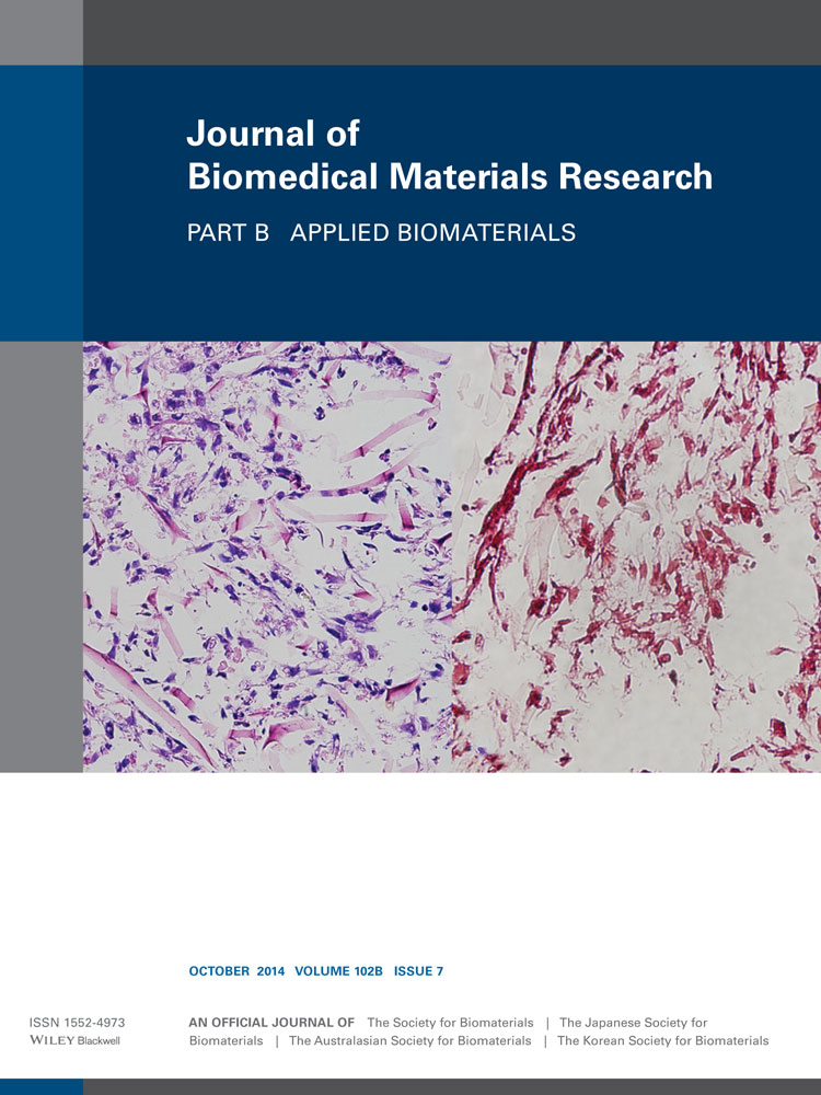
Osteogenic differentiation on DLC-PDMS-h surface
Corresponding Author
Antti Soininen
ORTON Research Institute, Helsinki, Finland
ORTON Orthopedic Hospital, Helsinki, Finland
Correspondence to: A. Soininen (e-mail: [email protected])Search for more papers by this authorEmilia Kaivosoja
Department of Medicine, Institute of Clinical Medicine, Biomedicum, Helsinki, Finland
Department of Electrical Engineering and Automation, Aalto University, Finland
Search for more papers by this authorTarvo Sillat
Department of Medicine, Institute of Clinical Medicine, Biomedicum, Helsinki, Finland
Search for more papers by this authorSannakaisa Virtanen
University of Erlangen Nuremberg, Nuremberg, Germany
Search for more papers by this authorYrjö T. Konttinen
ORTON Research Institute, Helsinki, Finland
ORTON Orthopedic Hospital, Helsinki, Finland
Department of Medicine, Institute of Clinical Medicine, Biomedicum, Helsinki, Finland
COXA Hospital for Joint Replacement, Tampere, Finland
Search for more papers by this authorVeli-Matti Tiainen
ORTON Research Institute, Helsinki, Finland
ORTON Orthopedic Hospital, Helsinki, Finland
Search for more papers by this authorCorresponding Author
Antti Soininen
ORTON Research Institute, Helsinki, Finland
ORTON Orthopedic Hospital, Helsinki, Finland
Correspondence to: A. Soininen (e-mail: [email protected])Search for more papers by this authorEmilia Kaivosoja
Department of Medicine, Institute of Clinical Medicine, Biomedicum, Helsinki, Finland
Department of Electrical Engineering and Automation, Aalto University, Finland
Search for more papers by this authorTarvo Sillat
Department of Medicine, Institute of Clinical Medicine, Biomedicum, Helsinki, Finland
Search for more papers by this authorSannakaisa Virtanen
University of Erlangen Nuremberg, Nuremberg, Germany
Search for more papers by this authorYrjö T. Konttinen
ORTON Research Institute, Helsinki, Finland
ORTON Orthopedic Hospital, Helsinki, Finland
Department of Medicine, Institute of Clinical Medicine, Biomedicum, Helsinki, Finland
COXA Hospital for Joint Replacement, Tampere, Finland
Search for more papers by this authorVeli-Matti Tiainen
ORTON Research Institute, Helsinki, Finland
ORTON Orthopedic Hospital, Helsinki, Finland
Search for more papers by this authorAbstract
The hypothesis was that anti-fouling diamond-like carbon polydimethylsiloxane hybrid (DLC-PDMS-h) surface impairs early and late cellular adhesion and matrix–cell interactions. The effect of hybrid surface on cellular adhesion and cytoskeletal organization, important for osteogenesis of human mesenchymal stromal cells (hMSC), where therefore compared with plain DLC and titanium (Ti). hMSCs were induced to osteogenesis and followed over time using scanning electron microscopy (SEM), time-of-flight secondary ion mass spectrometry (ToF-SIMS), immunofluorescence staining, quantitative real-time polymerase chain reaction (qRT-PCR), and hydroxyapatite (HA) staining. SEM at 7.5 hours showed that initial adherence and spreading of hMSC was poor on DLC-PDMS-h. At 5 days some hMSC were undergoing condensation and apoptotic fragmentation, whereas cells on DLC and Ti grew well. DAPI–actin–vinculin triple staining disclosed dwarfed cells with poorly organized actin cytoskeleton-focal complex/adhesion-growth substrate attachments on hybrid coating, whereas spread cells, organized microfilament bundles, and focal adhesions were seen on DLC and in particular on Ti. Accordingly, at day one ToF-SIMS mass peaks showed poor protein adhesion to DLC-PDMS-h compared with DLC and Ti. COL1A1, ALP, OP mRNA levels at days 0, 7, 14, 21, and/or 28 and lack of HA deposition at day 28 demonstrated delayed or failed osteogenesis on DLC-PDMS-h. Anti-fouling DLC-PDMS-h is a poor cell adhesion substrate during the early protein adsorption-dependent phase and extracellular matrix-dependent late phase. Accordingly, some hMSCs underwent anoikis-type apoptosis and failed to complete osteogenesis, due to few focal adhesions and poor cell-to-ECM contacts. DLC-PDMS-h seems to be a suitable coating for non-integrating implants/devices designed for temporary use. © 2014 Wiley Periodicals, Inc. J Biomed Mater Res Part B: Appl Biomater, 102B: 1462–1472, 2014.
REFERENCES
- 1 Antilla A, Tiainen VM, Kiuru M, Alakoski E, Arstila K. Preparation of diamond-like carbon polymer hybrid films using filtered pulsed arc discharge method. Surf Eng 2003; 19: 425–428.
- 2 Huikko K, Ostman P, Grigoras K, Tuomikoski S, Tiainen VM, Soininen A, Puolanne K, Manz A, Franssila S, Kostiainen R, Kotiaho T. Poly(dimethylsiloxane) electrospray devices fabricated with diamond-like carbon-poly(dimethylsiloxane) coated SU-8 masters. Lab Chip 2003; 3: 67–72.
- 3 Kiuru M, Alakoski E. Low sliding angles in hydrophobic and oleophobic coatings prepared with plasma discharge method. Mater Lett 2004; 58: 2213–2216.
- 4 Soininen A, Levon J, Katsikogianni M, Myllymaa K, Lappalainen R, Konttinen YT, Kinnari TJ, Tiainen VM, Missirlis Y. In vitro adhesion of staphylococci to diamond-like carbon polymer hybrids under dynamic flow conditions. J Mater Sci Mater Med 2011; 22: 629–636.
- 5 Kinnari TJ, Soininen A, Esteban J, Zamora N, Alakoski E, Kouri VP, Lappalainen R, Konttinen YT, Gomez-Barrena E, Tiainen VM. Adhesion of staphylococcal and Caco-2 cells on diamond-like carbon polymer hybrid coating. J Biomed Mater Res Part A 2008; 86A: 760–768.
- 6 Calzado-Martin A, Saldana L, Korhonen H, Soininen A, Kinnari TJ, Gomez-Barrena E, Tiainen VM, Lappalainen R, Munuera L, Konttinen YT and others. Interactions of human bone cells with diamond-like carbon polymer hybrid coatings. Acta Biomater 2010; 6: 3325–3338.
- 7 Myllymaa K, Levon J, Tiainen VM, Myllymaa S, Soininen A, Korhonen H, Kaivosoja E, Lappalainen R, Konttinen YT. Formation and retention of staphylococcal biofilms on DLC and its hybrids compared to metals used as biomaterials. Colloids Surf B Biointerf 2013; 101: 290–297.
- 8 Meazzini MC, Toma CD, Schaffer JL, Gray ML, Gerstenfeld LC. Osteoblast cytoskeletal modulation in response to mechanical strain in vitro. J Orthop Res 1998; 16: 170–180.
- 9 Kilian KA, Bugarija B, Lahn BT, Mrksich M. Geometric cues for directing the differentiation of mesenchymal stem cells. Proc Natl Acad Sci U S A 2010; 107: 4872–4877.
- 10 Oh S, Brammer KS, Li YSJ, Teng D, Engler AJ, Chien S, Jin S. Stem cell fate dictated solely by altered nanotube dimension. Proc Natl Acad Sci U S A 2009; 106: 2130–2135.
- 11 Kaivosoja E, Suvanto P, Barreto G, Aura S, Soininen A, Franssila S, Konttinen YT. Cell adhesion and osteogenic differentiation on three-dimensional pillar surfaces. J Biomed Mater Res Part A 2013; 101A: 842–852.
- 12 Green RM. Can we develop ethically universal embryonic stem-cell lines? Nat Rev Genet 2007; 8: 480–485.
- 13 Arvidson K, Abdallah BM, Applegate LA, Baldini N, Cenni E, Gomez-Barrena E, Granchi D, Kassem M, Konttinen YT, Mustafa K and others. Bone regeneration and stem cells. J Cell Mol Med 2011; 15: 718–746.
- 14 Gomez-Barrena E, Rosset P, Muller I, Giordano R, Bunu C, Layrolle P, Konttinen YT, Luyten FP. Bone regeneration: Stem cell therapies and clinical studies in orthopaedics and traumatology. J Cell Mol Med 2011; 15: 1266–1286.
- 15 Aspenberg P, Anttila A, Konttinen YT, Lappalainen R, Goodman SB, Nordsletten L, Santavirta S. Benign response to particles of diamond and SiC: Bone chamber studies of new joint replacement coating materials in rabbits. Biomaterials 1996; 17: 807–812.
- 16 Anselme K. Osteoblast adhesion on biomaterials. Biomaterials 2000; 21: 667–681.
- 17 Kalbacova M, Rezek B, Baresova V, Wolf-Brandstetter C, Kromka A. Nanoscale topography of nanocrystalline diamonds promotes differentiation of osteoblasts. Acta Biomater 2009; 5: 3076–3085.
- 18 Loder RT, Feinberg JR. Orthopaedic implants in children: Survey results regarding routine removal by the pediatric and nonpediatric specialists. J Pediatr Orthop 2006; 26: 510–519.
- 19 Suzuki M, Sakai Y. Effect of protein adsorption on cell attachment on solid-surfaces. Anim Cell Technol 1992; 4: 71–76.
- 20 Lodish HF. Molecular Cell Biology. New York: W.H. Freeman and Co.; 2013. xxxiii, 1154, 58 p.
- 21 Yamada KM, Geiger B. Molecular interactions in cell adhesion complexes. Curr Opin Cell Biol 1997; 9: 76–85.
- 22
McHargue CJ,
Kossowsky R,
Hofer WO, North Atlantic Treaty Organization. Scientific Affairs Division. Structure-Property Relationships in Surface-Modified Ceramics. Dordrecht; Boston: Kluwer Academic; 1989. x, 522 p.
10.1007/978-94-009-0983-0 Google Scholar
- 23 Alakoski E, Kiuru M, Tiainen VM, Anttila A. Adhesion and quality test for tetrahedral amorphous carbon coating process. Diamond Relat Mater 2003; 12: 2115–2118.
- 24 Anttila A, Salo J, Lappalainen R. High adhesion of diamond-like films achieved by the pulsed arc-discharge method. Mater Lett 1995; 24: 153–156.
- 25 Lifshitz Y. Diamond-like carbon - present status. Diamond Relat Mater 1999; 8: 1659–1676.
- 26 Anttila A, Hirvonen J-P, Koskinen J. Procedure and apparatus for the coating of materials by means of a pulsating plasma beam. US Patent 5078848 (1992).
- 27 Anttila A, Lappalainen R, Tiainen VM, Hakovirta M. Superior attachment of high-quality hydrogen-free amorphous diamond films to solid materials. Adv Mater 1997; 9: 1161–1164.
- 28 Lehto VP, Virtanen I. Vinculin in cultured bovine lens-forming cells. Cell Differ 1985; 16: 153–160.
- 29 Schmittgen TD, Livak KJ. Analyzing real-time PCR data by the comparative C-T method. Nat Protoc 2008; 3: 1101–1108.
- 30 Bregg RK. Horizons in Polymer Research. New York: Nova Science Publishers; 2005. ix, 199 p.
- 31 Homma H, Kuroyagi T, Izumi K, Mirley CL, Ronzello J, Boggs SA. Evaluation of surface degradation of silicone rubber using gas chromatography/mass spectroscopy. IEEE Trans Power Deliv 2000; 15: 796–803.
- 32 Kaivosoja E, Virtanen S, Rautemaa R, Lappalainen R, Konttinen YT. Spectroscopy in the analysis of bacterial and eukaryotic cell footprints on implant surfaces. Eur Cells Mater 2012; 24: 60–73.
- 33 Konttinen YT, Kaivosoja E, Stegaev V, Wagner HD, Levon J, Tiainen VM, Mackiewicz Z. Extracellular matrix and tissue regeneration. In: G Steinhoff, editor. Regenerative Medicine: From Protocol to Patient. Dordrecht; Heidelberg; New York; London: Springer; 2011, pp. 21–80.
- 34 Mathieu PS, Loboa EG. Cytoskeletal and focal adhesion influences on mesenchymal stem cell shape, mechanical properties, and differentiation down osteogenic, adipogenic, and chondrogenic pathways. Tissue Eng Part B-Rev 2012; 18: 436–444.
- 35 Ruiz SA, Chen CS. Emergence of patterned stem cell differentiation within multicellular structures. Stem Cells 2008; 26: 2921–2927.
- 36 McBeath R, Pirone DM, Nelson CM, Bhadriraju K, Chen CS. Cell shape, cytoskeletal tension, and RhoA regulate stem cell lineage commitment. Dev Cell 2004; 6: 483–495.
- 37 Goto T, Yoshinari M, Kobayashi S, Tanaka T. The initial attachment and subsequent behavior of osteoblastic cells and oral epithelial cells on titanium. Bio-Med Mater Eng 2004; 14: 537–544.
- 38 Barnthip N, Parhi P, Golas A, Vogler EA. Volumetric interpretation of protein adsorption: Kinetics of protein-adsorption competition from binary solution. Biomaterials 2009; 30: 6495–6513.
- 39 Roach P, Farrar D, Perry CC. Interpretation of protein adsorption: Surface-induced conformational changes. J Am Chem Soc 2005; 127: 8168–8173.
- 40 Roach P, Farrar D, Perry CC. Surface tailoring for controlled protein adsorption: Effect of topography at the nanometer scale and chemistry. J Am Chem Soc 2006; 128: 3939–3945.
- 41 Gray JJ. The interaction of proteins with solid surfaces. Curr Opin Struct Biol 2004; 14: 110–115.
- 42 Mauney J, Volloch V. Human bone marrow-derived stromal cells show highly efficient stress-resistant adipogenesis on denatured collagen IV matrix but not on its native counterpart: Implications for obesity. Matrix Biol 2010; 29: 9–14.
- 43 Kleinman HK, Klebe RJ, Martin GR. Role of collagenous matrices in the adhesion and growth of cells. J Cell Biol 1981; 88: 473–485.
- 44 Wahlgren M, Arnebrant T. Protein adsorption to solid surfaces. Trends Biotechnol 1991; 9: 201–208.
- 45 Meadows PY, Walker GC. Force microscopy studies of fibronectin adsorption and subsequent cellular adhesion to substrates with well-defined surface chemistries. Langmuir 2005; 21: 4096–4107.
- 46 Bacakova L, Filova E, Parizek M, Ruml T, Svorcik V. Modulation of cell adhesion, proliferation and differentiation on materials designed for body implants. Biotech Adv 2011; 29: 739–767.
- 47 Thevenot P, Hu WJ, Tang LP. Surface chemistry influences implant biocompatibility. Curr Top Med Chem 2008; 8: 270–280.
- 48 Killian MS, Krebs HM, Schmuki P. Protein denaturation detected by time-of-flight secondary ion mass spectrometry. Langmuir 2011; 27: 7510–7515.
- 49 Waugaman M, Sannigrahi B, McGeady P, Khan IM. Synthesis, characterization and biocompatibility studies of oligosiloxane modified polythiophenes. Eur Polym J 2003; 39: 1405–1412.
- 50 McGill G, Shimamura A, Bates RC, Savage RE, Fisher DE. Loss of matrix adhesion triggers rapid transformation-selective apoptosis in fibroblasts. J Cell Biol 1997; 138: 901–911.
- 51 Wehrle-Haller B. Assembly and disassembly of cell matrix adhesions. Curr Opin Cell Biol 2012; 24: 569–581.
- 52 Wehrle-Haller B. Structure and function of focal adhesions. Curr Opin Cell Biol 2012; 24: 116–124.
- 53 Roca-Cusachs P, del Rio A, Puklin-Faucher E, Gauthier NC, Biais N, Sheetz MP. Integrin-dependent force transmission to the extracellular matrix by alpha-actinin triggers adhesion maturation. Proc Natl Acad Sci U S A 2013; 110: E1361–E1370.
- 54 Faucheux N, Schweiss R, Lutzow K, Werner C, Groth T. Self-assembled monolayers with different terminating groups as model substrates for cell adhesion studies. Biomaterials 2004; 25: 2721–2730.
- 55 Kennedy SB, Washburn NR, Simon CG, Jr., Amis EJ. Combinatorial screen of the effect of surface energy on fibronectin-mediated osteoblast adhesion, spreading and proliferation. Biomaterials 2006; 27: 3817–3824.
- 56 Eisenbarth E, Velten D, Schenk-Meuser K, Linez P, Biehl V, Duschner H, Breme J, Hildebrand H. Interactions between cells and titanium surfaces. Biomol Eng 2002; 19: 243–249.
- 57 Linez-Bataillon P, Monchau F, Bigerelle M, Hildebrand HF. In vitro MC3T3 osteoblast adhesion with respect to surface roughness of Ti6A14V substrates. Biomol Eng 2002; 19: 133–141.
- 58 Webster TJ, Smith TA. Increased osteoblast function on PLGA composites containing nanophase titania. J Biomed Mater Res Part A 2005; 74A: 677–686.
- 59 Biggs MJP, Richards RG, McFarlane S, Wilkinson CDW, Oreffo ROC, Dalby MJ. Adhesion formation of primary human osteoblasts and the functional response of mesenchymal stem cells to 330 nm deep microgrooves. J R Soc Interf 2008; 5: 1231–1242.
- 60 Kunzler TP, Drobek T, Schuler M, Spencer ND. Systematic study of osteoblast and fibroblast response to roughness by means of surface-morphology gradients. Biomaterials 2007; 28: 2175–2182.
- 61 Kalbacova M, Broz A, Babchenko O, Kromka A. Study on cellular adhesion of human osteoblasts on nano-structured diamond films. Phys Status Solidi B-Basic Solid State Phys 2009; 246: 2774–2777.
- 62 Soininen A, Tiainen V-M, Konttinen Y, Sharma P, Busscher H, van der Mei HC. Physicochemical and microbial fouling characterization of novel, extremely hydrophobic, nanocomposite diamond like carbon polymer hybrid coatings. 2008; 8th World Biomaterials Congress 2008, Amsterdam, The Netherlands. 284 p.
- 63 Baier RE. The role of surface energy in thrombogenesis. Bull N Y Acad Med 1972; 48: 257–272.
- 64 Bitton G, Marshall KC. Adsorption of Microorganisms to Surfaces. New York: Wiley; 1980. x, 439 p.
- 65 Pereni CI, Zhao Q, Liu Y, Abel E. Surface free energy effect on bacterial retention. Colloids Surf B-Biointerf 2006; 48: 143–147.
- 66 Shin YN, Kim BS, Ahn HH, Lee JH, Kim KS, Lee JY, Kim MS, Khang G, Lee HB. Adhesion comparison of human bone marrow stem cells on a gradient wettable surface prepared by corona treatment. Appl Surf Sci 2008; 255: 293–296.
- 67 de Mel A, Cousins BG, Seifalian AM. Surface modification of biomaterials: A quest for blood compatibility. Int J Biomater 2012; 2012: 707863.
- 68 Altankov G, Richau K, Groth T. The role of surface zeta potential and substratum chemistry for regulation of dermal fibroblasts interaction. Materialwiss Werkst 2003; 34: 1120–1128.
- 69 Derjaguin B, Landau L. Theory of the stability of strongly charged lyophobic sols and of the adhesion of strongly charged particles in solutions of electrolytes. Acta Phys Chem URSS 1941; 14: 633–662.
- 70 Verwey EJW, Overbeek JTG. Theory of the Stability of Lyophobic Colloids. Amsterdam: Elsevier; 1948.
- 71 Marshall KC, Stout R, Mitchell R. Mechanism of initial events in sorption of marine bacteria to surfaces. J Gen Microbiol 1971; 68: 337–348.
- 72 Vanoss CJ, Good RJ, Chaudhury MK. The role of vanderwaals forces and hydrogen-bonds in hydrophobic interactions between bio-polymers and low-energy surfaces. J Colloid Interf Sci 1986; 111: 378–390.
- 73
Bongrand P,
Claesson PM,
Curtis ASG. Studying Cell Adhesion. Berlin, New York: Springer-Verlag; 1994. xiv, 250 p.
10.1007/978-3-662-03008-0 Google Scholar
- 74 Katsikogianni M, Missirlis YF. Concise review of mechanisms of bacterial adhesion to biomaterials and of techniques used in estimating bacteria-material interactions. Eur Cell Mater 2004; 8: 37–57.
- 75 Bos R, van der Mei HC, Busscher HJ. Physico-chemistry of initial microbial adhesive interactions - its mechanisms and methods for study. Fems Microbiol Rev 1999; 23: 179–230.


