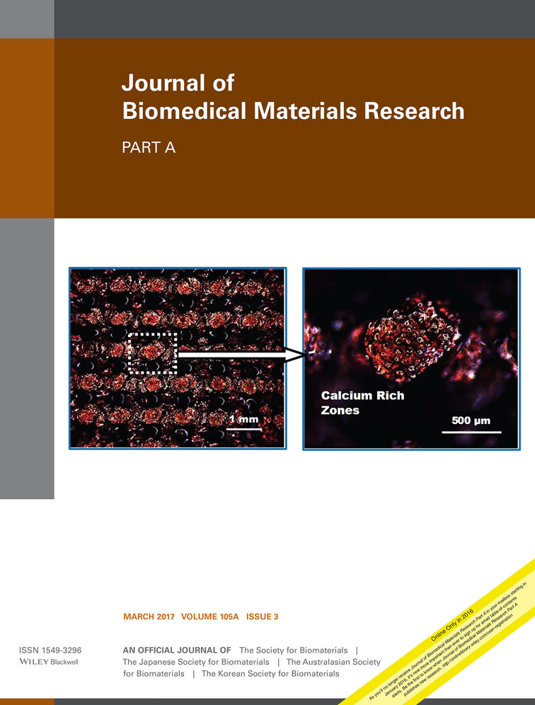
Skeletal muscle patch engineering on synthetic and acellular human skeletal muscle originated scaffolds
Birol Ay
Stem Cell Department, Kocaeli University, Institute of Health Sciences, Kocaeli, Turkey
Center for Stem Cell and Gene Therapies Research and Practice, Kocaeli University, Kocaeli, Turkey
Search for more papers by this authorErdal Karaoz
Stem Cell Department, Kocaeli University, Institute of Health Sciences, Kocaeli, Turkey
Center for Stem Cell and Gene Therapies Research and Practice, Kocaeli University, Kocaeli, Turkey
Search for more papers by this authorCumhur C. Kesemenli
Faculty of Medicine, Department of Orthopedics and Traumatology, Kocaeli University, Kocaeli, Turkey
Search for more papers by this authorCorresponding Author
Halime Kenar
Stem Cell Department, Kocaeli University, Institute of Health Sciences, Kocaeli, Turkey
BIOMATEN Center of Excellence in Biomaterials and Tissue Engineering, METU, Ankara, Turkey
Experimental and Clinical Research Center, Kocaeli University, Kocaeli, Turkey
Correspondence to: H. Kenar, Experimental and Clinical Research Center, Kocaeli University, Kocaeli, Turkey; e-mail: [email protected]Search for more papers by this authorBirol Ay
Stem Cell Department, Kocaeli University, Institute of Health Sciences, Kocaeli, Turkey
Center for Stem Cell and Gene Therapies Research and Practice, Kocaeli University, Kocaeli, Turkey
Search for more papers by this authorErdal Karaoz
Stem Cell Department, Kocaeli University, Institute of Health Sciences, Kocaeli, Turkey
Center for Stem Cell and Gene Therapies Research and Practice, Kocaeli University, Kocaeli, Turkey
Search for more papers by this authorCumhur C. Kesemenli
Faculty of Medicine, Department of Orthopedics and Traumatology, Kocaeli University, Kocaeli, Turkey
Search for more papers by this authorCorresponding Author
Halime Kenar
Stem Cell Department, Kocaeli University, Institute of Health Sciences, Kocaeli, Turkey
BIOMATEN Center of Excellence in Biomaterials and Tissue Engineering, METU, Ankara, Turkey
Experimental and Clinical Research Center, Kocaeli University, Kocaeli, Turkey
Correspondence to: H. Kenar, Experimental and Clinical Research Center, Kocaeli University, Kocaeli, Turkey; e-mail: [email protected]Search for more papers by this authorAbstract
The reconstruction of skeletal muscle tissue is currently performed by transplanting a muscle tissue graft from local or distant sites of the patient's body, but this practice leads to donor site morbidity in case of large defects. With the aim of providing an alternative treatment approach, skeletal muscle tissue formation potential of human myoblasts and human menstrual blood derived mesenchymal stem cells (hMB-MSCs) on synthetic [poly(l-lactide-co-caprolactone), 70:30] scaffolds with oriented microfibers, human muscle extracellular matrix (ECM), and their hybrids was investigated in this study. The reactive muscle ECM pieces were chemically crosslinked to the synthetic scaffolds to produce the hybrids. Cell proliferation assay WST-1, scanning electron microscopy (SEM), and immunostaining were carried out after culturing the cells on the scaffolds. The ECM and the synthetic scaffolds were effective in promoting spontaneous myotube formation from human myoblasts. Anisotropic muscle patch formation was more successful when human myoblasts were grown on the synthetic scaffolds. Nonetheless, spontaneous differentiation could not be induced in hMB-MSCs on any type of the scaffolds. Human myoblast-synthetic scaffold combination is promising as a skeletal muscle patch, and can be improved further to serve as a fast integrating functional patch by introducing vascular and neuronal networks to the structure. © 2016 Wiley Periodicals, Inc. J Biomed Mater Res Part A: 105A: 879–890, 2017.
Supporting Information
Additional Supporting Information may be found in the online version of this article.
| Filename | Description |
|---|---|
| jbma35948-sup-0001-suppinfo.tif9.1 MB | Supporting Information |
Please note: The publisher is not responsible for the content or functionality of any supporting information supplied by the authors. Any queries (other than missing content) should be directed to the corresponding author for the article.
REFERENCES
- 1 Bach AD, Beier JP, Stern-Staeter J, Horch RE. Skeletal muscle tissue engineering. J Cell Mol Med 2004; 8: 413–422.
- 2 Koning M, Harmsen MC, van Luyn MJ, Werker PM. Current opportunities and challenges in skeletal muscle tissue engineering. J Tissue Eng Regen Med 2009; 3: 407–415.
- 3 Mizuno H, Zuk PA, Zhu M, Lorenz HP, Benhaim P, Hedrick MH. Experimental myogenic differentiation by human processed lipoaspirate cells. Plast Reconstr Surg 2002; 109: 199–209.
- 4 Zuk PA, Zhu M, Ashjian P, De Ugarte DA, Huang JI, Mizuno H, Alfonso ZC, Fraser JK, Benhaim P, Hedrick MH. Human adipose tissue is a source of multipotent stem cells. Mol Biol Cell 2002; 13: 4279–4295.
- 5 Neumann T, Hauschka SD, Sanders JE. Tissue engineering of skeletal muscle using polymer fiber arrays. Tissue Eng 2003; 9: 995–1003.
- 6 Riboldi SA, Sampaolesi M, Neuenschwander P, Cossu G, Mantero S. Electrospun degradable polyesterurethane membranes: Potential scaffolds for skeletal muscle tissue engineering. Biomaterials 2005; 26: 4606–4615.
- 7 Wang L, Wu Y, Guo B, Ma PX. Nano fiber yarn/hydrogel core-shell scaffolds mimicking native skeletal muscle tissue for guiding 3D myoblast alignment, elongation, and differentiation. ACS Nano 2015; 9: 9167–9179.
- 8 Beier JP, Klumpp D, Rudisile M, Dersch R, Wendorff JH, Bleiziffer O, Arkudas A, Polykandriotis E, Horch RE, Kneser U. Collagen matrices from sponge to nano: New perspectives for tissue engineering of skeletal muscle. BMC Biotechnol 2009; 9: 34.
- 9 Valentin JE, Turner NJ, Gilbert TW, Badylak SF. Functional skeletal muscle formation with a biologic scaffold. Biomaterials 2010; 31: 7475–7484.
- 10 Choi JS, Lee SJ, Christ GJ, Atala A, Yoo JJ. The influence of electrospun aligned poly(ɛ-caprolactone)/collagen nanofiber meshes on the formation of self-aligned skeletal muscle myotubes. Biomaterials 2008; 29: 2899–2906.
- 11 Sato M, Ito A, Kawabe Y, Nagamori E, Kamihira M. Enhanced contractile force generation by artificial skeletal muscle tissues using IGF-I gene-engineered myoblast cells. J Biosci Bioeng 2011; 112: 273–278.
- 12 Criswell TL, Corona BT, Wang Z, Zhou Y, Niu G, Xu Y, Christ GJ, Soker S. The role of endothelial cells in myofiber differentiation and the vascularization and innervation of bioengineered muscle tissue in vivo. Biomaterials 2013; 34: 140–149.
- 13 Moon DG, Christ G, Stitzel JD, Atala A, Yoo JJ. Cyclic mechanical preconditioning improves engineered muscle contraction. Tissue Eng Part A 2008; 14: 473–482.
- 14 Boonen KJM, Langelaan MLP, Polak RB, van der Schaft DWJ, Baaijens FPT, Post MJ. Effects of a combined mechanical stimulation protocol: Value for skeletal muscle tissue engineering. J Biomech 2010; 43: 1514–1521.
- 15 Kaji H, Ishibashi T, Nagamine K, Kanzaki M, Nishizawa M. Electrically induced contraction of C2C12 myotubes cultured on a porous membrane-based substrate with muscle tissue-like stiffness. Biomaterials 2010; 31: 6981–6986.
- 16 Chen MC, Sun YC, Chen YH. Electrically conductive nanofibers with highly oriented structures and their potential application in skeletal muscle tissue engineering. Acta Biomater 2013; 9: 5562–5572.
- 17 Zhao C, Andersen H, Ozyilmaz B, Ramaprabhu S, Pastorin G, Ho HK. Spontaneous and specific myogenic differentiation of human mesenchymal stem cells on polyethylene glycol-linked multi-walled carbon nanotube films for skeletal muscle engineering. Nanoscale 2015; 7: 18239–18249.
- 18 Meng X, Ichim TE, Zhong J, Rogers A, Yin Z, Jackson J, Wang H, Ge W, Bogin V, Chan KW, Thébaud B, Riordan NH. Endometrial regenerative cells: A novel stem cell population. J Transl Med 2007; 5: 57.
- 19 Cui C-H, Uyama T, Miyado K, Terai M, Kyo S, Kiyono T, Umezawa A. Menstrual blood-derived cells confer human dystrophin expression in the murine model of Duchenne muscular dystrophy via cell fusion and myogenic transdifferentiation. Mol Biol Cell 2007; 18: 1586–1594.
- 20 Klumpp D, Rudisile M, Kühnle RI, Hess A, Bitto FF, Arkudas A, Bleiziffer O, Boos AM, Kneser U, Horch RE, Beier JP. Three-dimensional vascularization of electrospun PCL/collagen-blend nanofibrous scaffolds in vivo. J Biomed Mater Res Part A 2012; 100: 2302–2311.
- 21 Koffler J, Kaufman-Francis K, Shandalov Y, Yulia S, Egozi D, Dana E, Pavlov DA, Daria AP, Landesberg A, Levenberg S. Improved vascular organization enhances functional integration of engineered skeletal muscle grafts. Proc Natl Acad Sci U S A 2011; 108: 14789–14794.
- 22 De Coppi P, Bellini S, Conconi MT, Sabatti M, Simonato E, Gamba PG, Nussdorfer GG, Parnigotto PP. Myoblast-acellular skeletal muscle matrix constructs guarantee a long-term repair of experimental full-thickness abdominal wall defects. Tissue Eng 2006; 12: 1929–1936.
- 23 Borschel GH, Dennis RG, Kuzon WM. Contractile skeletal muscle tissue-engineered on an acellular scaffold. Plast Reconstr Surg 2004; 113: 595–602.
- 24 Porzionato A, Sfriso M, Pontini A, Macchi V, Petrelli L, Pavan P, Natali A, Bassetto F, Vindigni V, De Caro R. Decellularized human skeletal muscle as biologic scaffold for reconstructive surgery. Int J Mol Sci 2015; 16: 14808–14831.
- 25 Karaoz E, Aksoy A, Ayhan S, SarIboyacI AE, Kaymaz F, Kasap M. Characterization of mesenchymal stem cells from rat bone marrow: Ultrastructural properties, differentiation potential and immunophenotypic markers. Histochem Cell Biol 2009; 132: 533–546.
- 26 Burlingham WJ, Jankowska-Gan E, VanBuskirk A, Orosz CG, Lee J, Kusaka S. Loss of tolerance to a maternal kidney transplant is selective for HLA class II: Evidence from trans-vivo DTH and alloantibody analysis. Hum Immunol 2000; 61: 1395–1402.
- 27 Crisan M, Yap S, Casteilla L, Chen CW, Corselli M, Park TS, Andriolo G, Sun B, Zheng B, Zhang L, Norotte C, Teng PN, Traas J, Schugar R, Deasy BM, Badylak S, Bűhring HJ, Giacobino JP, Lazzari L, Huard J, Péault B. A perivascular origin for mesenchymal stem cells in multiple human organs. Cell Stem Cell 2008; 3: 301–313.
- 28 Dominici M, Le Blanc K, Mueller I, Slaper-Cortenbach I, Marini F, Krause D, Deans R, Keating A, Prockop D, Horwitz E. Minimal criteria for defining multipotent mesenchymal stromal cells. The International Society for Cellular Therapy position statement. Cytotherapy 2006; 8: 315–317.
- 29 Muratore M, Mitchell S, Waterfall M. Plasma membrane characterization, by scanning electron microscopy, of multipotent myoblasts-derived populations sorted using dielectrophoresis. Biochem Biophys Res Commun 2013; 438: 666–672.
- 30 Velleman SG. The role of the extracellular matrix in skeletal muscle development. Poult Sci 1999; 78: 778–784.
- 31 Merritt E, Cannon M, Hammers D, Le L, Gokhale R, Sarathy A, Song T, Tierney M, Suggs L, Walters T, Farrar R. Repair of traumatic skeletal muscle injury with bone-marrow-derived mesenchymal stem cells seeded on extracellular matrix. Tissue Eng Part A 2010; 16: 2871–2881.
- 32 Jeong SI, Kim B-S, Lee YM, Ihn KJ, Kim SH, Kim YH. Morphology of elastic poly(l-lactide-co-epsilon-caprolactone) copolymers and in vitro and in vivo degradation behavior of their scaffolds. Biomacromolecules 2004; 5: 1303–1309.
- 33 Shafiq M, Jung Y, Kim SH. In situ vascular regeneration using substance P-immobilised poly(l-Lactide-co-Ε caprolactone) scaffolds: Stem cell recruitment, angiogenesis, and tissue regeneration. Eur Cells Mater 2015; 30: 282–302.
- 34 Zhu Y, Chian KS, Chan-Park MB, Mhaisalkar PS, Ratner BD. Protein bonding on biodegradable poly(l-lactide-co-caprolactone) membrane for esophageal tissue engineering. Biomaterials 2006; 27: 68–78.
- 35 Schwab KE, Gargett CE. Co-expression of two perivascular cell markers isolates mesenchymal stem-like cells from human endometrium. Hum Reprod 2007; 22: 2903–2911.
- 36 Millay DP, O'Rourke JR, Sutherland LB, Bezprozvannaya S, Shelton JM, Bassel-Duby R, Olson EN. Myomaker is a membrane activator of myoblast fusion and muscle formation. Nature 2013; 499: 301–305.
- 37 Ott HC, Matthiesen TS, Goh SK, Black LD, Kren SM, Netoff TI, Taylor DA. Perfusion-decellularized matrix: Using nature's platform to engineer a bioartificial heart. Nat Med 2008; 14: 213–221.
- 38 Uygun BE, Soto-Gutierrez A, Yagi H, Izamis ML, Guzzardi MA, Shulman C, Milwid J, Kobayashi N, Tilles A, Berthiaume F, Hertl M, Nahmias Y, Yarmush ML, Uygun K. Organ reengineering through development of a transplantable recellularized liver graft using decellularized liver matrix. Nat Med 2010; 16: 814–820.
- 39 Panzavolta S, Gioffrè M, Focarete ML, Gualandi C, Foroni L, Bigi A. Electrospun gelatin nanofibers: Optimization of genipin cross-linking to preserve fiber morphology after exposure to water. Acta Biomater 2011; 7: 1702–1709.
- 40 Chieh HF, Su FC, Lin SC, Shen MR, Liao J. Der migration patterns and cell functions of adipose-derived stromal cells on self-assembled monolayers with different functional groups. J Biomater Sci Polym Ed 2013; 24: 94–117.
- 41 Demonbreun AR, Biersmith BH, McNally EM. Membrane fusion in muscle development and repair. Semin Cell Dev Biol 2015; 45: 48–56.
- 42 Hasan A, Memic A, Annabi N, Hossain M, Paul A, Dokmeci MR, Dehghani F, Khademhosseini A. Electrospun scaffolds for tissue engineering of vascular grafts. Acta Biomater 2014; 10: 11–25.
- 43 Gholobova D, Decroix L, Van Mulyder V, Desender L, Gerard M, Carpentier G, Vandenburgh H, Thorrez L. Endothelial network formation within human tissue-engineered skeletal muscle. Tissue Eng Part A 2015; 21: 2548–2558.
- 44 Guo X, Gonzalez M, Stancescu M, Vandenburgh HH, Hickman JJ. Neuromuscular junction formation between human stem cell-derived motoneurons and human skeletal muscle in a defined system. Biomaterials 2011; 32: 9602–9611.
- 45 Schultz SS, Lucas PA. Human stem cells isolated from adult skeletal muscle differentiate into neural phenotypes. J Neurosci Methods 2006; 152: 144–155.


