Correlation of amyloid and ameloblast-associated proteins to odontogenic cysts and tumors: A cross-sectional study
Abstract
Background and Aims
Odontogenic cysts and tumors often form hard and soft structures that resemble odontogenesis. It is well known that amyloid is produced in Pindborg tumors; however, it is still debatable whether it is also formed in other odontogenic tumors and cysts. This study aimed to detect the presence of amyloid in different odontogenic cysts and tumors in correlation to matrix proteins secreted during enamel formation; namely amelogenin and odontogenic ameloblast-associated protein.
Methods
This study included formalin fixed paraffin embedded tissue blocks of 106 different types of odontogenic cysts and tumors. Congo red and thioflavin T were performed to confirm the presence of amyloid; immunohistochemistry was used to detect amelogenin and odontogenic ameloblast-associated protein.
Results
Amyloid was detected in pindborg tumors (conventional), adenomatoid odontogenic tumors, odontogenic fibroma (Amyloid variant), follicular solid and unicystic ameloblastomas, radicular cysts, dentigerous cysts, dentinogenic ghost cell odontogenic tumor, ameloblastic fibroma, calcifying odontogenic cyst, and primordial Odontogenic tumor. Amelogenin was detected in 95.3% of the cases, while odontogenic ameloblast-associated protein was detected in 93.4% of the cases. The association between odontogenic ameloblast-associated protein and amyloid was highly significant at p < 0.01. However, there was no significant relationship between amelogenin and amyloid p > 0.05.
Conclusion
Although pindborg tumor is the bonafide example of amyloid deposition in odontogenic tumors, this study concluded that amyloid may be deposited in traces to massive amounts in various odontogenic cysts and tumors, and it is significantly linked to odontogenic ameloblast-associated protein but not amelogenin matrix protein, since all amyloid cases were odontogenic ameloblast associated protein positive.
1 INTRODUCTION
Odontogenesis is the process of teeth formation. Epithelial and connective tissue elements combine to form tooth germs in early tooth development. A tooth germ develops through the bud, cap, and bell stages.1 AMELX is a major protein component of the enamel matrix, and ameloblasts express it during the secretory stage,2 while ODAM is expressed during the maturation stage and has a wide and varied range of biological activities both physiological and pathological, mainly on the development of ameloblasts, enamel mineralization and the production of junctional epithelia.3, 4 Odontogenic cysts and tumors are a diverse group of pathologic lesions of the jaws and related soft tissues. These cysts and tumors often form structures that are normally produced in odontogenesis. One of these structures is amyloid.5 Amyloid is defined as an extracellular, proteinaceous fibrillar deposit that binds the Congo red dye and exhibits apple-green birefringence when polarized microscopy is used to examine this material. These features are the gold standard for amyloid detection.6 Amyloid may deposit in a physiological (functional) manner, as in tooth development,7 or pathologically, as in neoplastic conditions such as a pancreatic endocrine tumor,8 medullary thyroid carcinoma,9 mammary carcinoma,10 and calcifying epithelial odontogenic tumor.11 With the exception of the former tumor, few studies have been conducted concerning the presence of amyloid in other odontogenic lesions with conflicting results.11-17 This study evaluated the presence of amyloid in different odontogenic cysts and tumors in correlation to the immunohistochemical expression of enamel matrix proteins; Amelogenin (AMELX) and Odontogenic ameloblast-associated protein (ODAM).
2 MATERIALS AND METHODS
2.1 Data retrieval and tissue processing
The archives of the Oral and Maxillofacial Pathology Laboratory, Teaching Hospital/College of Dentistry, University of Baghdad, were searched for all available cases with a diagnosis of odontogenic cysts or tumors from September 2018 to December 2021. Ethical approval was obtained from the ethical committee in the college of dentistry/university of Baghdad (Ref: 125, 28/November/2019).
Paraffin-embedded tissue blocks with associated histopathological examination reports for 106 confirmed odontogenic cysts and tumor cases were retrieved from the Oral and maxillofacial pathology laboratory/college of dentistry, University of Baghdad. Immunohistochemistry was performed on tissue with a thickness of 4 µm, while Congo red and thioflavin T were used on an 8-µm tissue section for amyloid detection.
2.2 Histochemical and immunohistochemical staining
The primary antibodies used in this study were anti-Amelogenin (AMELX), and anti-ODAM, MyBioSource. Immunohistochemical positive controls were included in each run according to antibodies data sheets. The immunohistochemical expression of both matrix protein AMELX and ODAM were assessed based on the staining intensity as follows: (0) no staining, (1) weak staining, (2) moderate staining, and (3) strong staining. While Congo red special stain (Pathensitu) and Thioflavin T (Abcam) were performed to detect Amyloid. It is recommended to use both Congo red and thioflavin T in the detection of amyloid, combining the advantages of each technique.19 Only cases that showed positivity for both Congo red apple-green birefringence and thioflavin T fluorescence were considered amyloid positive.19 The scoring system was used for the assessment of odontogenic amyloid as follows: 0 (negative), 1+ (focal positive: 1–3 focus per low power field), 2+ (multifocal positive: more than 3 focus per low power field), and 3+ (diffuse positive). Both histochemical and immunohistochemical scoring were go through inter- and intra-observer calibrations.
2.3 Statistical analysis
Data management and analysis were performed using SPSS 21.0 package program (IBM Corp released in 2012, SPSS Statistics for Windows, Version 21.0. Armonk, NY: IBM Corp) Descriptive data were generated for all variables, Histochemical scoring for Amyloid and Immunohistochemical expression scores of the AMELX and ODAM were performed. The Chi-square test was used for determining the quantitative analysis of Amyloid score in odontogenic cysts and tumors. Spearman's rho Correlations coefficients were carried out to assess the relation between AMLEX and amyloid, ODAM, and amyloid. The significance level was set at p < 0.05.
3 RESULTS
The frequency of odontogenic cysts and tumors is illustrated in Table 1 for the 106 studied sample cases; solid ameloblastoma was the most frequent odontogenic lesion, accounting for approximately 15% of the total cases. The mean age was 28.24 ± 15.43 years, with males slightly more than females, and mandible to maxilla ratio about 2:1.
| Type of lesions | Number of cases | Mean of age ± SD | Gender | Site | ||
|---|---|---|---|---|---|---|
| Male | Female | Max. | Mand. | |||
| Odontogenic tumors, 58.5% | ||||||
| AB | 16 | 34.44 ± 14.36 | 8 | 8 | 2 | 14 |
| AOT | 10 | 27.6 ± 22.82 | 5 | 5 | 5 | 5 |
| UA | 9 | 29.22 ± 16.95 | 6 | 3 | 1 | 8 |
| AF | 8 | 19.0 ± 13.13 | 6 | 2 | 1 | 7 |
| CEOT | 8 | 25.13 ± 10.84 | 3 | 5 | 3 | 5 |
| O | 4 | 21.5 ± 9.0 | 3 | 1 | 3 | 1 |
| OF | 3 | 27.0 ± 15.1 | 1 | 2 | 2 | 1 |
| DGOT | 2 | 42.5 ± 3.54 | 1 | 1 | 0 | 2 |
| POT | 1 | 6 | 1 | 0 | 1 | 0 |
| CB | 1 | 14 | 1 | 0 | 0 | 1 |
| Odontogenic cysts, 41.5% | ||||||
| OKC | 12 | 29.0 ± 10.21 | 7 | 5 | 3 | 9 |
| RC | 10 | 30.0 ± 17.16 | 2 | 8 | 7 | 3 |
| DC | 10 | 16.8 ± 9.02 | 6 | 4 | 3 | 7 |
| COC | 7 | 28.57 ± 19.86 | 3 | 4 | 3 | 4 |
| GOC | 3 | 59 ± 7.94 | 1 | 2 | 0 | 3 |
| LPC | 1 | 50 | 0 | 1 | 0 | 1 |
| GC | 1 | 35 | 1 | 0 | 0 | 1 |
| Total | 106 | 28.24 ± 15.43 | 55 | 51 | 34 | 72 |
- Abbreviations: AB, solid ameloblastoma; AF, ameloblastic fibroma; AOT, adenomatoid odontogenic tumor; CB, cementoblastoma; COC, calcifying odontogenic cyst; COET, pindborg tumor; DC, dentigerous cyst; DGCT, dentinogenic ghost cell tumor; GC, gingival cyst; GOC, glandular odontogenic cyst; LPC, lateral periodontal cyst; O, odontoma; OF, odontogenic fibroma; OKC, odontogenic keratocyst; POT, primordial odontogenic tumor; RC, radicular cyst; UA, unicystic ameloblastoma.
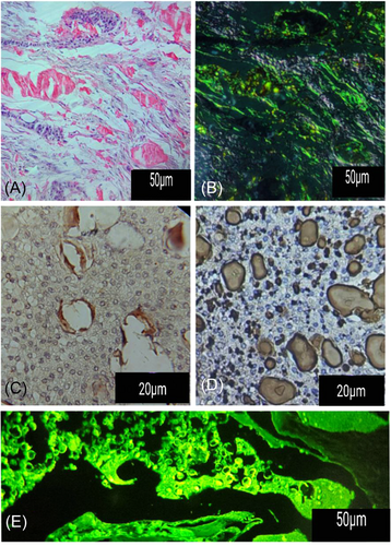
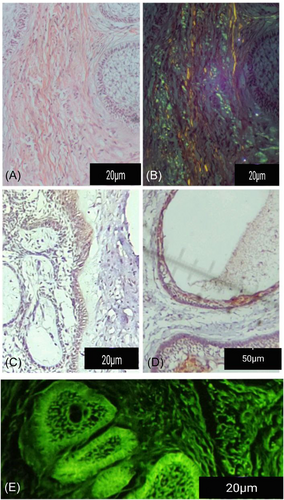
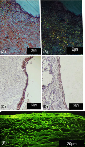
Most of the cases examined with thioflavin T fluoresced green-yellow, as seen in Figure 1. Not only does amyloid demonstrate positivity to thioflavin T, but keratin and calcifications also appear positive. Results obtained from thioflavin T were verified with Congo red stain and polarized lenses. In this study, Amyloid was detected in 33 cases (31.1% of the cases), pindborg tumor (conventional variant) was seen 100% affected, as shown in Table 2 and Figure 1, a number of other odontogenic tumors also produced amyloid in varying proportions like follicular solid ameloblastoma (Figure 2) and unicystic ameloblastoma, ameloblastic fibroma, calcifying odontogenic cyst, radicular cyst, dentigerous cyst (Figure 4), Dentinogenic ghost cell tumor, odontogenic fibroma (Amyloid variant). Additionally, a recently classified odontogenic tumor, a primordial odontogenic tumor, was shown to produce amyloid, as seen in Figure 3.
| Type of lesion | Total number of cases | Number of cases of amyloid negative | Amyloid distribution in positive cases | Percentage of amyloid positive | Chi-square | ||
|---|---|---|---|---|---|---|---|
| Focal | Multifocal | Diffuse | |||||
| AB | 16 | 11 | 1 | 4 | 0 | 31.25% | 0.074 |
| UA | 9 | 6 | 2 | 1 | 0 | 33.33% | 0.368 |
| AF | 8 | 7 | 0 | 1 | 0 | 12.50% | 0.368 |
| AOT | 10 | 4 | 3 | 2 | 1 | 60% | 0.607 |
| COC | 7 | 4 | 2 | 0 | 1 | 42.85% | 0.368 |
| KOC | 12 | 0 | 0 | 0 | 0 | 0% | |
| RC | 10 | 1 | 0 | 1 | 0 | 10% | 0.368 |
| DC | 10 | 3 | 0 | 3 | 0 | 30% | 0.050 |
| CEOT | 8 | 8 | 1 | 1 | 6 | 100% | 0.044* |
| O | 4 | 0 | 0 | 0 | 0 | 0% | |
| OF | 3 | 1 | 1 | 0 | 0 | 33.33% | 0.368 |
| GOC | 3 | 0 | 0 | 0 | 0 | 0% | |
| DGCT | 2 | 1 | 0 | 1 | 0 | 50% | 0.368 |
| GC | 1 | 0 | 0 | 0 | 0 | 0% | |
| LPC | 1 | 0 | 0 | 0 | 0 | 0% | |
| POT | 1 | 1 | 0 | 1 | 0 | 100% | 0.368 |
| CB | 1 | 0 | 0 | 0 | 0 | 0% | |
| Total | 106 | 73 | 10 | 15 | 8 | 31% | |
- Abbreviations: AB, solid ameloblastoma; AF, ameloblastic fibroma; AOT, adenomatoid odontogenic tumor; CB, cementoblastoma; COC, calcifying odontogenic cyst; COET, pindborg tumor; DC, dentigerous cyst; DGCT, dentinogenic ghost cell tumor; GC, gingival cyst; GOC, glandular odontogenic cyst; LPC, lateral periodontal cyst; O, odontoma; OF, odontogenic fibroma; OKC, odontogenic keratocyst; POT, primordial odontogenic tumor; RC, radicular cyst; UA, unicystic ameloblastoma.
- *Significant p < 0.05.
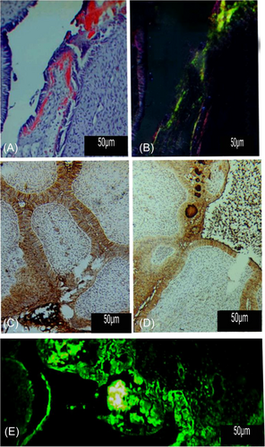
The immunohistochemical expression of AMELX was positive in 95.3% of the cases, while 93.4% of the cases were the expression of ODAM (Figure 5). Table 3 illustrates the Immunohistochemical expression scores of amyloid, AMELX, and ODAM. Of the total number of cases examined, 47 were positive for amyloid and ODAM, while 52 were positive for ODAM but negative for amyloid. All positive amyloid cases were also ODAM-positive. A positive correlation was found between Amyloid and ODAM p < 0.01. AMELX and Amyloid showed no significant relationship (Figure 5 and Table 3).
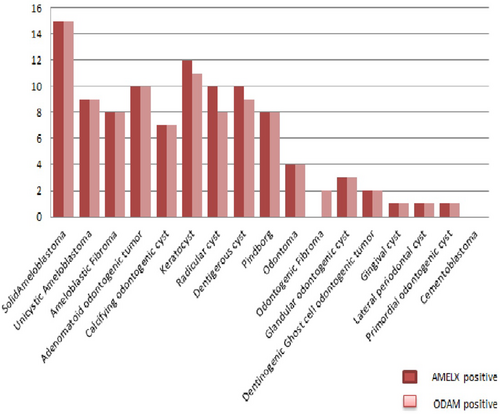
| Type of lesion | Total number of cases positive to AMELX | Total number of cases positive to ODAM | Total number of cases positive to amyloid | Number of cases with AMELX and amyloid positive | Number of cases with ODAM and amyloid positive |
|---|---|---|---|---|---|
| Solid ameloblastoma | 15 | 15 | 5 | 5 | 5 |
| Unicystic ameloblastoma | 9 | 9 | 3 | 3 | 3 |
| Ameloblastic fibroma | 8 | 8 | 1 | 1 | 1 |
| Adenomatoid odontogenic tumor | 10 | 10 | 6 | 6 | 6 |
| Calcifying odontogenic cyst | 7 | 7 | 3 | 3 | 3 |
| Keratocyst | 12 | 11 | 0 | 0 | 0 |
| Radicular cyst | 10 | 8 | 2 | 2 | 2 |
| Dentigerous cyst | 10 | 9 | 3 | 3 | 3 |
| Pindborg | 8 | 8 | 8 | 8 | 8 |
| Odontoma | 4 | 4 | 0 | 0 | 0 |
| Odontogenic fibroma | 0 | 2 | 1 | 0 | 1 |
| Glandular odontogenic cyst | 3 | 3 | 0 | 0 | 0 |
| Dentinogenic Ghost cell odontogenic tumor | 2 | 2 | 1 | 1 | 1 |
| Gingival cyst | 1 | 1 | 0 | 0 | 0 |
| Lateral periodontal cyst | 1 | 1 | 0 | 0 | 0 |
| Primordial odontogenic cyst | 1 | 1 | 1 | 1 | 1 |
| Cementoblastoma | 0 | 0 | 0 | 0 | 0 |
| Total | 101 | 99 | 34 | 33 | 34 |
| Parameter | ODAM | Amyloid | |||
|---|---|---|---|---|---|
| AMELX | Correlation | 0.303 | 0.135 | ||
| P value | 0.202 | 0.315 | |||
| ODAM | Correlation | 0.724 | |||
| P value | 0.000 | ||||
4 DISCUSSION
The present study represents the highest number of odontogenic cysts and tumors to evaluate the deposition of amyloid. Amyloid was detected for the first time in cases of unicystic ameloblastoma, ameloblastic fibroma, radicular cyst, dentigerous cyst, dentinogenic ghost cell tumor and, a recently classified odontogenic tumor named primordial odontogenic tumor. The study also detected amyloid in lesions proven previously: Pindborg tumor, adenomatoid odontogenic tumor, solid ameloblastoma,12 and odontogenic fibroma.17 Negative results were seen in all available cases of odontogenic keratocyst, lateral periodontal cyst, odontoma, cementoblastoma, glandular odontogenic cyst, and gingival cyst of the adults. These results may be due to the limited number of cases, or it could be due to other factors related to lesions' own behavior and level of differentiation, furthermore it is the same reason why other types of tumors associated with amyloid have negative results occasionally. In contrast to the present study, Buchner and David denied the presence of amyloid in lesions other than pindborg tumor.13 Most of these negative results could possibly be false negatives, and only a trace amount of amyloid could be present without being detected. The advantage of using thioflavin T stain is that it detects even small deposits of amyloid but is not completely specific for amyloid and stains other structures, including keratin and dentin. Therefore, these results should be confirmed by Congo red stain.19 The amyloid fibril Congo red complex has apple-green birefringence as an intrinsic property. The thickness of the section is essential (6–10 µm is recommended); if the section is too thick or thin, it may show miscellaneous birefringent colors. Using a modern microscope of high optical quality with color-corrected optics is essential, with the strongest possible light source that should be used to visualize faint birefringence. Only with these settings can even the smallest amyloid deposits can be detected.21, 22
The present study revealed a strong relationship between the pindborg tumor and to diffuse amount of amyloid, which is related to the diagnosis of the pindborg tumor. Meanwhile, the scattered focal and multifocal amounts of amyloid could be related to other odontogenic lesions.
In this study, the expression of AMLEX was positive in 95.3% of cases, with varying degrees of positivity. This result may be attributable to the fact that epithelial cells in these lesions maintain the ability to undergo ameloblastic differentiation from their original tissues. Still, other processes may prevent the epithelium from becoming completely differentiated. As a result, AMELX is likely an indication of epithelial cell differentiation in odontogenic lesions. This outcome matched the observations of previous studies23-26
In the amyloid deposits of pindborg tumors, ODAM was found as the predominant protein component.27 It was designated an odontogenic ameloblast-associated protein (ODAM) due to its high expression in enamel-associated epithelia.28, 29 In this study, the expression of ODAM was predominantly positive (93.4%), ranging from mild to intense positivity. These findings may be explained by the diverse biological activities of ODAM in the physiological and pathological processes of various odontogenic and non-odontogenic human tumors.18
ODAM is an Amyloid precursor protein and was formerly referred to as Amyloid pindborg (A pin).27 The identification of amyloid in tumors with different stages of differentiation supports its expression in most odontogenic lesions.
ODAM showed a highly significant relationship with amyloid at p < 0.01, this result is supported by previous studies which considered ODAM as an Amyloid precursor protein in Odontogenic lesions.7, 27 On the other hand, the results of the AMELX marker revealed a nonsignificant relationship with amyloid; as amyloid is not related to AMELX and is not related to the degree of aggressiveness of the lesion. According to the study by Murphy et al.7 which provided documentation for the ODAM-related nature of odontogenic amyloid associated with CEOTs, we assume that odontogenic amyloid may be both physiological tissue product since it was detected in the unerupted dental follicle,7 and pathological products since its precursor protein (ODAM) expressed in different odontogenic and non-odontogenic tumors as mentioned before. According to their differentiation level and the tumor's behavior, odontogenic lesions may produce amyloid in variable amounts.
4.1 Limitations of the study
- 1.
Most odontogenic lesions are rare, and the variability in types of odontogenic lesions from cystic, hamartomatous, benign, borderline, and malignant may limit the comprehensive evaluation of all odontogenic lesions.
- 2.
Lack of gene expression profiling using qPCR, being an immunohistochemical study, may result in limited findings for this study population.
AUTHOR CONTRIBUTIONS
Haider H. Al-Qazzaz: Conceptualization; data curation; methodology; resources; software; visualization; writing – original draft; writing – review and editing. Bashar H. Abdullah: Data curation; formal analysis; methodology; project administration; supervision; visualization; writing – review and editing. Omar S. Museedi: Data curation; software; validation; writing – review and editing.
ACKNOWLEDGMENTS
The authors would like to express our heartfelt gratitude to the oral and maxillofacial pathology laboratory teaching staff and technicians for their kind assistance throughout the study.
CONFLICT OF INTEREST
The authors declare no conflict of interest.
ETHICS STATEMENT
Ethical approval for this study was obtained in accordance with the declaration of Helsinki.
TRANSPARENCY STATEMENT
The lead author Haider H. Al-Qazzaz affirms that this manuscript is an honest, accurate, and transparent account of the study being reported; that no important aspects of the study have been omitted; and that any discrepancies from the study as planned (and, if relevant, registered) have been explained.
Open Research
DATA AVAILABILITY STATEMENT
The data that support the findings of this study are available from the corresponding author upon reasonable request.


