Glycemic Control Predicts Severity of Hepatocyte Ballooning and Hepatic Fibrosis in Nonalcoholic Fatty Liver Disease
Abstract
Background and Aims
Whether glycemic control, as opposed to diabetes status, is associated with the severity of NAFLD is open for study. We aimed to evaluate whether degree of glycemic control in the years preceding liver biopsy predicts the histological severity of NASH.
Approach and Results
Using the Duke NAFLD Clinical Database, we examined patients with biopsy-proven NAFLD/NASH (n = 713) and the association of liver injury with glycemic control as measured by hemoglobin A1c (HbA1c). The study cohort was predominantly female (59%) and White (84%) with median (interquartile range) age of 50 (42, 58) years; 49% had diabetes (n = 348). Generalized linear regression models adjusted for age, sex, race, diabetes, body mass index, and hyperlipidemia were used to assess the association between mean HbA1c over the year preceding liver biopsy and severity of histological features of NAFLD/NASH. Histological features were graded and staged according to the NASH Clinical Research Network system. Group-based trajectory analysis was used to examine patients with at least three HbA1c (n = 298) measures over 5 years preceding clinically indicated liver biopsy. Higher mean HbA1c was associated with higher grade of steatosis and ballooned hepatocytes, but not lobular inflammation. Every 1% increase in mean HbA1c was associated with 15% higher odds of increased fibrosis stage (OR, 1.15; 95% CI, 1.01, 1.31). As compared with good glycemic control, moderate control was significantly associated with increased severity of ballooned hepatocytes (OR, 1.74; 95% CI, 1.01, 3.01; P = 0.048) and hepatic fibrosis (HF; OR, 4.59; 95% CI, 2.33, 9.06; P < 0.01).
Conclusions
Glycemic control predicts severity of ballooned hepatocytes and HF in NAFLD/NASH, and thus optimizing glycemic control may be a means of modifying risk of NASH-related fibrosis progression.
Abbreviations
-
- AF
-
- advanced fibrosis
-
- BMI
-
- body mass index
-
- CMH
-
- Cochran-Mantel-Haenszel
-
- DUHS
-
- Duke University Health System
-
- GLP-1RA
-
- glucagon-like peptide-1 receptor agonist
-
- HB
-
- hepatocyte ballooning
-
- Hb1Ac
-
- hemoglobin A1c
-
- HF
-
- hepatic fibrosis
-
- IQR
-
- interquartile range
-
- LI
-
- lobular inflammation
-
- NAS
-
- NAFLD Activation Score
-
- RCS
-
- restricted cubic splines
-
- SGLT2i
-
- sodium-glucose cotransporter 2 inhibitor
-
- T1D
-
- type 1 diabetes
-
- T2D
-
- type 2 diabetes
NAFLD is a growing epidemic, affecting 1 in 4 persons worldwide(1, 2) and ~60% of patients with type 2 diabetes (T2D).(3, 4) The term NAFLD encompasses a disease spectrum with isolated steatosis on the most benign end, to NASH characterized by steatosis, inflammation, and ballooned hepatocytes, with or without fibrosis.(5) NASH increases the risk of fibrosis progression to cirrhosis with risk of hepatic decompensation and HCC, making NASH the leading indication for liver transplantation in the USA.(6) Higher grades of steatosis, inflammation, and ballooned hepatocytes render increased steatohepatitis severity, which, in turn, is strongly associated with progressive hepatic fibrosis (HF),(7) the primary predictor of liver-related morbidity and mortality.(8)
T2D is a well-established risk factor for the development of NAFLD, and is a strong predictor of advanced HF and complications of cirrhosis, such as HCC and liver-related mortality.(9-12) Despite the clear link between NAFLD and T2D, little is understood about how glycemic control impacts histological severity and associated risk for NAFLD progression. Although glucose-lowering drugs have been used as therapeutic approaches for NASH, it is unclear whether the therapeutic benefit is attributable to the glucose-lowering effect of such interventions. Furthermore, liver aminotransferases in patients with diabetes are particularly insensitive(13) and are normal in >70% of patients with biopsy-proven NASH,(14) limiting the ability to ascertain the effect of glucose lowering on NASH using noninvasive surrogates for disease activity. Therefore, studies which directly examine the association of glycemic control, as opposed to diabetes status alone, on histological features of liver injury in NAFLD/NASH are needed.
In order to address this evidence gap, we investigated the association between degree of glycemic control (assessed using mean hemoglobin A1c [HbA1c]), as well as prebiopsy trends in glycemic control in the years preceding a clinically indicated liver biopsy in a large cohort of well-phenotyped patients with biopsy-proven NAFLD/NASH.
Materials and Methods
Study Design and Data Source
We conducted a longitudinal cohort study using retrospectively/prospectively collected data from NAFLD subjects in the Duke University Health System (DUHS) NAFLD Biorepository (Duke University, Durham, NC). The DUHS NAFLD Clinical Database, established in 2007, is a prospective, open-enrolling, and well-annotated clinical database of patients who underwent clinical and histological evaluations of the suspected diagnosis of NASH as part of standard of care. The DUHS NAFLD Clinical Database was approved by the Duke University Institutional Review Board (Protocol #00102631) and conducted in accordance with the Declaration of Helsinki ethical guidelines. For the present study, NAFLD was defined as (1) presence of >5% hepatic steatosis on liver biopsy and (2) absence of histological and serological evidence for other chronic liver disease in a patient with risk factors for metabolic syndrome.
Demographic data (i.e., height, weight, BMI, age, sex, race, ethnicity, smoking status, and comorbid illnesses) and laboratory studies (i.e., lipids, glucose, HbA1c, liver aminotransferases, and measures of liver synthetic function) were obtained within 6 months of liver biopsy in all patients and extracted from the medical record, as otherwise available, for the preceding 5 years before liver biopsy for evaluation of NAFLD/NASH.
Study Population
We examined 713 patients enrolled into the DUHS NAFLD Clinical Database between January 2007 and October 2019, who met the following inclusion criteria for our study: (1) age ≥18 years, (2) histological diagnosis of NAFLD, and (3) at least one documented HbA1c value in the year preceding a clinically indicated liver biopsy for evaluation of NAFLD/NASH. Exclusion criteria included the following: (1) current alcohol consumption of ≥14 servings per week (for men) and at least seven servings per week (for women); (2) serological evidence of alternative forms of chronic liver disease (e.g., chronic viral hepatitis, primary biliary cirrhosis, autoimmune hepatitis, hemochromatosis, Wilson’s disease, and alpha-1-antitrypsin deficiency); (3) histological features suggesting coexisting liver diseases; (4) history of bariatric surgery; or (5) liver transplantation. No patients with decompensated or overt features of cirrhosis were included, given that none of these patients would have undergone a clinically indicated liver biopsy.
Diagnosis of diabetes was defined as an HbA1c ≥6.5% (≥48 mmol/mol), the presence of a diagnosis of diabetes as detailed in the medical history by a provider, at least two fasting glucose values ≥126 mg/dL (7.0 mmol/L) in excess of 6 months apart, or the use of glucose-lowering agents. The study scheme is detailed in Fig. 1.

HbA1c Measures
We used manual electronic health record chart review to extract additional HbA1c data from 5 years before to 90 days after liver biopsy. All patients meeting inclusion and exclusion criteria for the study were examined in the mean HbA1c analysis, which used HbA1c data over 1 year before to 90 days after biopsy. Only patients with three or more HbA1c levels from 5 years before to 90 days after liver biopsy were examined by group-based trajectory analysis. We allowed HbA1c data up to 90 days postbiopsy given that levels drawn in this time period (within 3 months) may still reasonably reflect glycemic control at time of biopsy.
Liver Histology
All liver biopsy specimens were stained with HE and Masson’s trichrome stains and reviewed and scored by a hepatopathologist according to the published Nonalcoholic Steatohepatitis Clinical Research Network (NASH CRN) grading and staging system.(15) The hepatopathologist was blinded to clinical phenotype of the patient and associated laboratory data. For the analyses, fibrosis stages 1a, 1b, and 1c were combined and treated as stage 1. To address our research interest in evaluating the association of portal inflammation with glycemic control, grades of portal inflammation were defined as grade 0 (absent) and grade 1 (present).
Primary Outcomes
The primary outcome was severity of HF stage as defined by the NASH CRN (stage 0-4).(15) Severity of individual histological features of steatohepatitis (grade of steatosis, lobular inflammation, and ballooned hepatocytes) and the composite assessment of severity of NASH as defined by NAFLD Activity Score (NAS) were analyzed as secondary outcomes. Presence of NASH was defined as NAS ≥4.
Statistical Analysis
Demographic, clinical, and hepatic histological data were summarized as a count (percent) or median (interquartile range; IQR). Wilcoxon’s rank-sum tests and Kruskal-Wallis’ tests were used to compare continuous variables between patients with and without diabetes or among different HbA1c trajectory groups, respectively. Cochran-Mantel-Haenszel (CMH) tests were used to compare categorical variables. A two-sided test with a P value of <0.05 was considered statistically significant for all analyses. Statistical analyses were conducted using SAS statistical software (version 9.4; SAS Institute Inc., Cary, NC).
Mean HbA1c Analysis
The association between mean HbA1c in the year preceding biopsy and hepatic histological features was investigated with ordinal logistic regression after testing for proportional odds assumption, multicolinearity, and linear association. The relationship between mean HbA1c and outcome measures was in some cases linear and in other cases nonlinear. For linear associations between mean HbA1c and severity of histological outcomes, ORs and 95% CIs were estimated using regular ordinal logistic regression analyses.
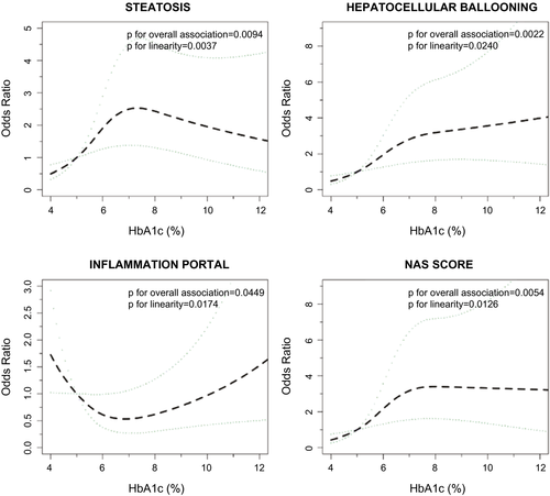
In order to ensure that the nonlinear relationship between HbA1c and certain histological outcomes was adequately captured, we used the restricted cubic spline (RCS) method for continuous variable transformation using R package rms followed by original logistic regression analysis.(16) RCS with three knots (corresponding to the 10th, 50th, and 90th percentiles of mean HbA1c on the basis of the distribution of our data) was used to estimate the dose-response relationship using an HbA1c reference of 5.0% (31 mmol/mol).(17) We chose a reference of 5.0% because this represents a low-normal HbA1c value against which to examine the impact of higher HbA1c values commonly observed in clinical practice. ORs and 95% CIs before and after the turning point (HbA1c of 7.0% [53 mmol/mol]), according to dose-response plots (Fig. 2), were estimated using an ordinal logistic regression model after transforming mean HbA1c values using the method described by Singer and Willett.(18) All analyses were adjusted for age, sex, race, body mass index (BMI), diabetes status, and hyperlipidemia status. The number of glucose-lowering drugs used by each patient did not impact the model and therefore was excluded as a covariate.

Group-Based Trajectory Analysis
To examine the association between hepatic histological features and trajectory pattern of HbA1c 5 years preceding (and up to 90 days after) biopsy, a group-based trajectory model(19) was used using SAS Proc Traj macro.(20) The Bayesian information criterion (BIC) was used to assess the optimal number of trajectory groups, where lower BIC values point to a better model. Other criteria for ascertaining the best fitting model included nonoverlapping CIs, reasonable sample sizes in each identified trajectory group (each group should include ≥5% of the subjects), and distinct average posterior probabilities across groups. Finally, a multivariable ordinal logistic regression model was applied to investigate the association between HbA1c trajectory groups and all histological features, adjusted for age, sex, race, BMI, diabetes status, and hyperlipidemia status.
In both mean HbA1c and group-based trajectory analyses, glucagon-like peptide-1 receptor agonist (GLP-1RA) use, thiazolidinedione use, and ethnicity were examined as covariates in the model (see Supporting Tables S2 and S3); however, they did not substantially impact results, so were excluded as covariates in the main analysis to avoid overfitting. Sodium-glucose cotransporter 2 inhibitor (SGLT2i) use was not examined as a covariate given its low prevalence in our cohort (0.4%).
Results
A total of 713 patients met inclusion and exclusion criteria for the study. Of the 713 patients, 49% (n = 348) had diabetes and 51% (n = 365) did not have diabetes. Table 1 describes patient characteristics of the cohort, with stratification by diabetes status. The median age of our cohort was 50 years, and most (59%; n = 417) were female. Patients with diabetes were older than those without (53 vs. 47 years; P < 0.0001) and were more likely to be female (67% vs. 50%; P < 0.0001). The majority of patients were White (84%) and non-Hispanic (72%), though there were proportionally more Black patients in the diabetes group (13% vs. 6.3%; P = 0.007). Cirrhosis (stage 4 fibrosis) was noted in 3.8%. Of patients with T2D, 66% were on metformin, and nearly 1 in 4 were on insulin therapy at time of liver biopsy. Median HbA1c was 6.9% (52 mmol/mol) in the diabetes group and 6.0% (42 mmol/mol) in the whole cohort (see Supporting Fig. S1 for distribution of HbA1c values). Compared to persons without diabetes, those with diabetes had higher median BMI (35 vs. 32 kg/m2; P < 0.0001) and triglycerides (166 vs. 144 mg/dL; P = 0.0027). The overwhelming majority, 98% of those with diabetes, were diagnosed with T2D whereas only 2% were diagnosed with type 1 or indeterminate/mixed-type diabetes (n = 8).
| Characteristics | Whole Cohort (n = 713) | No Diabetes (n = 365) | Diabetes (n = 348) | P Value |
|---|---|---|---|---|
| Age (years) | 50 (42, 58) | 47 (39, 56) | 53 (45, 59) | <0.0001* |
| Female sex (n, %) | 417 (58.5) | 184 (50.4) | 233 (67.0) | <0.0001† |
| Race (n, %) | ||||
| White | 598 (83.9) | 319 (87.4) | 279 (80.2) | 0.0070† |
| Black | 69 (9.7) | 23 (6.3) | 46 (13.2) | |
| Other | 46 (6.5) | 23 (6.3) | 23 (6.6) | |
| Ethnicity (n, %) | ||||
| Hispanic | 14 (2.0) | 8 (2.2) | 6 (1.7) | 0.0027† |
| Non-Hispanic | 511 (71.7) | 281 (77.0) | 230 (66.1) | |
| Unknown | 188 (26.4) | 76 (20.8) | 112 (32.2) | |
| Glucose-lowering drug use (n, %) | ||||
| Metformin | 232 (32.5) | 0 (0.0) | 232 (66.7) | <0.0001† |
| Sulfonylureas | 68 (9.5) | 0 (0.0) | 68 (19.5) | <0.0001† |
| Thiazolidinediones | 21 (3.0) | 0 (0.0) | 21 (6.0) | <0.0001† |
| DPP4 inhibitors | 25 (3.5) | 0 (0.0) | 25 (7.2) | <0.0001† |
| GLP-1RA | 22 (3.1) | 0 (0.0) | 22 (6.3) | <0.0001† |
| Insulin | 85 (11.9) | 0 (0.0) | 85 (24.4) | <0.0001† |
| SGLT2i | 3 (0.4) | 0 (0.0) | 3 (0.9) | 0.1158‡ |
| Other medications (n, %) | ||||
| Statins | 195 (27.4) | 61 (16.7) | 134 (38.5) | <0.0001† |
| Vitamin E | 43 (6.0) | 16 (4.4) | 27 (7.8) | 0.0586† |
| BMI (kg/m2) | 33.6 (30.3, 38.4) | 32.3 (29.6, 36.3) | 35.2 (31.6, 40.0) | <0.0001* |
| Systolic BP (mm Hg) | 132 (122, 141) | 131 (122, 140) | 133 (122, 142) | 0.3838* |
| Diastolic BP (mm Hg) | 78 (71, 85) | 79 (73, 86) | 76 (70, 83) | 0.0003* |
| Laboratory data | ||||
| HbA1c (%) | 6.0 (5.5, 6.9) | 5.6 (5.3, 5.9) | 6.9 (6.4, 8.1) | <0.0001* |
| HbA1c (mmol/mol) | 42 (37, 52) | 38 (34, 41) | 52 (46, 65) | <0.0001* |
| LDL (mg/dL) | 110 (83, 139) | 117 (89, 144) | 103 (77, 134) | <0.0001* |
| HDL (mg/dL) | 39 (32, 46) | 40 (34, 47) | 38 (31, 44) | 0.0047* |
| Triglycerides (mg/dL) | 155 (109, 225) | 144 (104, 217) | 166 (121, 236) | 0.0027* |
| eGFR (mL/min/1.73 m2) | 94.6 (80.8, 106.3) | 92.8 (80.7, 106.5) | 96.6 (81.0, 105.7) | 0.5153* |
| Steatosis, grade (n, %) | ||||
| 0 | 15 (2.1) | 9 (2.5) | 6 (1.7) | |
| 1 | 271 (38.0) | 128 (35.1) | 143 (41.1) | 0.2155† |
| 2 | 256 (35.9) | 135 (37.0) | 121 (34.8) | |
| 3 | 171 (24.0) | 93 (25.5) | 78 (22.4) | |
| HB, grade (n, %) | ||||
| 0 | 157 (22.1) | 107 (29.4) | 50 (14.4) | <0.00001† |
| 1 | 319 (44.9) | 169 (46.4) | 150 (43.2) | |
| 2 | 235 (33.1) | 88 (24.2) | 147 (42.4) | |
| LI, grade (n, %) | ||||
| 0 | 27 (3.9) | 16 (4.5) | 11 (3.3) | 0.5917† |
| 1 | 455 (65.6) | 235 (66.0) | 220 (65.1) | |
| 2 | 190 (27.4) | 92 (25.8) | 98 (29.0) | |
| 3 | 22 (3.2) | 13 (3.7) | 9 (2.7) | |
| Portal inflammation, grade (n, %) | ||||
| 0 | 420 (60.3) | 233 (65.6) | 187 (54.7) | 0.0032† |
| 1 | 277 (39.7) | 122 (34.4) | 155 (45.3) | |
| Fibrosis stage (n, %) | ||||
| 0 | 115 (16.1) | 79 (21.6) | 36 (10.3) | |
| 1 | 232 (32.5) | 135 (37.0) | 97 (27.9) | <0.0001† |
| 2 | 184 (25.8) | 87 (23.8) | 97 (27.9) | |
| 3 | 155 (21.7) | 56 (15.3) | 99 (28.5) | |
| 4 | 27 (3.8) | 8 (2.2) | 19 (5.5) | |
| NAS score (n, %) | ||||
| <4 (non-NASH) | 210 (30.4) | 125 (35.2) | 85 (25.2) | 0.0043† |
| ≥4 (definite NASH) | 482 (69.6) | 230 (64.8) | 252 (74.8) |
- Data presented as median (IQR), unless stated otherwise. Of patients with diabetes, 98% (n = 341) had T2D and 2% (n = 7) had T1D.
- The presence of NASH was defined as a NAS of >4.(14)
- * Wilcoxon’s rank-sum test.
- † CMH test.
- ‡ Fisher’s exact test.
- Abbreviations: DPP4, dipeptidyl peptidase 4; BP, blood pressure; eGFR, estimated glomerular filtration rate.
Mean HbA1c in the year preceding biopsy was significantly (linearly) associated with severity of liver fibrosis at time of biopsy, even after adjusting for age, sex, race, BMI, diabetes, and hyperlipidemia status. Every 1% increase in mean HbA1c was associated with 15% higher odds of increased HF, analyzed by the original logistic regression model (OR, 1.15; 95% CI, 1.01, 1.31; P = 0.039). The dose-response plot for HF, derived from RCS methods, also showed a linear trend (Supporting Fig. S2), though this did not reach statistical significance (P = 0.097). When assessing individual covariates from the model, presence of diabetes was associated with the highest odds of increased fibrosis severity (OR, 1.71; 95% CI, 1.20, 2.44; P = 0.003). Higher mean HbA1c preceding biopsy was also associated with higher grades of steatosis, ballooned hepatocytes, and portal inflammation, but not with lobular inflammation (LI; Table 2).
| Histological Outcomes (Linear Relationship) | OR (95% CI) | P Value |
|---|---|---|
| Fibrosis severity (stage 0-4) | 1.15 (1.01, 1.31) | 0.0390 |
| LI (score 0-3) | 1.12 (0.96, 1.30) | 0.1440 |
| Histological outcomes (nonlinear relationship) | HbA1c <7.0% (53 mmol/mol) | HbA1c >7.0% (53 mmol/mol) | ||
|---|---|---|---|---|
| OR (95% CI) | P Value | OR (95% CI) | P Value | |
| HB (score 0-2) | 1.62 (1.15, 2.28) | 0.0060 | 1.06 (0.87, 1.29) | 0.5843 |
| Steatosis (score 0-3) | 1.62 (1.15, 2.27) | 0.0054 | 0.89 (0.73, 1.08) | 0.2326 |
| Portal inflammation (score 0-1) | 0.67 (0.46, 0.99) | 0.0448 | 1.31 (1.04, 1.64) | 0.0196 |
| Definite NASH vs. no NASH (NAS ≥4 vs. <4)* | 1.86 (1.25, 2.78) | 0.0023 | 0.96 (0.75, 1.23) | 0.7526 |
- ORs represent odds of more severe histology for every 1% increase in HbA1c. An ordinal logistic regression model was used and was adjusted for age, sex, race, BMI, T2D, and hyperlipidemia. Restricted cubic spline regression was used to test the linear association between mean HbA1c and histological features. For outcomes with nonlinear relationship to HbA1c, ORs (95% CI) before and after an HbA1c of 7.0% were estimated using an ordinal logistic regression model after transforming mean HbA1c using the method described by Singer and Willett.(17) The choice of an HbA1c cutoff of 7.0% was data driven, based on dose-response plots (Fig. 2).
- * Presence of definite NASH was defined as a NAS of ≥4.(14)
Dose-response plots derived from RCS methods demonstrated that the associations were nonlinear and approximated inverted L-shaped curves for the relationships between mean HbA1c and steatosis, hepatocellular ballooning (HB), and NAS (Fig. 2). A V-shaped dose-response curve was observed for the association between HbA1c and portal inflammation (Fig. 2). For mean HbA1c <7.0% (53 mmol/mol), positive linear associations were detected for outcomes of steatosis, HB, and NAS; the significance of these linear associations was lost beyond an HbA1c of 7% (53 mmol/mol), and Table 2 reports ORs and 95% CIs for these histological outcomes after adjusting for age, sex, race, BMI, diabetes, and hyperlipidemia status.
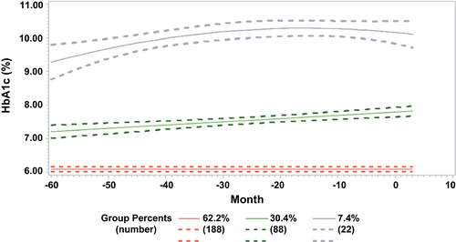
Group-based trajectory analysis was conducted for those patients with at least three measures of HbA1c values (n = 298); how this group compares to those with less than three HbA1c measures (i.e., those excluded from group-based trajectory analysis) can be seen in Supporting Table S1. Three HbA1c control groups were identified by group-based trajectory analysis: good (group 1), moderate (group 2), and poor (group 3) glycemic control (Fig. 3). A higher proportion of patients with poor glycemic control self-identified as “Other” race (31.8%) compared to the moderate (12.5%) and good (8.5%) glycemic control groups (Table 3). Median HbA1c values were 6.0% (42 mmol/mol), 7.6% (60 mmol/mol), and 10.0% (86 mmol/mol) in the good, moderate, and poor glycemic control groups, respectively. GLP-1RA use increased as glycemic control worsened (2.7% vs. 10.2%, vs. 13.6%, in groups 1, 2, and 3, respectively), as did insulin use (4.8% vs. 43.2%, vs. 77.3%, in groups 1, 2, and 3, respectively). Likewise, median BMI increased as glycemic control worsened (34.4 vs. 35.3 vs. 36.8 kg/m2, in groups 1, 2, and 3, respectively). Triglycerides were similar in moderate and poor control groups (median of 163 and 166 mg/dL, respectively) and were lower in the good glycemic control group (median 147.5 mg/dL), although not significant between groups.

| Characteristics | Group 1 Good Glycemic Control (n = 188) | Group 2 Moderate Glycemic Control (n = 88) | Group 3 Poor Glycemic Control (n = 22) | P Value |
|---|---|---|---|---|
| Age (years) | 52 (43, 60) | 54 (47, 60) | 51 (46, 57) | 0.4424* |
| Female sex (n, %) | 116 (61.7) | 58 (65.9) | 16 (72.7) | 0.5268† |
| Race (n, %) | ||||
| White | 152 (80.9) | 72 (81.8) | 14 (63.6) | 0.0718† |
| Black | 20 (10.6) | 5 (5.7) | 1 (4.6) | |
| Other | 16 (8.5) | 11 (12.5) | 7 (31.8) | |
| Ethnicity (n, %) | ||||
| Hispanic | 2 (1.1) | 3 (3.4) | 0 (0.0) | 0.0667† |
| Non-Hispanic | 137 (72.9) | 52 (59.1) | 12 (54.6) | |
| Unknown | 49 (26.1) | 33 (37.5) | 10 (45.5) | |
| Diabetes (n, %) | 112 (59.6) | 88 (100.0) | 22 (100.0) | <0.0001† |
| HbA1c (%) | 6.0 (5.6, 6.5) | 7.6 (7.2, 8.1) | 10.0 (9.4, 11.0) | <0.0001* |
| HbA1c (mmol/mol) | 42 (38, 48) | 60 (55, 65) | 86 (79, 97) | <0.0001* |
| Glucose-lowering drug use (n, %) | ||||
| Metformin | 73 (38.8) | 64 (72.7) | 15 (68.2) | <0.0001† |
| Sulfonylureas | 15 (8.0) | 30 (34.1) | 3 (13.6) | <0.0001† |
| Thiazolidinediones | 4 (2.1) | 7 (8.0) | 1 (4.6) | 0.0719† |
| DPP4 inhibitors | 4 (2.1) | 13 (14.8) | 1 (4.6) | 0.0002† |
| GLP-1RA | 5 (2.7) | 9 (10.2) | 3 (13.6) | 0.0104† |
| Insulin | 9 (4.8) | 38 (43.2) | 17 (77.3) | <0.0001† |
| SGLT2i | 0 (0.0) | 1 (1.14) | 1 (4.5) | 0.0390† |
| Other medications | ||||
| Statins | 69 (36.7%) | 46 (52.3%) | 10 (45.5%) | 0.0481† |
| Vitamin E | 18 (9.6%) | 2 (2.3%) | 0 (0.0%) | 0.0335† |
| BMI (kg/m2) | 34.4 (31.0, 39.5) | 35.3 (32.5, 40.5) | 36.8 (33.2, 41.6) | 0.1668* |
| Systolic BP (mm Hg) | 132 (123, 141) | 132 (121, 142) | 135 (127, 145) | 0.4037* |
| Diastolic BP (mm Hg) | 77 (69, 85) | 75 (70, 80) | 79 (73, 84) | 0.2632* |
| Laboratory data | ||||
| LDL (mg/dL) | 106 (82, 135) | 97 (73, 119) | 87 (69, 156) | 0.1986* |
| HDL (mg/dL) | 38 (33, 45) | 38 (30, 43) | 37 (30, 43) | 0.3036* |
| Triglycerides (mg/dL) | 147.5 (115.0, 219.0) | 163.0 (129,0, 218.0) | 166.0 (107.0, 233.0) | 0.4351* |
| eGFR (mL/min/1.73 m2) | 93.8 (77.3, 103.7) | 95.4 (82.1, 104.4) | 101.6 (77.0,116.0) | 0.2570* |
| Steatosis grade (n, %) | ||||
| 0 | 1 (0.5) | 2 (2.3) | 1 (4.6) | |
| 1 | 79 (42.0) | 37 (42.1) | 9 (40.9) | 0.4033† |
| 2 | 57 (30.3) | 33 (37.5) | 8 (36.4) | |
| 3 | 51 (27.1) | 16 (18.2) | 4 (18.2) | |
| HB, grade (n, %) | ||||
| 0 | 41 (21.9) | 9 (10.2) | 3 (13.6) | 0.0027† |
| 1 | 89 (47.6) | 36 (40.9) | 8 (36.4) | |
| 2 | 57 (30.5) | 43 (48.9) | 11 (50.0) | |
| LI, grade (n, %) | ||||
| 0 | 3 (1.6) | 6 (7.1) | 1 (4.6) | |
| 1 | 136 (73.5) | 50 (59.5) | 13 (59.1) | 0.7241† |
| 2 | 43 (23.2) | 25 (29.8) | 8 (36.4) | |
| 3 | 3 (1.6) | 3 (3.6) | 0 (0.0) | |
| Portal inflammation, grade (n, %) | ||||
| 0 | 110 (59.8) | 44 (50.6) | 11 (50.0) | 0.2992† |
| 1 | 74 (40.2) | 43 (49.4) | 11 (50.0) | |
| Fibrosis stage (n, %) | ||||
| 0 | 34 (18.1) | 8 (9.1) | 1 (4.6) | |
| 1 | 64 (34.0) | 19 (21.6) | 7 (31.8) | 0.0003† |
| 2 | 53 (28.2) | 20 (22.7) | 8 (36.4) | |
| 3 | 34 (18.1) | 31 (35.2) | 5 (22.7) | |
| 4 | 3 (1.6) | 10 (11.4) | 1 (4.6) | |
| NAS score (n, %) | ||||
| <4 (non-NASH) | 64 (34.8) | 15 (17.9) | 5 (22.7) | 0.0146† |
| ≥4 (definite NASH) | 120 (65.2) | 69 (82.1) | 17 (77.3) |
- Data presented as median (IQR), unless stated otherwise. The presence of NASH was defined as a NAS of >4.(14)
- * Kruskal-Wallis’ test.
- † CMH test.
- Abbreviations: DPP4, dipeptidyl peptidase 4; BP,,blood pressure; eGFR, estimated glomerular filtration rate.
Compared to the good glycemic control group, patients with moderate control had substantially higher odds of advanced versus mild fibrosis (OR, 4.59; 95% CI, 2.33, 9.06), as well as higher odds of more severe HB (OR, 1.74; 95% CI, 1.01, 3.01) and higher NAS score (OR, 2.49; 95% CI, 1.25, 4.95; Table 4). There was no significant difference between groups with good and moderate control as it related to severity of steatosis, portal inflammation, or LI (data not shown). Comparisons with the poor control group were limited by small sample size (n = 22).
| OR (95% CI) | P Value | |
|---|---|---|
| Fibrosis severity (advanced vs mild; stage 3-4 vs. 0-2) | ||
| Group 2 vs. group 1 (moderate vs. good control) | 4.59 (2.33, 9.06) | <0.0001* |
| Group 3 vs. group 1 (poor vs. good control) | 2.52 (0.81, 7.84) | 0.1117 |
| Group 3 vs. group 2 (poor vs. moderate control) | 0.55 (0.18, 1.65) | 0.2841 |
| HB (grade 0-2) | ||
| Group 2 vs. group 1 (moderate vs. good control) | 1.74 (1.01, 3.01) | 0.0479* |
| Group 3 vs. group 1 (poor vs. good control) | 1.79 (0.72, 4.43) | 0.2089 |
| Group 3 vs. group 2 (poor vs. moderate control) | 1.03 (0.41, 2.60) | 0.9525 |
| Definite NASH vs. no NASH (NAS ≥4 vs. <4) | ||
| Group 2 vs. group 1 (moderate vs. good control) | 2.49 (1.25, 4.95) | 0.0094* |
| Group 3 vs. group 1 (poor vs. good control) | 1.77 (0.60, 5.23) | 0.3037 |
| Group 3 vs. group 2 (poor vs. moderate control) | 0.71 (0.22, 2.56) | 0.5620 |
- Defined by HbA1c Trajectory.
- ORs represent a logistic regression analysis with adjustment for age, sex, race, BMI, T2D, and hyperlipidemia.
Discussion
This study examined, in a large cohort of well-phenotyped patients with biopsy-proven disease, the association between cumulative glucose exposure and glycemic trajectories on the histological severity of NAFLD/NASH. We found glycemic control preceding biopsy to be linearly associated with severity of fibrosis. HbA1c was also associated with severity of steatosis, HB, portal inflammation, and likelihood of NASH, but this relationship was nonlinear and varied depending on degree of glycemic control.
We found every 1% increase in mean HbA1c preceding biopsy to be associated with 15% higher odds of increase in fibrosis stage. Mean HbA1c was also associated with severity of ballooned hepatocytes (for HbA1c, <7.0% [53 mmol/mol]; OR, 1.62; 95% CI, 1.15, 2.28). A similar pattern was observed in group-based trajectory analysis, where patients with moderate glycemic control had higher odds of increased stage of HF and grade of ballooned hepatocytes than those with good control. The ability to discern an association between poor versus good/moderate glycemic control and the odds of more severe HF or ballooned hepatocytes in group-based trajectory analysis was limited by small sample size, though this is a question of great importance and should be explored in future studies. Notably, evidence suggests that the severity of ballooned hepatocytes is a strong predictor of HF.(7, 21) Therefore, the strong association of glycemic control with the severity of both ballooned hepatocytes and stage of HF supports existing knowledge of T2D being a strong predictor of progressive NAFLD/NASH.
A number of studies have reported on the link between the presence of T2D and increased fibrosis severity, though data are mixed, and the influence of HbA1c on fibrosis is unclear.(22-26) A recent epidemiological study by Tanaka et al. found glycemic control to be associated with advanced fibrosis (AF; as defined by the lab-based Fibrosis-4 index) up to an HbA1c of 7.9% (63 mmol/mol), but not beyond.(27) A similar finding was also reported in another study that used NAFLD fibrosis score to define AF, except that HbA1c was only predictive of AF in patients with HbA1c <6.5% (48 mmol/mol).(26)
We observed a linear association between HbA1c and fibrosis stage over a broad range of HbA1c, from ~5% to 11% (31-97 mmol/mol). However, the association between HbA1c and likelihood of NASH, as well as severity of other histological features of NASH (i.e., steatosis, ballooning), was weaker beyond an HbA1c level of 7% (53 mmol/mol), similar to the HbA1c cutoffs noted in the literature. The reason for this discrepancy, particularly between ballooned hepatocytes and fibrosis, is unclear. One possible explanation is the phenomenon of “burnt-out NASH,” whereby patients lose characteristic features of NASH with worsening fibrosis severity.(28, 29) Given that HbA1c preceding biopsy is linked with fibrosis severity, patients with poor glycemic control and severe fibrosis may have lost histological features of steatohepatitis, resulting in a nonlinear relationship between these histological features and HbA1c.
Another potential explanation for lack of linear association between steatosis and ballooning above an HbA1c of 7.0% is the greater utilization of glucose-lowering drugs with increasing HbA1c. Use of diabetes medications did not impact our results when included in the multivariable model, although change in medications over time was not captured in our study and may have still influenced this relationship between HbA1c and histological features. This is particularly relevant for steatosis, given that multiple glucose-lowering agents have been associated with reduction in hepatic steatosis.(30) Given that steatosis and HB severity were linearly associated with glycemic control up to an HbA1c of 7.0% (but not above), our results may also suggest that patients with prediabetes and mild or well-controlled diabetes (i.e., HbA1c <7.0%) are uniquely impacted by interventions to improve glycemic control and insulin resistance to avoid progression to severe NAFLD. To this end, several pharmacotherapies which lower blood glucose levels have demonstrated efficacy in achieving the endpoint of NASH resolution.(31-34)
Notably, an inverse association was observed between glycemic control and portal inflammation, whereby greater HbA1c <7.0% (53 mmol/mol) was associated with lower odds of portal inflammation, and greater HbA1c >7.0% (53 mmol/mol) was associated with higher odds of portal inflammation (Fig. 2). The reason for this finding is unclear. Evidence suggests that portal inflammation, unlike lobular, is not statistically linked to NASH, hence its exclusion from the NAFLD histological scoring system.(15) Nonetheless, moderate or severe portal inflammation has been linked to several features of advanced NAFLD,(35) and improvement in fibrosis has likewise been associated with improvement in portal, but not lobular, inflammation.(36) Therefore, our finding that poor glycemic control (above an HbA1c of 7.0% [53 mmol/mol]) was associated with greater odds of portal inflammation may be of clinical importance and warrants further investigation.
Very few studies have attempted to examine the influence of HbA1c on histological components of NASH, other than fibrosis, so we are limited in drawing comparisons to the literature. One longitudinal study by Hamaguchi et al. examined 39 patients with sequential liver biopsies and observed change in HbA1c to be associated with progression of liver fibrosis, but not liver inflammation.(22) These results parallel our findings, though they did not examine severity of steatosis or ballooned hepatocytes. These results should be interpreted with caution because of small sample size and lack of adjustment for change in weight over time(22) as a potential confounder to changes in histological features of NASH. Our study was of sufficient sample size (n = 713), and the analysis adjusted for BMI. However, our analysis was limited by smaller sample size for group-based trajectory analysis comparisons with the poor glycemic control group (n = 22). Larger population-based studies with prospectively collected and uniformly spaced HbA1c data paired with liver histology data would enhance our understanding of how glycemic control impacts the natural history of NAFLD disease progression and further validate the results and interpretation of our analysis.
Our study used liver histology as the gold standard by which to define the severity of NASH and stage HF. Earlier studies assessing the association of glycemic control and features of NAFLD used noninvasive approaches, such as lab-based scoring systems and/or imaging modalities to define disease activity. A recent study by Wang et al.(37) found glycemic measures to be associated with development and resolution of NAFLD by ultrasonography. Because of the lack of liver histology data, no conclusions could be rendered regarding the effect of glycemic control on severity of necroinflammation or fibrosis.(5)
For the first time in 2019, American Diabetes Association guidelines recommended review of laboratory and imaging data to proactively identify and risk stratify NAFLD in patients with T2D.(38) With growing appreciation of the overlap between T2D and NAFLD, it is becoming increasingly important to understand how glycemic control, as measured by HbA1c, impacts risk of NASH and fibrosis. Our study has demonstrated a 15% higher odds of increased fibrosis stage with every 1% increase in HbA1c level. Furthermore, odds of advanced (vs. mild) fibrosis were 4.5 times higher in those patients with moderate versus good glycemic control. Although these findings suggest HbA1c as a potential modifiable risk factor for NASH progression, we do not establish a causal relationship between glycemic control and NAFLD/NASH in this study. We likewise do not challenge the prevailing hypothesis that diabetes and NAFLD are both consequences of prolonged adipose tissue and hepatic insulin resistance, and that subclinical NAFLD likely precedes diabetes in most cases. As such, clinicians should continue to weigh the risks and benefits of lower HbA1c targets in their patients to ensure that they are not causing harm (e.g., attributable to hypoglycemia). Longitudinal studies are needed to better examine this complex interplay between glucose exposure, insulin resistance, and NAFLD and guide clinical care.
Our study has a few limitations. First, we adjusted for key variables at time of biopsy, such as age, BMI, and hyperlipidemia, but did not collect these data longitudinally so were unable to adjust for changes over time. Given that such covariates were our main method of capturing and adjusting for insulin resistance in multivariable analysis, our assessment of this important parameter could only be done cross-sectionally near the time of biopsy. With regard to BMI, we did exclude patients who underwent bariatric surgery to avoid confounding attributable to substantial weight loss during the study period. Although it was a small subset of the population, we may have missed unintentional weight loss in the poor glycemic control group given that this can occur with persistent, severe hyperglycemia. Second, we may not have accounted for all liver effects of glucose-lowering drugs. For instance, preliminary data suggest a potential benefit of GLP-1RA and SGLT2i for NAFLD beyond their effect on glucose and weight.(30, 39) However, use of SGLT2i was very low in this study (0.4%), and inclusion of GLP-1RA (and thiazolidinedione) use as covariates in the multivariable model did not materially impact our results (Supporting Fig. S3; Supporting Tables S2 and S3). Given that autoantibodies were not routinely checked in this cohort, it is possible we misclassified latent autoimmune diabetes of the adult as T2D, and though monogenic diabetes is rare, a small number of patients may have, in theory, been included in the analysis. Overall, NAFLD rarely occurs in the absence of insulin resistance, so any patients with hyperglycemia and NAFLD are likely to have T2D physiology, even if they have concurrent type 1 diabetes (T1D; n = 8 in this study), or other rare forms. As such, interpretation of our results were unlikely affected by misclassification of T2D or inclusion of T1D. Furthermore, the focus of this study was on the impact of cumulative glucose exposure on NAFLD, separate from other metabolic parameters (e.g., BMI, insulin resistance) that define clinical diabetes phenotypes.
In conclusion, this study a priori examined the effect of long-term glycemic control on histological outcomes of NAFLD/NASH. We found glycemic control preceding biopsy to be consistently associated with severity of HF and HB. This study provides key insights into the relationship between glycemic control and NASH, and further research in this area will have important implications in diabetes practice, both when counseling patients at high risk of NASH and when individualizing glycemic targets.
Author Contributions
A.S.A. designed the study, interpreted results, and drafted the manuscript. M.J.C., A.D.C., and M.F.A. designed the study, interpreted results, and edited the manuscript. Y.W. assisted in the design of the study, performed analyses, and edited the manuscript. C.A.M., C.D.G., R.H., D.L.P., K.A.S., R.S., D.D.P., A.M.D., and M.F.A. contributed to data contained in this analysis and edited the manuscript. All authors reviewed and approved the final manuscript for journal submission. There were no authors who contributed to the study but did not meet requirements for authorship.


