Hypometabolic patterns are related to post-surgical seizure outcomes in focal cortical dysplasia: A semi-quantitative study
Yuan Yao and Xiu Wang were first co-author.
Yuan Yao and Xiu Wang contributed equally to this work.
Abstract
Objective
Fluorine-18-fluorodeoxyglucose–positron emission tomography (FDG-PET) is routinely used for presurgical evaluation in many epilepsy centers. Hypometabolic characteristics have been extensively examined in prior studies, but the metabolic patterns associated with specific pathological types of drug-resistant epilepsy remain to be fully defined. This study was developed to explore the relationship between metabolic patterns or characteristics and surgical outcomes in type I and II focal cortical dysplasia (FCD) patients based on results from a large cohort.
Methods
Data from individuals who underwent epilepsy surgery from 2014 to 2019 with a follow-up duration of over 3 years and a pathological classification of type I or II FCD in our hospital were retrospectively analyzed. Hypometabolic patterns were quantitatively identified via statistical parametric mapping (SPM) and qualitatively analyzed via visual examination of PET-MRI co-registration images. Univariate analyses were used to explore the relationship between metabolic patterns and surgical outcomes.
Results
In total, this study included data from 210 patients. Following SPM calculations, four hypometabolic patterns were defined including unilobar, multi-lobar, and remote patterns as well as cases where no pattern was evident. In type II FCD patients, the unilobar pattern was associated with the best surgical outcomes (p = 0.014). In visual analysis, single gyrus (p = 0.032) and Clear-cut hypometabolism edge (p = 0.040) patterns exhibited better surgery outcomes in the type II FCD group.
Conclusions
PET metabolic patterns are well-correlated with the prognosis of type II FCD patients. However, similar correlations were not observed in type I FCD, potentially owing to the complex distribution of the epileptogenic region.
Plain Language Summary
In this study, we demonstrated that FDG-PET was a crucial examination for patients with FCD, which was a common cause of epilepsy. We compared the surgical prognosis for patients with different hypometabolism distribution patterns and found that clear and focal abnormal region in PET was correlated with good surgical outcome in type II FCD patients.
Highlights
- Interictal positron emission tomography (PET) analysis suggests that unilobar metabolic pattern has the best surgery outcome in FCD type II patients.
- In unilobar group of type II FCD, the single gyrus hypometabolic area distribution and clear hypometabolism edge indicate a better surgery prognosis.
- No prognosis difference was observed in different hypometabolic pattern groups in type I FCD patients.
1 INTRODUCTION
Focal cortical dysplasia (FCD) is the most common histopathological finding in children with drug-resistant epilepsy and the third most common etiology observed in adults undergoing epilepsy surgery.1-3 Surgical resection is an effective treatment modality for these patients. It is vital to perform a comprehensive presurgical evaluation based on a combination of multimodal neuroimaging, electrophysiologic, and semiology analyses to precisely localize the epileptogenic area and to clarify individual odds of a good prognosis.
As electroencephalography (EEG) exhibits low spatial resolution and some patients exhibit MRI-negative FCD,4, 5 [18F] fluorodeoxyglucose PET (FDG-PET) has been emphasized as a valuable tool for the noninvasive presurgical evaluation,6 as the hypometabolism may indicate the possibility of epileptogenic region in the corresponding area.7, 8 However, the functional deficit zone exhibiting PET hypometabolism does not always fully align with the epileptogenic zone (EZ).9, 10 As such, efforts to more fully understand metabolic patterns may aid value in precisely localizing the epileptogenic region and planning for surgical procedures. Studies have demonstrated that MRI-PET co-registration can significantly contribute to the detection of EZ, allowing for the detection of certain subtle hypometabolic changes not detectable via conventional PET imaging.11 It also can help to better clarify the relationship between the hypometabolic region and a given gyrus or sulcus. The integrated PET-MR technique involves the simultaneous acquisition of PET and MRI signals, enabling the true synchronization of information between these two imaging modalities. This approach also offers a novel perspective and holds clinical value for precise EZ localization and presurgical evaluation.12 Previous studies about metabolic patterns and their associations with prognostic outcomes have demonstrated that favorable surgical outcomes tend to occur in individuals exhibiting focal hypometabolic patterns13, 14 or with a low volume of remote hypometabolism.15 While FCD is a common pathological type of intractable epilepsy, the association between metabolic patterns and different FCD pathological subtypes, as defined by the International League Against Epilepsy (ILAE),16 has yet to be fully clarified. Accordingly, the present study utilized a large cohort of FCD patients with a long follow-up interval to explore these hypometabolic patterns in specific FCD subtypes and their associations with surgical outcomes to advance the current understanding of the value of PET examination in FCD patients.
2 MATERIALS AND METHODS
2.1 Data collection and patients
A single institutional database was used in this study of 210 total patients from Beijing Tian Tan hospital who underwent resection surgery from January 2014 to December 2019 and met the inclusion criteria. The medical records of all patients were reviewed in detail, and all patients or their legally authorized representatives provided informed consent to participate. This study was performed as per the Declaration of Helsinki and approved by the Institutional Review Board of Beijing Tiantan Hospital (protocol code: KYSQ 2021-366-01). Inclusion criteria were as follows1: patients exhibited drug-resistant epilepsy defined by continual seizures following treatment with the maximum tolerated dose of at least two antiseizure medications,2 pathological examination yielded a definitive classification of type I or type II FCD,3 patients had not undergone prior central nervous system surgery, and4 patients had undergone follow-up for at least 3 years. Patients were excluded if1 they had experienced seizures within 6 h of or during PET examination,2 patients diagnosed as FCD type III who exhibited other pathological findings including hippocampal sclerosis (HS), glioneuronal tumors, vascular malformations, and ischemic injury early in life because these pathological changes may influence the metabolism condition, or3 histopathological types were not clearly defined.
Surgical recommendations were made during discussions by a multidisciplinary team of epileptologists, neurosurgeons, and pediatricians (for patients <14 years of age). The presumed EZ was defined based on anatomo-electro-clinical correlations using semiology, neuroimaging, and video-electroencephalogram (EEG) data. Typically, the area of abnormal imaging change was considered as EZ in MRI-positive patients, including changes in gyri and sulci morphology, hyperintense T2/FLAIR signal, transmantle sign, blurring of gray/white matter interface, cortical thickening, etc. In MRI-negative patients, the determination of the EZ is based primarily on intracranial EEG findings. The semiology and scalp-EEG information were also carefully considered to locate EZ. When the presumed EZ was not sufficiently clear or near the eloquent cortex, intracranial EEG was used to precisely define the resection area. Following the integration of these preoperative data, microsurgical techniques were used to completely remove the preoperatively determined EZ. In this study, the pathology was defined according to pathology reports. The referenced diagnostic criteria for FCD were the 2011 ILAE classification.17 Patients underwent follow-up at 3, 6, and 12 months postoperatively, and annually thereafter. The Engel classification was used to evaluate patient prognosis.18
2.2 Magnetic resonance imaging
All MRI scans were performed with a 3-T Siemens Verio scanner (Siemens), including Three-Dimensional Magnetization Prepared Rapid Acquisition Gradient Echo sequence [T1WI-3D-MPRAGE, repetition time (TR) = 2200 ms, echo time (TE) = 2.26 ms, slice thickness = 1 mm, voxel size = 1 mm × 1 mm × 1 mm], Three-Dimensional Fluid Attenuated Inversion Recovery sequence (3D-FLAIR, TR = 6500 ms, TE = 431 ms, slice thickness = 1 mm, voxel size = 1 mm × 1 mm × 1 mm).
2.3 Definition of hypometabolic regions
Interictal PET scanning was routinely performed for all patients and the 52 healthy controls also received the same protocol. PET examinations were conducted under standard resting conditions with the GE Discovery ST PET-CT system (300 mm field of view [FOV], matrix 192*192, 3.27 mm slice thickness). 18-FDG was intravenously injected at a mean dose of 310 MBq/70 kg body weight. Before the PET scan, all patients were fasted at least 6 h and the glucose level was measured to ensure glycemia <8 mmol/L.19 After the tracer was injected, the duration of uptake time was 1 h for all patients. Patients were watched by their parental and an assistant doctor during uptake, the seizure event was recorded according to their report and patients themselves.
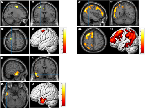
- Unilobar: The hypometabolic area was concordant with the EZ and confined to a single lobe.
- Multi-lobar: The hypometabolic area was concordant with the EZ, but extended beyond a single brain lobe (≥2 lobes).
- Remote: The hypometabolic region was not concordant with the EZ, appearing outside the EZ.
- None: No statistically significant hypometabolic area was identified in whole-brain analyses (Figure 1).

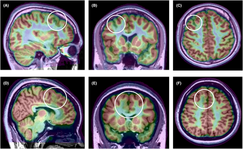
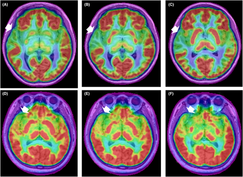
Given the high sensitivity of PET-MRI co-registration images and the advantages of detecting subtle metabolic changes compared to SPM,6, 21, 22 to further explore factors associated with surgical outcomes, patient fusion images were reviewed. The FDG-PET images were co-registered to corresponding 3D-MPRAGE T1-weighted images with the co-registration algorithm in SPM12 and were displayed in MRIcron (http://people.cas.sc.edu/rorden/mricron/index.html), after which they were overlaid on the T1 images with a spectrum setting at 60%–80% transparency. Surgical outcomes were further compared in the unilobar group according to the relationship between hypometabolism and gyrus, the edge of the hypometabolism area, lobe distributions, and pathological subtypes. The relationship between hypometabolism and the cerebral gyrus is divided into the following two patterns1: Single gyrus (SG): The hypometabolic area was located within a single gyrus, be it the bottom of the sulcus or an entire gyrus.2 ≥2 gyri: Hypometabolism was observed in more than one gyrus but within one lobe (Figure 2). The edge of the hypometabolic area was also divided into two patterns1: Clear-cut: There was a sharp edge between the hypometabolic area and normal metabolic brain tissue.2 Non-clear-cut: The edge of the hypometabolic area was blurry, and it was hard to distinguish the boundary of the hypometabolic area from normal brain tissue (Figure 3).


All patients' standardized PET analysis results and fusion images were subject to review by a senior epileptologist and a senior radiologist. When there was a disagreement, the decision was made in consultation with the director of epilepsy center. They were all blinded to the post-surgical outcomes.
2.4 Surgery
We collected the surgical procedures information in FCD I and II groups. To further determine the extent of surgical resection, the binary resection masks of patients were manually created using VOI tools in MRIcron based on the postoperative CT scan. The binary mask representing the hypometabolic area was subsequently extracted from the t-test result matrix, utilizing the same threshold applied in PET pattern analysis. The proportion of the resected hypometabolic region was defined as the ratio between the volume of the resected area overlapping with the hypometabolic region and the overall hypometabolic volume. The resective volume and the proportion were then calculated using custom MATLAB scripts (MATLAB 2021a, MathWorks).
2.5 Statistical analysis
SPSS (v 26.0; SPSS Inc) was used for data analysis. Data are given as means ± standard deviation or medians (interquartile range [IQR]) when normally and non-normally distributed, respectively. Categorical data were compared with chi-square tests or Fisher's exact test. p < 0.05 was the significance threshold.
3 RESULTS
3.1 Demographic data
In total, this study enrolled 210 patients (109 female, 51.9%) of whom 62 and 148 were affected by type I and type II FCD, respectively. The mean age of these patients at the time of PET scan was 22.1 ± 10.6 years. The median age at scan and interquartile range of patients was 23.2 and 16.6–30.2 years, with a mean epilepsy duration of 10.7 ± 8.2 years, and a mean age at seizure onset of 8.0 ± 6.7 years. Of these patients, 173 (82.4%) underwent the stereo-electroencephalography (SEEG) examination prior to surgical treatment. MRI-detectable lesions were identified in 87 (41.4%) patients, including gray-white matter blurring, changes in cortical thickness, signal increases (primarily FLAIR), and transmantle sign.23 With respect to prognostic outcomes, the overall Engel class I rates in the type II and type I FCD groups were 85.5% and 58.1%, respectively, with an overall rate of 77.6%. All patients underwent follow-up for over 3 years (4.5 ± 1.3 years), and additional patient demographic characteristics are summarized in Table 1.
| FCD type I (n = 62) | FCD type II (n = 148) | Total (n = 210) | |
|---|---|---|---|
| Gender (female) | 35 (56.5%) | 74 (50%) | 109 (51.9%) |
| Age of scan (years, mean ± SD) | 21.6 ± 10.2 | 17.8 ± 10.7 | 22.1 ± 10.6 |
| Age of onset (years, mean ± SD) | 10.1 ± 7.7 | 7.2 ± 6.0 | 8.0 ± 6.7 |
| Duration of epilepsy (years, mean ± SD) | 10.3 ± 6.9 | 10.5 ± 8.8 | 10.7 ± 8.2 |
| Underwent SEEG | 58 (93.5%) | 115 (77.7%) | 173 (82.4%) |
| MRI Lesional | 8 (12.9%) | 79 (53.4%) | 87 (41.4%) |
| Lateralization (left) | 30 (48.4%) | 66 (44.6%) | 96 (45.7%) |
| Follow-up time (years, mean ± SD) | 4.6 ± 1.3 | 4.3 ± 1.5 | 4.5 ± 1.3 |
| Prognosis | |||
| Engel I | 36 (58.1%) | 127 (85.8%) | 163 (77.6%) |
| Engel II-IV | 26 (41.9%) | 21 (14.2%) | 47 (22.4%) |
- Abbreviations: SD, standard deviation; SEEG, stereoelectroencephalography.
3.2 Surgical information
We compared surgery-related information between FCD I and II. The Surgical procedure was classified as lesionectomy, lobectomy, multilobar resection, and hemispherectomy. Lesionectomy was the most common procedure in FCD I and FCD II groups, accounting for 51.6% and 58.1%, respectively, with no statistically significant difference in surgical procedure between the two groups (p = 0.083). Resective volume of patients who have done the resection surgery was calculated. In the FCD I group, the volume was 15.7 ± 7.1 cm3 (mean ± standard deviation); In FCD II, it was 13.9 ± 8.3 cm3. The difference was not significant between the two groups (p = 0.152). The proportion of the resected hypometabolic region was 0.64 ± 0.14 (mean ± standard deviation) in the FCD I group, which was lower than 0.72 ± 0.19 in the FCD II group (p = 0.005) (Table 2).
| FCD type I | FCD type II | p | |
|---|---|---|---|
| Surgical procedures | 0.083 | ||
| Lesionectomy | 32 (51.6%) | 86 (58.1%) | |
| Lobectomy | 10 (16.1%) | 37 (25.0%) | |
| Multilobar resection | 15 (24.2%) | 18 (12.2%) | |
| Hemispherectomy | 5 (8.1%) | 7 (4.7%) | |
| Resective volume, cm3 | 15.7 ± 7.1 | 13.9 ± 8.3 | 0.152 |
| Proportion of resected hypometabolic region | 0.64 ± 0.14 | 0.72 ± 0.19 | 0.005* |
- * p < 0.05.
3.3 Hypometabolic patterns and prognosis
In quantitative analyses, four metabolic patterns were defined and prognostic factors were analyzed among groups. Of the 148 type II FCD patients, 104 (70.3%) were categorized as exhibiting the unilobar pattern, while 26 (17.6%) and 11 (7.4%), respectively, exhibited the multi-lobar and remote patterns, and 7 (4.7%) did not exhibit any significant hypometabolic area as defined by SPM. The unilobar pattern was associated with the best surgical outcomes, with an Engel class I rate of 90.4% (94/104, p = 0.035), as compared to respective rates of 80.8%, 72.7%, and 57.1% in the remaining three groups. However, owing to limited sample numbers in these groups, intergroup comparisons did not reveal any significant differences following Bonferroni correction (p < 0.008) (Table S1). The type I FCD lesions were detected in 29, 26, 3, and 4 patients in the unilobar, multi-lobar, remote, and none groups, respectively. Patients in the unilobar group exhibited a better prognosis than those in the other groups, whereas no significant differences were observed among these four groups (p = 0.637). The rates of Engel I outcome in the unilobar, multi-lobar, remote, and none groups were 65.5%, 53.8%, 33.3%, and 50%, respectively (Table 3).
| Total | Engel I | Engel II-IV | p | |
|---|---|---|---|---|
| FCD I | 62 | 36 (58.1%) | 26 (41.9%) | 0.637 |
| Unilobar | 29 (46.8%) | 19 (65.5%) | 10 (34.5%) | |
| Multi-lobar | 26 (41.9%) | 14 (53.8%) | 12 (46.2%) | |
| Remote | 3 (4.8%) | 1 (33.3%) | 2 (66.7%) | |
| None | 4 (6.5%) | 2 (50%) | 2 (50%) | |
| FCD II | 148 | 127 (85.8%) | 21 (14.2%) | 0.035* |
| Unilobar | 104 (70.3%) | 94 (90.4%) | 10 (9.6%) | |
| Multi-lobar | 26 (17.6%) | 21 (80.8%) | 5 (19.2%) | |
| Remote | 11 (7.4%) | 8 (72.7%) | 3 (27.3%) | |
| None | 7 (4.7%) | 4 (57.1%) | 3 (42.9%) |
- * p < 0.05.
Further unilobar group categorization was performed. In this group, hypometabolic regions were most often observed in the frontal lobe and the temporal lobe in type II and I FCD, respectively, no differences in prognosis were observed among different lobes (FCD I: p = 0.967, FCD II: p = 0.509). Further differentiation by pathological subtypes was also performed, revealing that 57 and 47 patients were respectively diagnosed with type IIa and type IIb FCD, while 14, 10, and 5 patients were respectively diagnosed with type Ia, Ib, and Ic FCD. No differences in surgical outcomes were observed among these subgroups (FCD I: p = 0.344, FCD II: p = 0.748).
Visual review of PET-MRI fusion images led to the further definition of two patterns in the unilobar group of type II FCD patients. Specifically, 74 and 30 of these patients were classified in the SG and ≥2 gyri groups, respectively, and there were statistical difference in seizure outcomes between the two groups (p = 0.032). Patients in the SG group exhibited better surgical outcomes, with a 94.6% (70/74) Engel class I rate as compared to 80% (24/30) in the ≥2 gyri group. (Table 4). No similar differences in these patterns were observed in the type I FCD group as all unilobar patients were assigned to the ≥2 gyri pattern (Table S2). Patients who have a sharp hypometabolic edge incline gain a better surgery outcome (p = 0.046) in type II FCD group.
| Total | Engel I | Engel II-IV | p | |
|---|---|---|---|---|
| Lobe | 104 | 94 | 10 | 0.509 |
| Frontal | 59 | 55 | 4 | |
| Temporal | 15 | 13 | 2 | |
| Occipital | 10 | 8 | 2 | |
| Parietal | 11 | 10 | 1 | |
| Insular | 9 | 8 | 1 | |
| Pathological type | 0.748 | |||
| IIa | 57 | 52 | 5 | |
| IIb | 47 | 42 | 5 | |
| PET/MRI | 0.032* | |||
| Single gyrus | 74 | 70 | 4 | |
| ≥2 gyri | 30 | 24 | 6 | |
| Hypometabolism edge | 0.046* | |||
| Clear-cut | 86 | 80 | 6 | |
| Non-clear-cut | 18 | 14 | 4 |
- * p < 0.05.
4 DISCUSSION
In this study, we performed a semi-quantitative analysis of the hypometabolic status of 210 patients who were histopathologically diagnosed as type I or type II FCD. This led to the establishment of four hypometabolic patterns (unilobar, multi-lobar, remote, and none). Of these patterns, the unilobar pattern was associated with a significantly better prognosis among type II FCD patients although the same was not true in type I FCD. We also found that in the unilobar group of type II FCD patients, the SG and Clear-cut edge group exhibited better post-surgical seizure outcomes.
In prior studies, individuals affected by drug-resistant epilepsy have been shown to benefit from FDG-PET as an approach to EZ localization.24-26 However, extended or remote hypometabolic areas beyond the EZ were also common finding in such imaging interpretation,15, 27 potentially complicating efforts to accurately localize the EZ. By definition, the hypometabolism area represents a functional deficit zone (FDZ) which has the potential to be larger or smaller than a real EZ.9 Accordingly, the pattern of the hypometabolic area and how it relates to patient prognosis should be figured out. One study found extratemporal hypometabolism to have a negative impact on the prognosis of TLE patients.13 Some studies have also demonstrated that remote hypometabolism is a risk factor in both TLE and ETLE.15, 28 In 2019, Tomás et al. reported that SPM-based focal hypometabolic patterns were associated with more favorable postoperative outcomes than other patterns.14 This is partially consistent with the present results because we only detected this association in type II FCD patients. However, some studies have reported the opposite findings. For example, Jayalakshmi et al. reported that focal hypometabolic activity did not correlate with good prognostic outcomes in patients with FCD,29, 30 although they only employed non-quantitative methods. These inconsistencies among prior studies may also be attributable to insufficient differentiation among pathological types.
In type I FCD cases, dyslamination occurs together with the disruption of tissue architecture, whereas neurons and glial cells appear normal. Type II FCD is associated with the activation of mTOR signaling as a result of genetic mutations and is generally accompanied by dysplastic, megalocytic neurons intermixed with normal neurons or balloon cells in type IIb FCD cases.16 In addition to their distinct pathological characteristics, these two forms of FCD also exhibit distinct clinical features. In type I FCD patients, the epileptogenic region is often relatively large with a wide discharge range and severe seizure burden and neurological deficits being common.31, 32 This concept was also revealed by effective connectivity analysis.33 Even following invasive evaluation, patients may be unable to receive resective surgery in some cases. In contrast, diagnosing and treating type II FCD can generally be done more readily as the semiology, EEG, and imaging data are generally more consistent with localized lesions, particularly in type IIb FCD cases.34, 35 Bottom-of-sulcus dysplasia (BOSD) corresponds to a form of type II FCD with favorable surgical outcomes. MRI abnormality signs could highly indicate its location, including cortical thickening at the sulcal bottom, blurring of the gray-white matter boundary, abnormal signal intensity, and transmantle sign. However, when MRI is negative or doubtful, PET could be a crucial examination to make confirmation and enhance the detection rate.36-38 On the one hand, PET hypometabolism itself could indicate epileptogenicity.39 On the other hand, it can assist in the judgment of MRI signs. Meanwhile, if a hypometabolic brain region is accompanied by a definite MRI abnormal sign, it is very likely to be considered as EZ. In this condition, direct resection surgery could be performed without intracranial EEG.34 As such, the prognostic outcomes of type II FCD cases were notably better than those of type I FCD, even going through the same preoperative evaluation process. In our analysis, SPM-defined PET metabolic patterns offer different levels of predictive value for surgical outcomes in these two subtypes of FCD. We hypothesize that in type I FCD cases the actual EZ range is larger than the FDZ revealed by PET, whereas in type II FCD the EZ may be relatively localized and confined to the FDZ. As such, when type I FCD is strongly suspected based on anatomical and electroclinical data, surgical options and patient prognosis should be taken into careful consideration even if the hypometabolic area detected via PET is limited. In type II FCD patients, hypometabolic regions are often closely associated with the EZ. Chassoux et al. demonstrated that in over half of analyzed diagnosed Taylor-type FCD cases which correspond to type II FCD under the most recent classification guidelines,40 the EZ range is highly consistent with hypometabolic regions.22 Our results similarly highlight this feature of type II FCD.
Surgery information was also considered in this study. The surgical procedure and resective volume showed no significant difference between the two groups. Although the PET hypometabolic pattern of type II FCD tends to be focal, the extent of surgical resection and the choice of surgical procedure were also based on the patients' anatomical-electro-clinical characteristics. The resection region will be moderately extended compared to the extent PET suggests if the anatomical-electro-clinical relationship was complex or other strong evidence existed. This may be the reason why there was no difference in the surgical procedure and resection volume between the two groups. We also calculated the proportion of resected hypometabolic region. This may indicate the correlation between the FDG-PET hypometabolism and surgical resection. According to our results, the resection rate was higher in the FCD II group. This result was consistent with our findings. The hypometabolic regions in FCD II were more likely to be resected completely because they tended to be focal and had a clear-cut hypometabolism edge. If the lesion suggested by anatomical-electro-clinical characteristics was consistent with the PET hypometabolic region, we prefer to resect this region as completely as possible. Nevertheless, in FCD I, it is hard to entirely resect the hypometabolic region because it more likely tended to be large and had a non-clear-cut distinction with the normal brain.
Prior reports have indicated that PET-MRI co-registration can outperform SPM when detecting small or deeply located FCD lesions despite the latter technique being more objective.21 Owing to the co-registration and coordination conversion involved in SPM calculation processes, the association between hypometabolic regions and cerebral sulci or gyri was difficult to define with precision at the individual level, particularly in type II FCD lesions located at the bottom of the sulcus. As such, visual analysis was performed following quantitative calculations to better differentiate among these metabolic patterns. First, the SG group exhibited better prognostic outcomes in line with prior reports.22 BOSD, which corresponds to a form of type II FCD with good surgical outcomes, was included in this group.41 Second, patients with clear-cut hypometabolic edge had better prognosis in type II FCD. This may be attributed to the clear hypometabolic edge that could facilitate surgical planning and total EZ area excision, resulting in a better surgical prognosis. In contrast, no SG pattern was successfully defined in type I FCD patients and no prognosis difference was observed between different hypometabolic edge groups, providing further evidence of the difficulties associated with detecting FCD lesions via PET alone.
Based on these results, PET can be effectively employed for diagnosis and presurgical evaluation in cases of type II FCD. Those patients exhibiting SPM-defined unilobar hypometabolism experienced better surgical outcomes, with individuals in the SG and clear-cut edge group exhibiting a better prognosis. When semiology, electrophysiology, and multimodal neuroimaging (particularly PET) are consistent when clarifying the EZ in type II FCD cases, there is a high chance of good surgical outcomes. In contrast, predicting the surgical prognosis of type I FCD patients via SPM or visually based on hypometabolism distribution patterns is challenging, and the benefits and risks of surgical treatment in these patients should be weighed carefully.
4.1 Limitations
This study is subject to some limitations. For one, this was a retrospective analysis that is susceptible to potential selection bias. In addition, these results are not fully quantitative given that following SPM calculations, metabolic pattern classification was performed artificially. Due to limits in current computing capabilities and sometimes the lack of subjects with similar ages in the control group, some subtle hypometabolic patterns cannot be detected by SPM alone. Pathological specimens were not re-reviewed in this study. Therefore, patients' pathology was defined only according to the pathology reports. Another limitation of this study was that we only analyzed the metabolism at the macroscopical level by PET instead of microscopic field, which could also provide important information on metabolism in epilepsy.42 Lastly, patients with ambiguous pathological test results were not included in this study owing to limitations to the current detection level. These excluded patients may primarily include FCD types Ib and Ic, given that they can be challenging to define based on current definitions.
5 CONCLUSIONS
In summary, PET hypometabolism patterns exhibit different levels of predictive value with respect to surgical outcomes in type I and type II FCD patients. In cases of type II FCD, a more favorable prognosis was observed for the unilobar, SG, and Clear-cut groups, offering value for the localization of the EZ and the formulation of appropriate surgical plans. In contrast, metabolic patterns in type I FCD were not correlated with surgical outcomes, highlighting a more complex relationship with EZ.
AUTHOR CONTRIBUTIONS
Conception and design: Chao Zhang, Yuan Yao. Acquisition of data: Yuan Yao, Xiu Wang, Baotian Zhao. Analysis and interpretation of data: Yuan Yao, Jiajie Mo, Bowen Yang, Zhihao Guo, Zilin Li. Drafting the article: Yuan Yao. Critically revising the article: Wenhan Hu, Xiaoqiu Shao. Reviewed submitted version of manuscript: all authors. Approved the final version of the manuscript on behalf of all authors: Kai Zhang. Administrative/technical/material support: Xiuliang Fan, Du Cai, Lin Sang, Zhong Zheng. Study supervision: Kai Zhang.
FUNDING INFORMATION
National Key R&D Program of China (2021YFC2401201), Capital's Funds for Health Improvement and Research (2022-1-1071 and 2020-2-1076), and National Natural Science Foundation of China (82071457, 82271495, and 82201603).
CONFLICT OF INTEREST STATEMENT
No conflict of interest exists in the submission of this manuscript. We confirm that we have read the Journal's position on issues involved in ethical publication and affirm that this report is consistent with those guidelines.
Open Research
DATA AVAILABILITY STATEMENT
The data presented in this study are available on request from the corresponding author. The data are not publicly available due to privacy regulations for patients.


