Taking a walk on the heart failure side: comparison of metabolic variables during walking and maximal exertion
Massimo Mapelli and Elisabetta Salvioni are sharing first author privileges.
Abstract
Aims
Although cardiopulmonary exercise testing (CPET) is the gold standard to assess exercise capacity, simpler tests (i.e., 6-min walk test, 6MWT) are also commonly used. The aim of this study was to evaluate the relationship between cardiorespiratory parameters during CPET and 6MWT in a large, multicentre, heterogeneous population.
Methods
We included athletes, healthy subjects, and heart failure (HF) patients of different severity, including left ventricular assist device (LVAD) carriers, who underwent both CPET and 6MWT with oxygen consumption measurement.
Results
We enrolled 186 subjects (16 athletes, 40 healthy, 115 non-LVAD HF patients, and 15 LVAD carriers). CPET-peakV̇O2 was 41.0 [35.0–45.8], 26.2 [23.1–31.0], 12.8 [11.1–15.3], and 15.2 [13.6–15.6] ml/Kg/min in athletes, healthy, HF patients, and LVAD carriers, respectively (P < 0.001). During 6MWT they used 63.5 [56.3–76.8], 72.0 [57.8–81.0], 95.5 [80.3–109], and 95.0 [92.0–99.0] % of their peakV̇O2, respectively. None of the athletes, 1 healthy (2.5%), 30 HF patients (26.1%), and 1 LVAD carrier (6.7%), reached a 6MWT-V̇O2 higher than their CPET-peakV̇O2. Both 6MWT-V̇O2 and walked distance were significantly associated with CPET-peakV̇O2 in the whole population (R2 = 0.637 and R2 = 0.533, P ≤ 0.001) but not in the sub-groups. This was confirmed after adjustment for groups.
Conclusions
The 6MWT can be a maximal effort especially in most severe HF patients and suggest that, in absence of prognostic studies related to 6MWT metabolic values, CPET should remain the first method of choice in the functional assessment of patients with HF as well as in sport medicine.
Background
Maximal cardiopulmonary exercise testing (CPET) is the most accurate method to measure exercise performance in different populations, including healthy subjects, athletes, and patients with heart failure (HF).1, 2 In this latter condition, peak exercise oxygen uptake (peakV̇O2) provides relevant prognostic information.3 However, due to the limited availability of CPET, exercise performance is usually assessed by simpler tests such as the 6-min walk test (6MWT). As a matter of facts, peakV̇O2 and 6MWT distance are considered interchangeable as regards HF performance evaluation and prognosis. However, this is not the case being 6MWT affected not only by CPET determinants such as respiratory, circulatory, and metabolic functions, but also by lower limb muscle strength and balance ability.4, 5 In any case, the 6MWT is generally considered a submaximal (and therefore safer) test, recent trials6-8 demonstrated how daily-life activities, including a brisk walk, represent maximal exercises from a metabolic point of view for many HF patients.9 This is particularly evident for the most severe patients, in which the V̇O2 measured during 6MWT exceed the peakV̇O2 in almost half of the cases.6 To date, few studies limited to HF patients with unconclusive results10, 11 address the relationship between the oxygen uptake during a 6MWT and a maximal exercise in different subjects are still lacking.
Aims
The aim of this study was to assess the relationship between the cardiorespiratory parameters collected during maximal CPET and 6MWT in a large, multicentre, heterogeneous group of subjects including athletes, healthy subjects, patients with HF of different severity including left ventricular assist device (LVAD) carriers.
Methods
HF patients (including LVAD carriers), healthy subjects, and athletes were recruited at Heart Failure Units of Centro Cardiologico Monzino IRCCS, Istituti Clinici Scientifici Maugeri IRCCS, and Department of Cardiology, University of Copenhagen, Rigshospitalet. None of the healthy subjects or athletes was on treatment with any drugs possibly affecting the cardiorespiratory system. In all study locations, subjects underwent the same exercise protocol and data analysis, for both CPET and 6MWT. HF patients were clinically stable with no recent admissions for worsening HF. Inclusion criteria were as follows: age 18–80 years and New York Heart Association I–III. All HF patients were evaluated by echocardiography to determine left ventricular ejection fraction (LVEF, Simpson biplane method).6 As part of our routine HF assessment, all patients underwent at least one previous CPET and 6MWT at our laboratory, which confirmed that patients were familiar with the procedures and setting.12, 13 Exclusion criteria were the use of long-term oxygen therapy, previous heart transplantation, neuromuscular co-morbidities affecting the possibility to perform exercise tests, and concomitant moderate or severe chronic obstructive pulmonary disease.14 CPETs were performed by means of a stationary ergospirometer (Quark PFT, COSMED, Rome, Italy) using an electronically braked cycle ergometer and peakV̇O2 was measured as previously described.3, 15, 16 According to their CPET V̇O2, non-LVAD HF patients were divided in three groups (Group1, <12; Group2, 12–16; and Group3, >16 mL/kg/min. The 6MWTs were performed between one and two working days from the CPET and at the same time of the day of CPET using a dedicated hospital corridor. The metabolic values during the 6MWT were collected and assessed using a wearable ergospirometer (K5, COSMED), as previously described.6, 17-19
Statistical analysis
Continuous variables are reported as median and interquartile range as appropriate for non-normally distributed parameters. Categorical variables are presented as n and percentage. Between groups comparison was done by Mann–Whitney U test or Kruskal–Wallis as appropriate, while chi-squared test was used for categorical variables.
Association of variables are shown as linear regressions. R2 were also calculated by multivariable linear regressions with group variable (athletes, healthy subjects, HF patients and LVAD patients) as covariate.
A P-value <0.05 was used to define statistical significance. All statistical analyses were performed using IBM SPSS Statistics v. 27.
Results
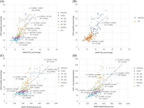
One hundred and eighty-six subjects were enrolled (16 athletes, 40 healthy, 115 non-LVAD HF patients, and 15 LVAD carriers) and completed the study procedures with no significant adverse events. Table 1 shows the main characteristics of the population and their metabolic values with significance between healthy subjects and HF patients. According to exercise limitation severity, HF patients showed higher V̇E/V̇CO2 slope (Table 1). In Table 2, results are presented according to the four study groups (athletes, healthy, HF patients, and LVAD carriers). CPET-peakV̇O2 was 41.0 [35.0, 45.8], 26.2 [23.1, 31.0], 12.8 [11.1, 15.3], and 15.2 [13.6, 15.6] ml/Kg/min (corresponding to 145 [127, 163], 102 [91.0, 122], 57.0 [49.5, 67.0], and 57.0 [49.5, 67.0]% of the predicted) in athletes, healthy, HF patients, and LVAD carriers, respectively (P < 0.001). During 6MWT athletes, healthy, HF patients, and LVAD carriers used 63.5 [56.3, 76.8], 72.0 [57.8, 81.0], 95.5 [80.3, 109], and 95.0 [92.0, 99.0] % of their peakV̇O2, respectively. The correlations between CPET-peakV̇O2 and 6MWT-V̇O2 are shown in Figure 1. Panel A shows relationship between the two values in the whole population (R2 = 0.6366, P ≤ 0.001) and in the different groups of subjects, including HF patient subgroups divided according to the CPET-peakV̇O2. Panel B shows the correlation in non-HF patients (healthy and athletes combined) in comparison with HF patients (both LVAD and non-LVAD carriers) (R2 = 0.2895 and 0.3716, respectively). None of the athletes, 1 healthy (2.5%), 30 HF patients (26.1%), and 1 LVAD carrier (6.7%), reached a V̇O2 during 6MWT higher than 110% of their measured peak V̇O2. The value of 110% of peak V̇O2 at CPET was chosen to consider the differences in the metabolism cost of walking (6MWT) versus cycling (CPET). Further, at multivariate analysis the association between the V̇O2 at CPET and at 6MWT was still significant after adjustment for groups, but with lower R2 within each group analysed except for LVAD patients (R2 = 0.335 for HF patients, 0.798 for LVAD, −0.024 for athletes, 0.122 for healthy subjects, 0.633 for the entire population).
| Healthy | HF | All | ||
|---|---|---|---|---|
| (N = 56) | (N = 130) | (N = 186) | P | |
| Height (m) | 174 [168, 179] | 172 [166, 178] | 173 [167, 178] | 0.304 |
| Weight (kg) | 75.1 [69.8, 85.3] | 75.0 [68.0, 86.8] | 75.0 [69.0, 86.0] | 0.87 |
| BMI (kg/m2) | 24.8 [23.4, 27.8] | 25.5 [23.9, 28.1] | 25.3 [23.7, 28.1] | 0.31 |
| Age (years) | 60.5 [55.8, 64.0] | 70.0 [64.0, 75.0] | 67.0 [59.3, 73.0] | <0.001 |
| Females | 13 (23.2%) | 23 (17.7%) | 36 (19.4%) | 0.502 |
| Males | 43 (76.8%) | 107 (82.3%) | 150 (80.6%) | |
| Haemoglobin (g/dL) | 14.8 [14.5, 15.02] | 13.3 [12.3, 14.2] | 13.5 [12.4, 14.3] | <0.001 |
| LVEF (%) | NA | 26.4[32–39.4] | NA | - |
| PAPs (mmHg) | NA | 29 [35–45] | NA | - |
| CPET data | ||||
| CPET-peakV̇O2 (mL/min) | 2,300 [1820, 3,090] | 1,010 [820, 1,250] | 1,200 [888, 1720] | <0.001 |
| CPET-peakV̇O2 (mL/min/kg) | 28.4 [24.6, 39.4] | 13.1 [11.3, 15.4] | 15.2 [12.0, 23.8] | <0.001 |
| CPET-peakV̇O2 (% of pred) | 112 [92.5, 137] | 56.2 [49.0, 66.0] | 64.0 [52.0, 92.5] | <0.001 |
| VE/VCO2 slope | 28.8 [26.2, 31.4] | 36.0 [31.4, 43.6] | 33.9 [28.5, 40.7] | <0.001 |
| Peak heart rate (b.p.m.) | 154 [145, 166] | 98.0 [85.0, 123] | 116 [90.0, 148] | <0.001 |
| Peak workload (Watt) | 189 [150, 224] | 75.0 [60.0, 99.0] | 93.5 [67.0, 133] | <0.001 |
| 6MWT data | ||||
| Rest heart rate (b.p.m.) | 78.0 [69.0, 90.0] | 70.0 [61.0, 80.0] | 72.0 [65.0, 82.0] | <0.001 |
| End heart rate (b.p.m.) | 112 [100, 125] | 86.0 [76.0, 100] | 96.0 [79.5, 111] | <0.001 |
| 6MWT-V̇O2 (mL/min) | 1,550 [1,250, 1920] | 971 [792, 1,190] | 1,090 [844, 1,380] | <0.001 |
| 6MWT-V̇O2 (mL/min/kg) | 21.8 [17.2, 23.9] | 12.9 [10.8, 15.1] | 14.5 [11.7, 17.8] | <0.001 |
| Task V̇O2 (%of peak) | 0.710 [0.578, 0.810] | 0.950 [0.820, 1.08] | 0.900 [0.730, 1.03] | <0.001 |
| 6MWT VE/VCO2 | 30.8 [29.5, 32.7] | 43.2 [39.2, 49.4] | 39.7 [32.3, 46.7] | <0.001 |
| Distance (m) | 524 [473, 631] | 397 [326, 450] | 430 [362, 509] | <0.001 |
- 6MWT, 6-min walking test; 6MWT-V̇O2, oxygen uptake at 6MWT; BMI, body mass index; CPET, cardiopulmonary exercise test; CPET-peakV̇O2, oxygen uptake at peak exercise at CPET; Task V̇O2, oxygen uptake expressed as percentage of the oxygen uptake at peak exercise at CPET; VE/VCO2, ventilation versus CO2 production; HF, heart failure.
| Athletes | Healthy | HF (no LVAD) | LVAD | All | P | |
|---|---|---|---|---|---|---|
| (N = 16) | (N = 40) | (N = 115) | (N = 15) | (N = 186) | ||
| Height (m) | 179 [175, 182] | 171 [164, 178] | 170 [165, 176] | 183 [179, 189] | 173 [167, 178] | <0.001 |
| Weight (kg) | 87.8 [76.2, 94.8] | 73.0 [68.0, 80.0] | 74.0 [67.0, 83.0] | 93.0 [81.5, 105] | 75.0 [69.0, 86.0] | <0.001 |
| BMI (kg/m2) | 26.7 [24.1, 27.7] | 24.7 [23.2, 27.4] | 25.3 [23.7, 28.0] | 27.7 [25.0, 30.4] | 25.3 [23.7, 28.1] | 0.0632 |
| Age (years) | 60.5 [59.0, 64.5] | 60.5 [54.8, 63.3] | 71.0 [65.0, 76.0] | 65.0 [56.5, 68.0] | 67.0 [59.3, 73.0] | <0.001 |
| Females | 0 (0%) | 13 (32.5%) | 23 (20.0%) | 0 (0%) | 36 (19.4%) | |
| Males | 16 (100%) | 27 (67.5%) | 92 (80.0%) | 15 (100%) | 150 (80.6%) | 0.00774 |
| Haemoglobin (g/dL) | 14.8 [14.5, 15.02] | NA | 13.3 [12.3, 14.2] | 13.8 [12.3, 14.6] | 12.6 [10.7, 14.0] | <0.001 |
| LVEF (%) | NA | NA | 26.4[32–39.4] | NA | - | - |
| PAPs (mmHg) | NA | NA | 29 [35–45] | NA | - | |
| CPET data | ||||||
| CPET-peakV̇O2 (mL/min) | 3,440 [3,050, 3,690] | 1910 [1,660, 2,370] | 966 [793, 1,190] | 1,450 [1,220, 1,530] | 1,200 [888, 1720] | <0.001 |
| CPET-peakV̇O2 (mL/min/kg) | 41.0 [35.0, 45.8] | 26.2 [23.1, 31.0] | 12.8 [11.1, 15.3] | 15.2 [13.6, 15.6] | 15.2 [12.0, 23.8] | <0.001 |
| CPET-peakV̇O2 (% of pred) | 145 [127, 163] | 102 [91.0, 122] | 57.0 [49.5, 67.0] | 55.8 [46.3, 57.4] | 64.0 [52.0, 92.5] | <0.001 |
| VE/VCO2 slope | 31.8 [30.0, 34.3] | 26.9 [24.3, 30.0] | 35.8 [31.0, 43.9] | 37.9 [34.6, 42.7] | 33.9 [28.5, 40.7] | <0.001 |
| Peak heart rate (b.p.m.) | 152 [146, 157] | 155 [145, 167] | 95.0 [83.5, 114] | 131 [119, 151] | 116 [90.0, 148] | <0.001 |
| Peak workload (Watt) | 225 [175, 275] | 175 [132, 201] | 72.0 [57.5, 99.0] | 75.0 [75.0, 99.0] | 93.5 [67.0, 133] | <0.001 |
| Rest heart rate (b.p.m.) | 75.0 [67.0, 91.0] | 80.0 [69.8, 90.0] | 70.0 [61.0, 78.8] | 80.0 [76.3, 80.0] | 72.0 [65.0, 82.0] | <0.001 |
| End heart rate (b.p.m.) | 125 [121, 130] | 107 [97.0, 121] | 84.0 [75.0, 97.5] | 126 [103, 138] | 96.0 [79.5, 111] | <0.001 |
| 6MWT-V̇O2 (mL/min) | 2,180 [1930, 2,580] | 1,370 [1,190, 1,610] | 937 [772, 1,100] | 1,320 [1,130, 1,400] | 1,090 [844, 1,380] | <0.001 |
| 6MWT-V̇O2 (mL/min/kg) | 24.8 [22.5, 27.5] | 18.8 [15.8, 22.2] | 12.7 [10.6, 15.0] | 14.4 [13.0, 15.1] | 14.5 [11.7, 17.8] | <0.001 |
| Task V̇O2 (%of peak) | 0.635 [0.563, 0.768] | 0.720 [0.578, 0.810] | 0.955 [0.803, 1.09] | 0.950 [0.920, 0.990] | 0.900 [0.730, 1.03] | <0.001 |
| 6MWT VE/VCO2 | 29.3 [27.7, 29.8] | 31.6 [30.3, 33.7] | 44.2 [39.8, 49.9] | 38.7 [35.5, 40.6] | 39.7 [32.3, 46.7] | <0.001 |
| Distance (m) | 765 [686, 825] | 499 [466, 525] | 391 [325, 449] | 420 [390, 470] | 430 [362, 509] | <0.001 |
- 6MWT, 6-min walking test; 6MWT-V̇O2, oxygen uptake at 6MWT; BMI, body mass index; CPET, cardiopulmonary exercise test; CPET-peakV̇O2, oxygen uptake at peak exercise at CPET; HF, heart failure; Task V̇O2, oxygen uptake expressed as percentage of the oxygen uptake at peak exercise at CPET; VE/VCO2, ventilation versus CO2 production.

We also analysed in all the study groups the regressions between walked distance at 6MWT and peakV̇O2 (panel C) or workload (panel D) at CPET.
Conclusion
Our study demonstrated for the first time in a broad population, that V̇O2 values obtained during a standard 6MWT are significantly related to the peakV̇O2 measured with a maximal CPET, generally considered the gold standard method to assess functional capacity. On average 6MWT-V̇O2 can elicit 75.3 ± 33.8% of the CPET V̇O2, but this ratio can be extremely variable depending on the population considered. In fact, as previously described in three recent trials,6-8 the more severe the patient with HF, the more they consume their ‘V̇O2 reserve’, going so far as to erode it in 42% of cases in patients with CPET V̇O2 below 12 mL/kg/min. Interestingly, LVAD carriers showed metabolic behaviour similar to the one of their non-LVAD HF patients counterpart since, with an exercise capacity similar to the one of severe HF patients (peakV̇O2 14.9 mL/kg/min, 53.8% of the predicted, and V̇E/V̇CO2 slope 38.6) their 6MWT-V̇O2 corresponds to 95.9% of CPET V̇O2, a value similar to the one of ‘moderate’ HF subjects (see Table 1, Group 2). Of note, both LVAD and HF group 1 patients have a flat correlation between peakV̇O2 at CPET and V̇O2 at 6MWT, suggesting that walking is much better tolerated than cycling in these patients possibly due the overall greater muscle mass utilized with walking. On the other hand, in highly trained subjects (athletes group showed an average peakV̇O2 of 39.6 mL/kg/min), the 6MWT-V̇O2 corresponded to 66.7% of their CPET V̇O2, demonstrating how far from a maximal exercise they are while walking. The different V̇O2 behaviour in HF and healthy during maximal CPET and 6MWT deserves some comments. Despite a general good correlation of the two parameters in the whole cohort (R2 = 0.6366), dividing the population in non-HF and HF subjects (Figure 1) results in a significant reduction in the goodness of correlation (R2 < 0.4 for both), even if a significant group effect at the multivariate analysis was not present. In other words, the ability to predict peakV̇O2 based on the V̇O2 values obtained at 6MWT appears to be rather limited, particularly in patients with HF. Also in the athletes' subgroup, the correlation between 6MWT-V̇O2 and CPET V̇O2 is low, probably due to an even wider delta between the V̇O2 elicited in the two different exercises related to the extreme fitness of some muscle groups and to the intrinsic characteristics of 6MWT: a fixed execution time, the frequent and sudden direction change and the physical constrains related to type of exercise (a walk), making hard for a well-trained individual to reach a truly maximal effort.
Similar considerations come from the analysis of the regressions between walked distance at 6MWT and peakV̇O2 (panel C) or workload (panel D) at CPET, suggesting that estimating the exercise capacity from 6MWT can lead to a non-negligible error. In fact, considering subjects of the same type (HF patients, athletes, etc.) the variability is very high. Finally, our study compared metabolic values obtained during different types of exercise: walking during 6MWT and cycling during CPET. Indeed, few are the reasons for a difference in the metabolic cost of walking/running versus cycling including the walking/running greater muscle mass at work and the metabolic cost of up-lifting the abdomen which is relevant in case of overweight subjects. We considered these differences by using 110% of predicted V̇O2 cycling value as suggested by Wasserman.16, 20-22 However, we opted for cycloergometer instead of treadmill mainly for safety reason, being the cycloergometer generally better tolerated especially in frail subjects like severe HF patients and LVAD carriers.
In conclusion, these data confirm that 6MWT can be a maximal effort especially in most severe HF patients and suggest that, in absence of prognostic studies related to 6MWT metabolic values, CPET should remain the first method of choice in the functional assessment of patients with HF as well as in sport medicine.
Conflict of interest
The authors certify that they have no conflict of interest.
Funding
This work was supported by the Italian Ministry of Health, Rome, Italy (Ricerca Corrente, Centro Cardiologico Monzino IRCCS).


