Efficacy of shear wave elastography for assessment of liver function in patients with heart failure
Abstract
Aims
Liver dysfunction is important for prognosis in heart failure (HF). Shear wave elastography (SWE), which is a novel ultrasound technique for charactering tissues, has been used in liver diseases. However, clinical implication of SWE, including dispersion slope, remains unknown in heart diseases. This study aimed to evaluate the efficacy of SWE assessing liver function in the severity of HF.
Methods and results
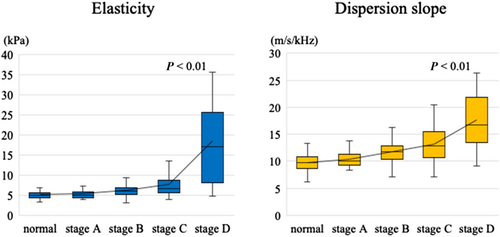
We enrolled 316 consecutive patients with or suspected heart diseases, who were classified according to the American College of Cardiology Foundation/American Heart Association stage of HF, including 37 with Stage A, 139 with Stage B, 114 with Stage C, and 26 with Stage D, and 45 normal subjects. Elasticity and dispersion slope of shear wave were assessed according to the HF stage. Elasticity and dispersion slope were not elevated in normal subjects and patients with Stage A. Elasticity was slightly increased from Stage A to Stage C and was remarkably elevated in Stage D (normal: 5.2 ± 1.1 kPa, Stage A: 5.4 ± 1.2 kPa, Stage B: 6.4 ± 1.8 kPa, Stage C: 7.8 ± 3.5 kPa, and Stage D: 17.7 ± 12.7 kPa), whereas dispersion slope was gradually increased from Stage A to Stage D (normal: 9.7 ± 1.7m/s/kHz, Stage A: 10.4 ± 1.6m/s/kHz, Stage B: 11.7 ± 2.4m/s/kHz, Stage C: 13.2 ± 3.4m/s/kHz, and Stage D: 17.6 ± 5.6 m/s/kHz). In the early HF stage, dispersion slope was elevated. In the advanced HF stage, both elasticity and dispersion slope were elevated. Liver function test abnormalities were observed only from Stage C or Stage D.
Conclusions
Dispersion slope could detect early liver damage, and the combination of elasticity and dispersion slope could clarify the progression of liver dysfunction in HF. SWE may be valuable to manage therapeutic strategies in patients with HF.
Introduction
Heart failure (HF) is a clinical syndrome that leads to multiple organ injuries, including liver dysfunction. Cardiohepatic interaction has been reported to associate with a poor prognosis in patients with HF.1-3 Passive hepatic congestion due to increased central venous pressure causes liver dysfunction, which presents as an abnormality called nutmeg liver on pathological examination.4-6 The management of HF in the early stage, prior to the progression of liver dysfunction, is important to improve clinical outcomes.
Shear wave elastography (SWE) is a novel ultrasound technique that is used to assess tissue characteristics based on shear wave propagation velocity, which provides quantitative estimates of tissue elasticity and viscosity.7-10 Shear wave is generated by inducing a push pulse of ultrasound wave, which deforms a part of the tissue. The velocity of shear wave within the tissue is detected by tracking pulse. SWE can be used to measure two parameters such as elasticity of shear wave, which is related to the tissue hardness, and dispersion slope of shear wave, which reflects the tissue viscosity.11, 12
Shear wave elastography has been used to evaluate liver diseases, including fatty liver, hepatitis, and cirrhosis. Elasticity is correlated with the degree of fibrosis.13, 14 Dispersion slope is associated with inflammation, necrosis, and steatosis.15-17 A few recent studies in the field of heart diseases have reported that elasticity measured on the liver is related to adverse outcomes in patients with HF.18 However, the clinical implication of SWE has not been well investigated in patients with HF. Especially, the clinical significance of dispersion slope remains unknown.
Hepatic congestion results in compression of the bile canaliculi and ductules, causing necrosis of liver cells. Persistence of HF extends the liver cells necrosis, contributing to liver fibrosis.19 Therefore, we hypothesized that dispersion slope, which reflects the viscosity, is sensitively elevated in the early stage of HF to reflect hepatic congestion and that the combination of elasticity and dispersion slope is useful to clarify the progression of liver dysfunction in patients with HF. The aim of this study was to evaluate the efficacy of SWE for assessment of liver function in patients with different severities of HF.
Methods
Study population
We prospectively enrolled 316 consecutive patients with confirmed or suspected heart diseases and 45 normal subjects who underwent SWE in Okayama University Hospital from March 2018 to May 2020. Patients were classified according to the American College of Cardiology Foundation/American Heart Association stage of HF.20 The stage of HF was diagnosed based on medical history, symptoms, physical examinations, and clinical examinations, including electrocardiogram, echocardiography, biomarker measurements, and chest X-ray. Normal subjects were defined as those with no history of heart diseases and no abnormal findings on clinical examinations. Patients with a history or signs of liver diseases such as fatty liver, hepatitis, cirrhosis, and/or hepatic tumours and a history of alcohol abuse (≥20 g/day) were excluded. Patients undergoing dialysis and those with congenital heart diseases were also excluded. All patients gave informed consent to undergo examination. The study was approved by the ethical committee of our institution.
Shear wave elastography
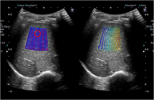
Shear wave elastography measurements for assessment of liver function were performed using Aplio i900 with a 3.5 to 5.0 MHz convex probe (Canon Medical Systems, Otawara, Japan) at the time of outpatient clinic or at discharge from HF treatment. SWE was obtained on the right lobe of the liver through the intercostal spaces at the end-expiratory period in the supine position. Patients were requested to hold their breath during the acquisition. The grey-scale image was optimized for the best acoustic window and best gain setting. A sample box of 2.0 × 2.0 cm was placed on the grey-scale image at a depth of 1.0 to 1.5 cm from the liver capsule to avoid reverberation artefacts and intrahepatic vessels. A 1.0-cm-diameter circular region of interest was placed on the propagation map and exhibited smooth and parallel lines (Figure 1).11, 21 SWE measurements were performed 10 times in each patient, and the average elasticity and dispersion slope of shear wave were calculated.

Clinical assessments
Transthoracic echocardiography was performed at the same time as the SWE measurements. Left ventricular (LV) end-diastolic diameter, LV end-systolic diameter, LV ejection fraction, LV mass index, left atrial volume index, early diastolic mitral inflow velocity to mitral annular velocity ratio (E/e′), tricuspid regurgitation pressure gradient, and inferior vena cava diameter were measured. Furthermore, we evaluated right atrial (RA) size by measuring RA area in the apical four-chamber view. Liver function tests were also performed, including measurement of aspartate aminotransferase (AST), alanine aminotransferase (ALT), alkaline phosphatase (ALP), γ-glutamyl transpeptidase (GGT), bilirubin, cholinesterase, albumin, and prothrombin time levels. Haemoglobin, plasma B-type natriuretic peptide, and serum creatinine levels were also measured.
Cardiac catheterization was performed in 94 of the 316 patients, based on the judgement of physicians. Pulmonary artery wedge pressure, mean pulmonary artery pressure, systolic right ventricular pressure, RA pressure, cardiac output, and cardiac index were obtained. The measurements were determined at the end-expiratory period, with an average of five cycles. Cardiac output and cardiac index were calculated using the Fick equation.
Variability
Inter-observer and intra-observer differences were analysed in 20 randomly selected images. SWE was evaluated by two blinded observers and by a single observer at two different times. Reliability was calculated using Pearson's correlation coefficient. Variability was calculated as the percentage error of each measurement and derived as the difference between the measurements divided by the mean value.
Statistical analysis
Data are presented as mean ± standard deviation for continuous variables and as number and percentage for categorical variables. The analysis of variance or the Wilcoxon rank-sum test was used to explore differences for continuous variables between the groups. The χ2 test was used to compare for categorical variables. The cut-off values of elasticity and dispersion slope for the HF stage were estimated using receiver operating characteristic curve. Pearson's correlation coefficient was calculated to identify the relationship between SWE and clinical parameters. Statistical analysis was performed with statistical software (JMP Version 14.0; SAS Institute Inc., Cary, NC, USA), and significance was defined as a P value of <0.05.
Results
Clinical characteristics
The study population consisted of 316 patients, including 37 with Stage A, 139 with Stage B, 114 with Stage C, and 26 with Stage D, and 45 normal subjects. The mean age of all patients was 66 ± 16 years. Comparisons of clinical characteristics between the groups are shown in Table 1. As the HF stage advanced, age was increased, and body mass index was decreased. The prevalence of hypertension and dyslipidaemia was higher in Stages A–C. Atrial fibrillation was observed more frequently in Stage D. LV end-diastolic diameter and LV end-systolic diameter were increased, and LV ejection fraction was decreased in Stages C and D. E/e′ ratio and left atrial volume index were increased in Stages B–D. Tricuspid regurgitation pressure gradient and inferior vena cava diameter were higher in Stage D.
| Variables | Normal | Stage A | Stage B | Stage C | Stage D | P |
|---|---|---|---|---|---|---|
| (n = 45) | (n = 37) | (n = 139) | (n = 114) | (n = 26) | ||
| Age (years) | 51 ± 17 | 64 ± 13 | 66 ± 16 | 71 ± 11 | 71 ± 9 | <0.01 |
| Male | 18 (40%) | 18 (49%) | 74 (53%) | 53 (47%) | 16 (62%) | 0.37 |
| Body mass index (kg/m2) | 22.4 ± 3.4 | 22.8 ± 4.0 | 22.6 ± 3.5 | 22.7 ± 4.1 | 19.8 ± 2.6 | <0.01 |
| Hypertension | 0 (0%) | 24 (65%) | 62 (45%) | 47 (41%) | 5 (19%) | <0.01 |
| Dyslipidaemia | 0 (0%) | 23 (62%) | 62 (45%) | 52 (46%) | 8 (31%) | <0.01 |
| Diabetes mellitus | 0 (0%) | 11 (30%) | 32 (23%) | 32 (28%) | 8 (31%) | <0.01 |
| Atrial fibrillation | 0 (0%) | 0 (0%) | 22 (19%) | 18 (20%) | 10 (38%) | <0.01 |
| Heart diseases | ||||||
| Ischaemic heart disease | 0 (0%) | 0 (0%) | 40 (29%) | 15 (13%) | 5 (19%) | <0.01 |
| Cardiomyopathy | 0 (0%) | 0 (0%) | 25 (18%) | 71 (62%) | 13 (50%) | <0.01 |
| Valvular disease | 0 (0%) | 0 (0%) | 16 (12%) | 21 (18%) | 4 (15%) | <0.01 |
| Echocardiography | ||||||
| LV end-diastolic diameter (mm) | 44 ± 3 | 44 ± 4 | 46 ± 6 | 53 ± 10 | 58 ± 10 | <0.01 |
| LV end-systolic diameter (mm) | 28 ± 2 | 28 ± 4 | 31 ± 8 | 42 ± 12 | 46 ± 15 | <0.01 |
| LV ejection fraction (%) | 64 ± 4 | 64 ± 5 | 60 ± 11 | 43 ± 17 | 40 ± 13 | <0.01 |
| LV mass index (g/m2) | 70 ± 13 | 73 ± 15 | 94 ± 27 | 122 ± 35 | 131 ± 48 | <0.01 |
| E/e′ ratio | 8.1 ± 2.1 | 9.6 ± 2.9 | 12.6 ± 6.5 | 17.3 ± 8.2 | 28.4 ± 13.1 | <0.01 |
| Left atrial volume index (mL/m2) | 28 ± 5 | 29 ± 7 | 42 ± 23 | 56 ± 25 | 90 ± 53 | <0.01 |
| RA size (cm2) | 12 ± 2 | 13 ± 3 | 17 ± 5 | 20 ± 9 | 25 ± 10 | <0.01 |
| Tricuspid regurgitation gradient (mmHg) | 20 ± 5 | 20 ± 5 | 22 ± 7 | 26 ± 9 | 32 ± 15 | <0.01 |
| Inferior vena cava diameter (mm) | 11 ± 3 | 10 ± 3 | 12 ± 5 | 13 ± 5 | 19 ± 8 | <0.01 |
| Laboratory | ||||||
| AST (U/L) | 22 ± 9 | 21 ± 9 | 23 ± 8 | 25 ± 9 | 35 ± 23 | <0.01 |
| ALT (U/L) | 21 ± 11 | 19 ± 10 | 19 ± 11 | 20 ± 12 | 26 ± 16 | 0.21 |
| ALP (U/L) | 226 ± 89 | 214 ± 82 | 235 ± 86 | 243 ± 119 | 303 ± 83 | <0.01 |
| GGT (U/L) | 28 ± 31 | 34 ± 21 | 30 ± 22 | 50 ± 52 | 103 ± 75 | <0.01 |
| Total bilirubin (mg/dL) | 0.8 ± 0.6 | 0.6 ± 0.3 | 0.7 ± 0.3 | 0.8 ± 0.3 | 1.4 ± 0.9 | <0.01 |
| Cholinesterase (U/L) | 310 ± 72 | 316 ± 94 | 294 ± 48 | 271 ± 80 | 224 ± 80 | 0.04 |
| Albumin (g/dL) | 3.9 ± 0.7 | 3.9 ± 0.5 | 4.0 ± 0.5 | 3.9 ± 0.5 | 3.6 ± 0.4 | 0.36 |
| Prothrombin time (s) | 10.4 ± 0.6 | 10.4 ± 0.5 | 10.6 ± 1.2 | 11.0 ± 1.4 | 11.0 ± 0.6 | 0.16 |
| Haemoglobin (g/dL) | 12.6 ± 2.0 | 12.8 ± 1.1 | 12.8 ± 1.8 | 12.7 ± 2.0 | 11.8 ± 1.6 | 0.88 |
| Creatinine (mg/dL) | 0.7 ± 0.2 | 0.8 ± 0.2 | 0.9 ± 0.5 | 1.1 ± 0.5 | 1.6 ± 1.5 | <0.01 |
| B-type natriuretic peptide (pg/mL) | 17 ± 13 | 30 ± 19 | 121 ± 137 | 456 ± 580 | 762 ± 789 | <0.01 |
- ALP, alkaline phosphatase; ALT, alanine aminotransferase; AST, aspartate aminotransferase; E/e′, early diastolic mitral inflow velocity to mitral annular velocity ratio; GGT, γ-glutamyl transpeptidase; LV, left ventricular; RA, right atrial.
- Data are presented as mean ± standard deviation or number (%) of patients.
Shear wave elastography
The values of SWE for each group were shown in Figure 2. Elasticity of shear wave was 5.2 ± 1.1 kPa in normal subjects, 5.4 ± 1.2 kPa in patients with Stage A, 6.4 ± 1.8 kPa in patients with Stage B, 7.8 ± 3.5 kPa in patients with Stage C, and 17.7 ± 12.7 kPa in patients with Stage D. Dispersion slope of shear wave was 9.7 ± 1.7 m/s/kHz in normal subjects, 10.4 ± 1.6 m/s/kHz in patients with Stage A, 11.7 ± 2.4 m/s/kHz in patients with Stage B, 13.2 ± 3.4 m/s/kHz in patients with Stage C, and 17.6 ± 5.2 m/s/kHz in patients with Stage D. Elasticity and dispersion slope were not elevated in normal subjects or those with Stage A. Elasticity was slightly increased from Stage A to Stage C and was remarkably elevated in Stage D. Dispersion slope was gradually increased from Stage A to Stage D. In the early stage of HF, dispersion slope was elevated. In the advanced stage of HF, both elasticity and dispersion slope were increased. The cut-off value of dispersion slope between Stages A and B was 11.5 m/s/kHz, with area under the curve of 0.68, sensitivity of 56%, and specificity of 79%, and the cut-off value between Stages B and C was 12.9 m/s/kHz, with area under the curve of 0.62, sensitivity of 50%, and specificity of 76%. The cut-off value of elasticity between Stages C and D was 14.6 kPa, with area under the curve of 0.79, sensitivity of 50%, and specificity of 95%.

Liver function parameters
Liver function test parameters, including AST, ALT, ALP, GGT, and bilirubin levels, were normal value in patients with Stage B, whereas an elevation of dispersion slope was observed. Only GGT level was increased from Stage C. In patients with Stage D, ALP level was increased, and cholinesterase was decreased (Table 1).
Haemodynamic measurements
Cardiac catheterization was performed in 21 patients with Stage B, 56 with Stage C, and 17 with Stage D (Table 2). Pulmonary artery wedge pressure, mean pulmonary artery pressure, systolic right ventricular pressure, and RA pressure were increased from Stage B to Stage D. RA pressure was weakly correlated with elasticity (r = 0.36, P < 0.01).
| Variables | Stage B | Stage C | Stage D | P |
|---|---|---|---|---|
| (n = 21) | (n = 56) | (n = 17) | ||
| Pulmonary artery wedge pressure (mmHg) | 11 ± 4 | 12 ± 7 | 21 ± 7 | <0.01 |
| Mean pulmonary artery pressure (mmHg) | 17 ± 4 | 20 ± 9 | 28 ± 10 | <0.01 |
| Systolic right ventricular pressure (mmHg) | 27 ± 7 | 31 ± 12 | 45 ± 14 | <0.01 |
| Right atrial pressure (mmHg) | 4 ± 3 | 6 ± 5 | 9 ± 5 | <0.01 |
| Cardiac output (L/min) | 3.7 ± 1.5 | 3.6 ± 1.5 | 3.2 ± 0.8 | 0.58 |
| Cardiac index (L/min/m2) | 2.4 ± 0.8 | 2.3 ± 0.8 | 2.0 ± 0.4 | 0.35 |
- Data are presented as mean ± standard deviation.
Relationships of shear wave elastography with clinical parameters
Elasticity and dispersion slope were correlated with LV end-diastolic diameter, LV end-systolic diameter, LV ejection fraction, LV mass index, left atrial volume index, E/e′, tricuspid regurgitation pressure gradient, and RA size. Plasma B-type natriuretic peptide and serum creatinine levels were also correlated with elasticity and dispersion slope (Table 3).
| Variables | Elasticity | Dispersion slope | ||
|---|---|---|---|---|
| r | P value | r | P value | |
| Echocardiography | ||||
| LV end-diastolic diameter | 0.19 | <0.01 | 0.16 | <0.01 |
| LV end-systolic diameter | 0.20 | <0.01 | 0.19 | <0.01 |
| LV ejection fraction | 0.22 | <0.01 | 0.24 | <0.01 |
| LV mass index | 0.25 | <0.01 | 0.27 | <0.01 |
| Left atrial volume index | 0.55 | <0.01 | 0.36 | <0.01 |
| RA size | 0.45 | <0.01 | 0.44 | <0.01 |
| E/e′ | 0.22 | <0.01 | 0.32 | <0.01 |
| Tricuspid regurgitation pressure gradient | 0.22 | <0.01 | 0.25 | <0.01 |
| Laboratory | ||||
| Creatinine | 0.04 | 0.04 | 0.16 | <0.01 |
| B-type natriuretic peptide | 0.23 | <0.01 | 0.22 | <0.01 |
- E/e′, early diastolic mitral inflow velocity to mitral annular velocity ratio; LV, left ventricular; RA, right atrial.
Reproducibility
There was good agreement in the measurements of elasticity between the two blinded observers (r = 0.91, P < 0.01) and for the intra-observer (r = 0.96, P < 0.01). The inter-observer and intra-observer variabilities for the value of elasticity were 6.6% and 5.8%, respectively.
Discussion
The major findings of the present study were as follows: (i) elasticity of shear wave was remarkably elevated in patients with Stage D, whereas dispersion slope of shear wave was gradually increased from Stage A to Stage D; and (ii) an elevation of dispersion slope was observed prior to the occurrence of liver function tests abnormalities. SWE, including elasticity and dispersion slope, can be used to effectively detect liver damage in the early stage of HF and to assess the progression of liver dysfunction in patients with HF. To the best of our knowledge, this is the first study to show the efficacy of SWE with combined measurement of elasticity and dispersion slope in patients with HF.
Liver dysfunction in heart failure
Heart failure causes liver dysfunction, which results in worse outcomes. Liver function parameters are used to assess three conditions: cholestasis, which is reflected by ALP, GGT, and bilirubin; hepatocyte integrity, which is reflected by AST and ALT; and liver function mass, which is reflected by cholinesterase, albumin, and prothrombin time levels. Liver function tests play an important role in the prediction of mortality in patients with HF,1, 2 but abnormalities in the previously mentioned parameters are often not observed unless HF is advanced.22 In the present study, almost liver function parameters were not changed in Stages A–C. An increase in bilirubin level and a decrease in cholinesterase level were observed in Stage D. In the liver, 70% of the blood supply is dependent on the portal system, and 30% is delivered by the hepatic artery. Because blood is supplied from both systems, liver function test abnormalities are less likely to occur.19 Evaluation of liver damage using liver function parameters is difficult in the early stage of HF. Few methods are available to effectively diagnose early liver damage in patients with HF.
Shear wave elastography
Shear wave elastography provides information regarding elasticity of shear wave, which is calculated by shear wave speed, as well as dispersion slope of shear wave, which reflects the frequency dependence of both speed and attenuation of shear waves in the viscous component.11, 12, 21 SWE has been recognized as a useful method for assessing liver diseases.13-16, 23, 24 Elasticity is significantly increased in cirrhosis.13, 14 Dispersion slope is more sensitively increased in hepatitis and fatty liver.15, 16 In the field of heart disease, several studies have reported that elasticity is correlated with central venous pressure in acute phase of HF, which is associated with cardiac events.25-28 However, the usefulness of SWE has not been fully investigated in patients with HF. Additionally, because these studies have focused on only elasticity, the assessments of the clinical significance of dispersion slope are lacking.
The present study investigated the efficacy of both elasticity and dispersion slope in patients with different severities of HF, including patients without HF and asymptomatic patients with HF. Thus, this study was able to clarify the graduated changes in elasticity and dispersion slope according to the stage of HF. Dispersion slope was elevated in the early stage of HF, such as Stage B. In the advanced stage of HF, such as Stage D, both elasticity and dispersion slope were increased with the remarkable elevation of elasticity. Interestingly, dispersion slope was elevated prior to the appearance of liver function test abnormalities. When liver function test abnormalities occurred, both elasticity and dispersion slope were already increased. In patients with HF, venous congestion initially leads to compress bile canaliculi and ductules in the liver. The persistence of this state causes necrosis of liver cells due to hypoperfusion,19 resulting in liver function test abnormalities. Dispersion slope may be able to detect the initial state of venous congestion in the liver. Therefore, SWE has a potential to diagnose liver damage more sensitively than liver function test. Based on these findings, the present study suggests that dispersion slope can be used to effectively detect early liver damage and that the combination of elasticity and dispersion slope distinguishes the degree of liver damage in patients with HF. In the present study, the sensitivity and specificity of SWE for identifying the stage of HF were not high. However, we consider that SWE is useful to estimate the severity of HF, including the other clinical parameters.
Previous studies have reported that elasticity is strongly correlated with central venous pressure in patients with acute decompensated HF.25-27 The liver is enveloped by a non-elastic capsule. Increased central venous pressure causes an enlarged and firm liver. Because elasticity reflects the tissue hardness, high value of elasticity is reasonable in the acute phase of HF. In the present study, elasticity was correlated with RA pressure, but the correlation was weak. As the reason, we consider that elasticity was measured at the time of outpatient clinic or at discharge from HF treatment, which was the chronic phase of HF. Elasticity is reflective of central venous pressure and HF-induced liver fibrosis.29-31 In the chronic phase of HF, elasticity may be more strongly affected by liver fibrosis complicated by long-standing HF.
Clinical implications
Because liver dysfunction is a prognostic factor in patients with HF, it is important to diagnose early liver damage and the degree of liver dysfunction in clinical practice. SWE is a simple, objective, and non-invasive modality to assess liver function. The present study suggests that SWE can evaluate liver dysfunction from early stage to advanced stage of HF. Furthermore, SWE can become a novel technique with which to differentiate the stage of HF. Especially, dispersion slope may be effective for identifying Stage B. Because transition from Stage B to Stage C portends an increase in mortality risk,32 the determination of Stage B is important for the prevention of HF.33 In the clinical setting, SWE can provide information for the progression of HF and may be effective for establishing therapeutic strategies of HF. Furthermore, SWE may be useful to judge the effects of HF therapies.
Study limitations
There are several limitations in the present study. First, the number of patients was relatively small for assessment of the usefulness of SWE in patients with HF. The number of patients with each stage of HF was different. Larger studies are required to confirm our findings. Second, cardiac catheterization was not performed in all patients. Third, this study excluded patients with liver disease, but this might have been insufficient. Liver biopsy was not performed for ethical reasons. SWE measurements might be affected by liver disease. Finally, the relationships between SWE parameters and histological findings were not assessed.
Conclusions
Elasticity of shear wave was remarkably elevated in Stage D, whereas dispersion slope of shear wave was gradually increased from Stage A to Stage D. An elevation of dispersion slope occurred prior to the appearance of liver function tests abnormalities. Assessment of dispersion slope may be able to detect early liver damage in patients with HF. The combination of elasticity and dispersion slope has the potential to clarify the progression of liver dysfunction. SWE may be valuable for therapeutic management in patients with HF.
Conflict of interest
None declared.
Funding
This paper was written with no external funding.


