Primary diagnosis of angiosarcoma by fine needle aspiration: Lessons learned from 3 cases
Abstract
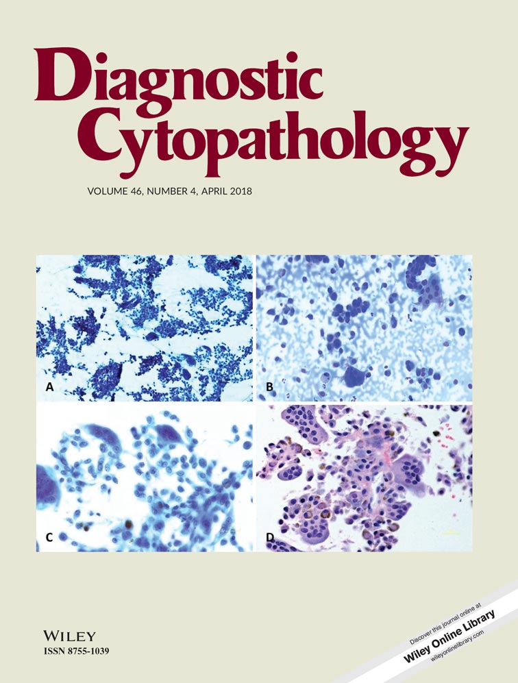
Angiosarcomas are rare malignancies arising from cells of endothelial origin and are aggressive sarcomas that can occur in any anatomic site. They are reported to have predilection for the scalp, extremities and breasts. The incidence of these tumors is increasing, which has been suggested to be attributable to the growing use of radiotherapy to treat breast and other malignancies. There is currently limited literature describing the primary cytologic diagnosis of angiosarcoma on fine needle aspirate material. We describe the findings of three cases of angiosarcoma diagnosed by fine needle aspiration. Our three cases offer distinct radiologic, clinical and cytopathologic points-of-view: a thyroid angiosarcoma, a mediastinal angiosarcoma and a skin angiosarcoma. The cytomorphology of angiosarcoma is characterized by large highly atypical spindle to epithelioid cells with abundant cytoplasm in dispersed single cells or loose aggregates. The nuclei are large and pleomorphic with vesicular chromatin and prominent nucleoli. Mitoses are readily identified. The background can be bloody and/or necrotic. Occasional intracytoplasmic lumens are a helpful morphologic feature suggesting vascular differentiation. HHV-8 immunostaining may aid in the differential diagnosis with Kaposi sarcoma while epithelioid hemangioendothelioma can be distinguished based on morphologic features. Given the metastatic potential and high mortality rate associated with these tumors, this entity is an important consideration in the contexts herein described.
CONFLICT OF INTEREST
The authors have no conflict(s) of interest to disclose.