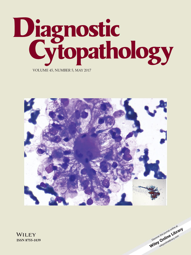
Volume 45, Issue 5 pp. 406-417
Original Article

Differential diagnosis of well-differentiated squamous cell carcinoma from non-neoplastic oral mucosal lesions: New cytopathologic evaluation method dependent on keratinization-related parameters but not nuclear atypism

Hitoshi Hara PHD

Hitoshi Hara PHD

Pathology Division, Yamanashi Prefectural Central Hospital, Kofu, Japan

Search for more papers by this author
Tsuneo Misawa DDS, PHD

Tsuneo Misawa DDS, PHD

Oral Surgery Division, Yamanashi Prefectural Central Hospital, Kofu, Japan

Search for more papers by this author
Eri Ishii CT

Eri Ishii CT

Pathology Division, Yamanashi Prefectural Central Hospital, Kofu, Japan

Search for more papers by this author
Miki Nakagawa CT

Miki Nakagawa CT

Pathology Division, Yamanashi Prefectural Central Hospital, Kofu, Japan

Search for more papers by this author
Saki Koshiishi CT

Saki Koshiishi CT

Pathology Division, Yamanashi Prefectural Central Hospital, Kofu, Japan

Search for more papers by this author
Kenji Amemiya CT

Kenji Amemiya CT

Pathology Division, Yamanashi Prefectural Central Hospital, Kofu, Japan

Search for more papers by this author
Toshio Oyama MD

Toshio Oyama MD

Pathology Division, Yamanashi Prefectural Central Hospital, Kofu, Japan

Search for more papers by this author
Kazuya Tominaga DDS, PHD

Kazuya Tominaga DDS, PHD

Department of Oral Pathology, Osaka Dental University, Hirakata, Japan

Search for more papers by this author
Jun Cheng MD, PHD

Jun Cheng MD, PHD

Division of Oral Pathology, Department of Tissue Regeneration and Reconstruction, Niigata University Graduate School of Medical and Dental Sciences, Niigata, Japan

Search for more papers by this author
Akio Tanaka DDS, PHD

Akio Tanaka DDS, PHD

Department of Oral Pathology, Osaka Dental University, Hirakata, Japan

Search for more papers by this author
Takashi Saku DDS, PHD

Corresponding Author

Takashi Saku DDS, PHD

Department of Oral Pathology, Osaka Dental University, Hirakata, Japan

Division of Oral Pathology, Department of Tissue Regeneration and Reconstruction, Niigata University Graduate School of Medical and Dental Sciences, Niigata, Japan

Correspondence to: Takashi Saku, DDS, PhD, Osaka Dental University, 8-1 Kuzuha Hanazono-cho, Hirakata 573-1121, Japan. E-mail: [email protected]Search for more papers by this author
First published: 16 February 2017
Citations: 4

Conflicts of Interest Disclosure: The authors have no conflicts of interest to declare.

Abstract

Background

The cytology of oral squamous cell carcinoma (SCC) is challenging because oral SCC cells tend to be well differentiated and lack nuclear atypia, often resulting in a false negative diagnosis. The purpose of this study was to establish practical cytological parameters specific to oral SCCs.

Methods

We reviewed 123 cases of malignancy and 53 of non-neoplastic lesions of the oral mucosa, which had been diagnosed using both cytology and histopathology specimens. From those, we selected 12 SCC and 4 CIS cases that had initially been categorized as NILM to ASC-H with the Bethesda system, as well as 4 non-neoplastic samples categorized as LSIL or ASC-H as controls, and compared their characteristic findings. After careful examinations, we highlighted five cytological parameters, as described in Results. Those 20 cytology samples were then reevaluated by 4 independent examiners using the Bethesda system as well as the 5 parameters.

Results

Five cytological features, (i) concentric arrangement of orangeophilic cells (indicating keratin pearls), (ii) large number of orangeophilic cells, (iii) bizarre-shaped orangeophilic cells without nuclear atypia, (iv) keratoglobules, and (v) uneven filamentous cytoplasm, were found to be significant parameters. All malignant cases contained at least one of those parameters, while none were observed in the four non-neoplastic cases with nuclear atypia. In reevaluations, the Bethesda system did not help the screeners distinguish oral SCCs from non-neoplastic lesions, while use of the five parameters enabled them to make a diagnosis of SCC.

Conclusion

Recognition of the present five parameters is useful for oral SCC cytology. Diagn. Cytopathol. 2017;45:406–417. © 2017 Wiley Periodicals, Inc.

The full text of this article hosted at iucr.org is unavailable due to technical difficulties.