The absence of evidence is not the evidence of absence: A case report on the challenges in diagnosing ostial left main stenosis
Ayesha Shaik MD
Department of Internal Medicine, University of Connecticut Health Center, Farmington, Connecticut, USA
Search for more papers by this authorWassim Mosleh MD
Division of Cardiology, University of Connecticut Health Center, Farmington, Connecticut, USA
Search for more papers by this authorKhagendra Dahal MD
Division of Cardiology, University of Connecticut Health Center, Farmington, Connecticut, USA
Search for more papers by this authorChristopher Pickett MD
Division of Cardiology, University of Connecticut Health Center, Farmington, Connecticut, USA
Search for more papers by this authorCorresponding Author
Michael Azrin MD
Division of Cardiology, University of Connecticut Health Center, Farmington, Connecticut, USA
Correspondence
Michael Azrin, MD, Division of Cardiology, University of Connecticut Health Center, John Dempsey Hospital, 263 Farmington Ave, Farmington, Connecticut, 06030, USA.
Email: [email protected]
Search for more papers by this authorAyesha Shaik MD
Department of Internal Medicine, University of Connecticut Health Center, Farmington, Connecticut, USA
Search for more papers by this authorWassim Mosleh MD
Division of Cardiology, University of Connecticut Health Center, Farmington, Connecticut, USA
Search for more papers by this authorKhagendra Dahal MD
Division of Cardiology, University of Connecticut Health Center, Farmington, Connecticut, USA
Search for more papers by this authorChristopher Pickett MD
Division of Cardiology, University of Connecticut Health Center, Farmington, Connecticut, USA
Search for more papers by this authorCorresponding Author
Michael Azrin MD
Division of Cardiology, University of Connecticut Health Center, Farmington, Connecticut, USA
Correspondence
Michael Azrin, MD, Division of Cardiology, University of Connecticut Health Center, John Dempsey Hospital, 263 Farmington Ave, Farmington, Connecticut, 06030, USA.
Email: [email protected]
Search for more papers by this authorAbstract
Because left main (LM) coronary artery stenosis is known to have higher mortality and morbidity compared to lesions in other territories, an early diagnosis and management are crucial to prevent worse outcomes. Due to limitations of coronary angiography (CA), the diagnosis of ostial LM stenosis solely based on CA may result in underdiagnosis of such lesions. Therefore, additional testing is often needed either by pressure wire or intravascular ultrasound (IVUS) to make appropriate diagnosis. We, hereby, present a case of left main ostial stenosis in a 56-year-old male that was missed on multiple coronary angiograms, and highlights many of the considerations in the diagnosis of LM disease.
Supporting Information
| Filename | Description |
|---|---|
| ccd29191-sup-0001-VideoS1.avivideo/avi, 14.8 MB | VIDEO S1 Cinematography of the second coronary angiogram, right anterior oblique-caudal view of the left coronary system with the deep engagement of the left main without adequate reflux flow of contrast beyond the ostium. Bifurcation of left main into left anterior descending and left circumflex artery with no evidence of obstructive disease visualized. Use of 5 Fr Jacky catheter with side-hole prevented dampening upon engagement of left main ostium. |
| ccd29191-sup-0002-VideoS2.avivideo/avi, 25.8 MB | VIDEO S2 Cinematography of the second coronary angiogram, right anterior oblique-cranial view of left coronary system with the deep engagement of the left main and no evidence of obstructive disease visualized in the left main, left anterior descending or left circumflex arteries. |
| ccd29191-sup-0003-VideoS3.avivideo/avi, 20 MB | VIDEO S3 Cinematography of the third coronary angiogram, rotational coronary angiography of the left coronary system showing the absence of significant obstructive disease in the left main, left anterior descending or left circumflex arteries. |
| ccd29191-sup-0004-VideoS4.avivideo/avi, 20.3 MB | VIDEO S4 Cinematography of the third coronary angiogram [2013], right anterior oblique-caudal view of the left coronary system with the deep engagement of the left main adequate reflux flow of contrast, however, no clear evidence of ostial disease. Bifurcation of left main into left anterior descending and left circumflex artery with no evidence of obstructive disease visualized. |
| ccd29191-sup-0005-VideoS5.avivideo/avi, 18.3 MB | VIDEO S5 Cinematography of the third coronary angiogram [2013], left anterior oblique-caudal view of the left coronary system with no clear evidence of ostial disease due to foreshortening of the left main artery ostium in this projection. Bifurcation of left main into left anterior descending and left circumflex artery with no evidence of obstructive disease visualized. |
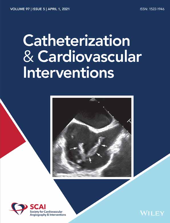
| ccd29191-sup-0006-VideoS6.avivideo/avi, 96.3 MB | VIDEO S6 Intravascular ultrasound images of the fourth coronary angiogram, an eccentric intimal plaque with calcification is noted in the ostial left main (frames 660–735) showing a minimal luminal area of <6 mm2. |
Please note: The publisher is not responsible for the content or functionality of any supporting information supplied by the authors. Any queries (other than missing content) should be directed to the corresponding author for the article.
REFERENCES
- 1Ramadan R, Boden WE, Kinlay S. Management of left main coronary artery disease. J Am Heart Assoc. 2018; 7:e008151.
- 2Mehta N. Ostial Lesions: Tips and Tricks. CSI Cardiology Update 2015. India: Jp Medical Ltd; 2016.
10.5005/jp/books/12785_111 Google Scholar
- 3Rathore S, Terashima M, Katoh O, et al. Predictors of angiographic restenosis after drug eluting stents in the coronary arteries: contemporary practice in real world patients. EuroIntervention. 2009; 5: 349-354.
- 4Schrem SS, Tunick PA, Slater J, Kronzon I. Transesophageal echocardiography in the diagnosis of ostial left coronary artery stenosis. J Am Soc Echocardiogr. 1990; 3(5): 367-373. https://doi.org/10.1016/S0894-7317(14)80136-3.
- 5Feld H, Fisher M, Shani J. Coronary angiography with 5 French diagnostic catheters may miss an ostial left main stenosis. J Interv Cardiol. 1993; 6: 131-136. https://doi.org/10.1111/j.1540-8183.1993.tb00845.
- 6Bourdillon PDV. Use of sideholes for diagnostic coronary arteriography by judkins technique in patients with ostial coronary stenosis. Cathet Cardiovasc Diagn. 1992; 27: 259-266. https://doi.org/10.1002/ccd.1810270404.
- 7Fujino Y, Bezerra HG, Attizzani GF, et al. Frequency-domain optical coherence tomography assessment of unprotected left main coronary artery disease-a comparison with intravascular ultrasound. Catheter Cardiovasc Interv. 2013; 82: E173-E183.
- 8Achenbach S, Rudolph T, Rieber J, et al. Performing and interpreting fractional flow reserve measurements in clinical practice: an expert consensus document. Interv Cardiol. 2017; 12: 97-109.
- 9Ghekiere O, Salgado R, Buls N, Leiner T, Mancini I, Vanhoenacker P, Dendale P, Nchimi A Image quality in coronary CT angiography: challenges and technical solutions. Br J Radiol 2017; 90(1072): 1-13. https://doi.org/10.1259/bjr.20160567.
- 10Fine JJ, Hopkins CB, Hall PA, Delphia RE, Attebery TW, Newton FC. Noninvasive coronary angiography: agreement of multi-slice spiral computed tomography and selective catheter angiography. Int J Cardiovasc Imaging. 2004; 20: 549-552.
- 11Juwana YB, Wirianta J, Suryapranata H, de Boer MJ. Left main coronary artery stenosis undetected by 64-slice computed tomography: a word of caution. Neth Heart J. 2007; 15(7–8): 255-256. https://doi.org/10.1007/BF03085993.
- 12Shin D, Huang K, Sunjic I, Berlowitz M, Prida X. Delayed development of coronary ostial stenosis following surgical aortic valve replacement: a case report of unusual presentation. Case Rep Cardiol. 2018; 2018:8512584. https://doi.org/10.1155/2018/8512584.