Basal neutrophil function in human aging: Implications in endothelial cell adhesion
Abstract
Much attention has been drawn to the pro-inflammatory condition that accompanies aging. This study compared parameters from non-stimulated neutrophils, obtained from young (18–30 years old [y.o.]) and elderly (65–80 y.o.) human volunteers. Measured as an inflammatory marker, plasmatic concentration of hs-CRP was found higher in elderly individuals. Non-stimulated neutrophil production of ROS and NO was, respectively, 38 and 29% higher for the aged group. From the adhesion molecules evaluated, only CD11b expression was elevated in neutrophils from the aged group, whereas no differences were found for CD11a, CD18, or CD62. A 69% higher non-stimulated in vitro neutrophil/endothelial cell adhesion was observed for neutrophils isolated from elderly donors. Our results suggest that with aging, neutrophils may be constitutively producing more reactive species in closer proximity to endothelial cells of vessel walls, which may both contribute to vascular damage and reflect a neutrophil intracellular disrupted redox balance, altering neutrophil function in aging.
Abbreviations
-
- hs-CRP
-
- high sensitivity c reaction protein
-
- RNS
-
- nitrogen-derived species
-
- ROS
-
- reactive oxygen-derived species
-
- TLR
-
- toll-like receptor
-
- y.o.
-
- years old
Introduction
Although much has yet to be elucidated on the subject, aging has been intrinsically related to increased oxidative damage along an organism's life course, contributing, when not directly causing, the general physiological and functional decline that characterizes the aging process. This relationship is frequently more pronounced in age-related degenerative diseases (Junqueira et al., 2004, 2008). Oxidative damage results from an imbalanced pro and antioxidant condition in the organism, favoring oxidant species, namely reactive oxygen- and nitrogen-derived species (ROS and RNS, respectively). However, classical oxidative damage to biomolecules may not be the sole contribution of ROS and RNS to aging. The association of these reactive species to inflammatory developments has led to the hypothesis of “molecular inflammation,” stated as the molecular activation of pro-inflammatory genes by altered redox-sensitive cellular signal pathways that would eventually lead to fully expressed inflammatory tissues and organs, which is exacerbated by aging (Chung et al., 2006). Indeed, the term “inflamm-aging” was coined in order to describe the progressive pro-inflammatory condition found in elderly humans, even in the absence of an immune challenge, rendering the older subject more susceptible to age-related diseases, especially those with an inflammatory pathogenesis (Franceschi et al., 2000).
Both adaptive and innate immunity components are affected by aging, and although the concept of immunosenescence has originally focused on the adaptive immune system, there is growing evidence on its impact on innate immunity as well (Solana et al., 2012). Nevertheless, a pivotal step to acute inflammatory response is the recruitment of effector immune cells from blood and their migration through endothelium to tissues. Neutrophil infiltration in inflamed tissue is a multi-step process involving rolling, activation, adhesion, and transmigration. The recruitment process begins with the neutrophil capture by the vessel wall, followed by rolling over the endothelial layer. Marginalization also allows higher exposure of neutrophils to appropriate stimulation, which is crucial to neutrophil firm adhesion and transmigration. This process involves a series of adhesive interactions between circulating neutrophils and endothelial cells. Neutrophil adhesion to vascular cells is followed by diapedesis, a process of complex movements of neutrophils between the endothelial cells toward the subendothelial area. Cell movements are mainly driven by the presence of a chemotactic gradient generated by the inflammatory sites (Kuijpers et al., 1992; Yuan et al., 2012). Once in the target location, neutrophils are usually activated for intense microbicidal activity, including elevated production of ROS and RNS in a process called oxidative burst. Moreover, ROS and RNS also work in concert, regulating neutrophil function itself, by modulating cell signaling (Fialkow et al., 2007). This stimulated neutrophil response, as well as the efficiency of the phagocytic activity, has been found altered in human aging (Chan et al., 1998). In fact, other neutrophil function indicators are usually found modified with age, such as diminished microbial killing capacity, delayed chemotaxis, apoptosis, and ROS production upon stimulation, and increased recruitment following systemic trauma (Mahbub et al., 2011). These effects are a reflection of altered neutrophil signal transduction response, such as toll-like receptor (TLR) signaling pathways. Although TLR2 and TLR4 expression was found unaltered on neutrophils by aging, recruitment and distribution of these receptors due to modification on lipid raft content on neutrophil membranes was observed (Fulop et al., 2004).
Vascular damage is an important factor associated with aging. Even in the absence of traditional risk factors, aged-induced pathophysiological alterations render the cardiovascular system more prone to disease. Increased reactive species production and a general pro-inflammatory condition are important factors in vascular aging (Ungvari et al., 2010). Considering that neutrophil adhesion profile may change over the years and that its production of reactive species can be deleterious to the endothelial microenvironment, it is important to evaluate how neutrophils from aged individuals are playing their role and how they can interfere with vascular damage over the years. Therefore, the aim of the present work was to study the impact of aging on the adhesion properties of human neutrophils to cultured endothelial cells, as well as on related parameters, such as reactive species production (ROS and NO) and adhesion molecules expression (CD11b, CD11a, CD18, or CD62), by comparing cells isolated from elderly or young donors.
Materials and methods
Volunteer selection
Forty volunteers were selected for this study. Twenty-seven healthy young individuals (16 males and 11 females), aged between 18 and 30 years old (y.o.), and 13 healthy elderly individuals (3 males and 8 females), aged between 65 and 80 y.o., were selected excluding individuals reporting recent or chronic use of anti-inflammatory, statins, supplementation with antioxidant vitamins, tobacco, intensive exercise practice, and presence of anti-immune disease, severe cardiovascular disease, hypertension, and diabetes mellitus. Young volunteers were recruited among undergraduate and graduate students from Federal University of São Paulo (UNIFESP). No young volunteer reported hospitalization in the 12-month period prior sample collection. For the aging group, volunteers were selected from the EPIDOSO Project at Centro de Estudos do Envelhecimento, Preventive Medicine Department, UNIFESP (Ramos et al., 1998). All volunteers declared themselves aware about the study and consented their samples according to a Consentient Term, signed by them. This study was submitted and approved by UNIFESP's Research Ethics Committee (CEP 0131/09).
Sample collection
Blood was collected by peripheral vein puncture in heparinized vacuum tubes (10 mL). Plasma was separated for high sensitivity C-reactive protein (hs-CRP) evaluation. Whole blood was used in flow cytometry techniques and neutrophils were isolated for adhesion assays.
High sensitivity C-reactive protein (hs-CRP)
hs-CRP was measured by solid-phase competitive chemiluminescent enzyme immunoassay. During incubation, hs-CRP in samples competed with analogue hs-CRP in the solid phase. Alkaline phosphatase marked with anti-hs-CRP was applied and the test unit was incubated into a new 30-min cycle. Non-linked enzyme was removed by sequential washes. Analysis was made in IMMULITE® (Siemens).
Surface adhesion molecules
Whole blood samples were incubated with monoclonal antibodies for surface adhesion molecules, marked with the following fluorochromes: anti cluster of differentiation molecule (CD) 66b (Fluorescein—FITC) (BD PharMingen™); anti CD18 (Phycoerythrin—PE) (BD™); anti CD62 (PE) (BD™), anti CD11a (Allophycocyanin—APC) (BD™), or anti CD11b (APC) (BD™). After a 15-min incubation in the dark at room temperature, erythrocytes were lysed (BD FACS Lysing Solution™). Cells were washed with phosphate-buffered saline (PBS) solution and resuspended in PBS 1% NaN3, pH 7.2 (Brunialti et al., 2002). Five thousand neutrophils were acquired in each tube from the assay, based on its labeling with CD66b-FITC. Acquisition was carried out in flow cytometer (BD FACS Calibur™) using CellQuest software for acquisition and analysis.
Oxidative metabolism
Total blood was incubated with 2′7′-dichlorofluorescein diacetate (DCFH-DA) (Sigma–Aldrich). During a 30-min incubation in a shaking bath, at 37°C in the dark, DCFH is oxidized to DCF, a polar compound that remains inside the cell and gives off high levels of green fluorescence (Hasui et al., 1989). After incubation, addition of EDTA 3 mM interrupted the reaction. Cells were centrifuged and erythrocytes lysed by hypotonicity (three alternate sequences of NaCl 0.2% and NaCl 1.6% addition for 30 s each, followed by centrifugation). Cells were resuspended in PBS containing EDTA 3 mM and immediately taken for acquisition at a flow cytometer (BD FACS Calibur™).
Nitric oxide production
The procedure was similar to the one described for the oxidative metabolism assay. Incubation took place with 4,5-diaminofluorescein diacetate (DAF-DA) (Invitrogen), a stable non-fluorescent compound that inside the cell is hydrolyzed to 4,5-diaminofluorescein (DAF). DAF is then oxidized in the presence of nitric oxide, emitting green fluorescence (Nagata et al., 1999).
Neutrophil isolation
Heparinized venous blood was obtained from young and elderly volunteers, and neutrophils were isolated by Ficoll-Hystopaque centrifugation followed by dextran sedimentation. Residual erythrocytes were lysed with two cycles of hypotonic saline. Cells were then washed in PBS solution and finally suspended in PBS added with CaCl2 (100 mM), MgCl2 (50 mM), and glucose (100 mg/mL) (Klempner and Gallin, 1978). Cell viability was obtained by counting on a Neubauer chamber and verified by Trypan Blue (0.6%) exclusion; viability was considered acceptable above 95%.
Cell adhesion assay
For these experiments, stabilized human umbilical vein endothelial cells (HUVECs) were cultured with RPMI medium (Gibco®), 10% FBS, in a 24-well plate (104 cells/well) 24 h before the experiment. Prior to the assay, the endothelial layer was washed once and then it was co-incubated with neutrophils (2.5 × 10⁵ cells/well) for 60 min, at 37°C, in a total volume of 200 μL. Non-adherent neutrophils were removed by aspiration and each well, washed twice. Culture medium was removed and 100 μL of a 0.25% Rose Bengal solution was added for 5 min, at room temperature. The staining was removed and each well was washed twice with RPMI. 200 μL of an ethanol/PBS solution (1:1 v/v) was added. After 30 min, when cells liberated all staining, optic density was determined at 570 nm in a plate reader. Optic density was calculated from the density of each well minus the optic density from a well containing only HUVECs (Gamble and Vadas, 1988). In order to avoid daily variations in staining intensities, adhesion results for the aged group are expressed as relative values to the results obtained for the young group on each experimental day.
Statistical analysis
T-student test for independent samples was used to compare results from groups of old and young subjects, considering P < 0.05 for statistical significance.
Results
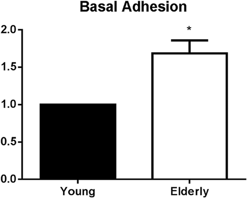
Normalized results for neutrophil adhesion to cultured endothelial cells (HUVECs) revealed a higher degree of cell adhesion for neutrophils isolated from elderly donors, when compared to the young group (Figure 1), even in the absence of any further stimuli. Non-stimulated adhesion was 69% higher for the elderly group and this difference was statistically significant (P < 0.01).
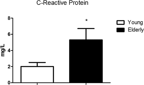
In order to better establish if aging could alter some of the conditions that influence neutrophil adhesion, a pro-inflammatory parameter was assessed. Higher levels of hs-CRP were found in the plasma of elderly subjects (5.31 ± 1.41 mg/L), when compared to young subjects (2.01 ± 0.49 mg/L) (Figure 2), indicating a general pro-inflammatory condition in the earlier group.
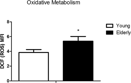
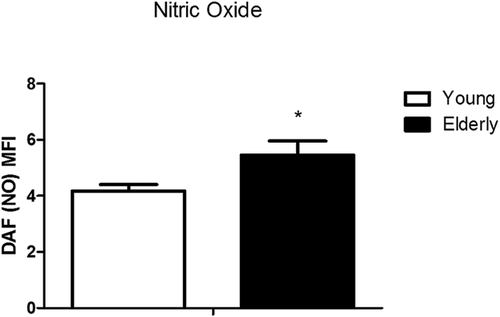
Regarding isolated neutrophils, aging also represented a difference in their basal function. In the absence of any further inflammatory stimulus, neutrophils from aged subjects exhibited higher ROS (P = 0.0372) and NO (P = 0.0140) production than neutrophils from young individuals, as shown by DCFH and DAF oxidation, respectively. Basal neutrophil ROS production was almost 38% higher for the aged group (Figure 3), while NO production showed a 29% difference (Figure 4), thus revealing a higher steady state neutrophil production of reactive species with aging.
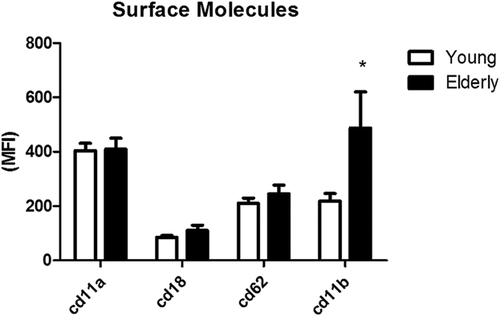
The expression of adhesion molecules could also lead to altered neutrophil adhesion to endothelial cells, so their presence on the surface of isolated neutrophils was assessed by flow cytometry, using fluorochrome marked antibodies. Indeed, neutrophils from elderly donors showed increased CD11b surface expression than cells from the younger group (Figure 5). No differences were found for any of the other neutrophil adhesion molecules assessed in this study (CD11a, CD18, and CD62) (Figure 5).
Discussion
Usually, studies that investigate leukocyte function focus on possible alterations in their ability to respond when facing an inflammatory challenge. In the current study, we chose to study the effect of aging on non-stimulated neutrophils, aiming to evaluate the functioning of these cells closer to a physiological resting situation, instead of simulating the promptitude of an inflammatory response. The hypothesis is that as there is a persistent inflammatory scenario in aged organisms, in addition to an unbalanced pro and anti-oxidant status, neutrophils could exhibit a somewhat pre-activated state in response to their original microenvironment. This could lead to altered neutrophil response to certain stimuli in an inflammatory scenario, but also it could inflict a different behavior in adhesion, transmigration, and eventual dysfunction in ROS and RNS production, even in the absence of such stimuli. Our results suggest that the human innate immune defense, represented by its largest cell population, could be altered by the influence of the pro-inflammatory state found in the organism of an old person.
In an inflammatory response, neutrophils are recruited from the blood to the affected areas. These neutrophils receive signals from the endothelium to firmly adhere and then pass from the circulation to the tissue. Neutrophil adhesion to endothelial cells requires a regulated expression of adhesion molecules in one or both cell types. Some molecules are expressed by neutrophils and others are complementarily expressed on the endothelial surface. The firm adhesion to endothelial cells can be mediated by members of the immunoglobulin superfamily, like intercellular adhesion molecule 1 (ICAM-1), ICAM-2 and vascular cell adhesion molecule 1 (VCAM-1) and receptors for advanced glycation endproducts (RAGE) in endothelial surface, but the main responsible are the integrins, like Mac-1 (CD11b/CD18) (Zimmerman et al., 1992). During neutrophil activation, CD11b molecules are stocked in secretory granules that are exposed on the cell surface, rapidly responding to external stimuli (Borregaard et al., 1994).
Different results have been shown for CD11b levels in aged subjects, compared to young subjects. Differences may be related to health, nutrition, and lifestyle conditions of the studied subjects, but even then comparisons may generate different responses. For instance, CD11b was found lower in neutrophils isolated from nursing home residents when compared to community dwellers (Juthani Mehta et al., 2014), while frailty was associated with CD11b and TNF-α increase in another study (Verschoor et al., 2015). In a cohort study with healthy aged individuals, no differences in neutrophil CD11b expression were found when compared to young subjects (Butcher et al., 2001). Interestingly, increased CD11b levels have been associated with hypertension, a condition of high prevalence in the elderly population (Rea et al., 2013). Also, a study conducted with postmenopausal women showed a strong positive correlation of carotid intima-media thickness with CD11b expression and membrane-bound TNF-α (Figueroa-Vega et al., 2015), strengthening the connection between CD11b levels and pro-inflammatory parameters, leading to cardiovascular risk factors. Overall, increased CD11b is associated with higher adhesive response of stimulated neutrophils, even though the effectiveness of their migration and function may not be preserved with age (Wessels et al., 2010). In the present work, the analysis of the main surface molecules involved in cell adhesion revealed a significant increase in CD11b expression by non-stimulated neutrophils of aged donors in comparison to young donors. This is in accordance with our adhesion results, as elevated CD11b may contribute to the higher rates of cell adhesion found for the aged group.
CRP plasmatic concentration was used as an inflammatory marker in the current study. This is an acute phase protein produced mainly by hepatocytes in response to pro-inflammatory diseases. Expected values for young population go up to 3.0 mg/L. Small elevations in CRP may indicate chronic inflammation and are present in a wide variety of age-associated diseases (McDade et al., 2010). Accordingly, our results show a 2.6-fold increase in the concentration of this protein in the plasma of aged subjects, indicating a pro-inflammatory state in this group. Interestingly, Woollard and collaborators demonstrated that CRP can modulate monocytary adhesion to primary human endothelial cells. In an in vitro study with monocytes, they observed an increase on CD11b expression mediated by CRP (Woollard et al., 2002). Thus, the higher levels of plasmatic CRP in aged subjects may be related to the increased CD11b expression by their neutrophils, adding elements to the observed increased endothelial cell adhesion observed in aged neutrophils.
Oxidants produced by neutrophils also promote leukocyte adhesion to vascular endothelium. In vitro studies suggest an interaction between leukocytes and oxygen reactive metabolites and show that H2O2 promotes the adhesion of neutrophils to endothelial cell monolayers (Lewis et al., 1988). On the other hand, Shappel et al. (1990) showed that CD11b/CD18-mediated neutrophil adhesion to vascular endothelium induces H2O2 production by human neutrophils. Grisham et al. (1998) proposed an interaction between ROS and RNS in leukocyte/endothelium adhesion in an ischemia/reperfusion model. They suggest that O2•− and its secondary products have a pro-adhesive effect, increasing levels of NFκB, cytokines, adhesion molecules, and other pro-inflammatory mediators. On the other hand, NO would provide a modulating role in this process, by reacting with O2•− and lowering the availability of this pro-adhesive reactive species, as well as by increasing IκB and thus decreasing NFκB levels. Pullar et al. (2002) also showed the capacity of HOCl modulating the neutrophil/endothelium adhesion by the increase of ICAM-1 expression.
NO is an important factor for the health and function of endothelial cells. O2•− is a highly efficient scavenger for NO, causing formation of very reactive and deleterious ONOO− in near diffusion-controlled reaction (Beckman and Koppenol, 1996). An increase in O2•−, and subsequent ONOO− formation, reduces NO bioavailability and leads to endothelial dysfunction, one of the major causes for the progression of vascular disease and senescence (Van der Loo et al., 2009). Increased ROS production leading to endothelial dysfunction in aging has been observed both in laboratory animals and in humans (Ungvari et al., 2010).
A pre-activated state of neutrophils in aging can be an important factor in the immune response of aged individuals. If neutrophils are more prone to marginalizing and transmigrating, even in absence of chemotactic factors triggered by a local inflammatory process, they can contribute to a pro-inflammatory condition in the tissues. In the presence of a weak stimulus, these neutrophils can contribute to tissue damage due to exacerbated, although inefficient, respiratory burst. On the other hand, even though oxidative stress is clearly important in the age-related alterations in endothelial dysfunction and, consequently, vascular function, neutrophil-derived ROS are usually considered only in the occurrence of oxidative burst. Our work suggests that neutrophils from elderly individuals may produce more ROS and RNS constitutively and that this production, associated with a closer proximity of the neutrophils to the endothelial cells, may contribute to vascular damage in aging organisms. It may also reflect a neutrophil intracellular disrupted redox balance, which may influence redox-sensitive cell processes, altering neutrophil function in aging. Further studies must be conducted in order to draw a parallel between neutrophil activation in aging, regulation of the involved cell signaling, and altered cell response.
Acknowledgments and funding
The authors acknowledge the help of the staff from Centro de Estudos do Envelhecimento for the contact and clinical assessment of the elderly volunteers for this work. We would also like to thank Reinaldo Salomão, MD, PhD, and his laboratory team for the help using the flow cytometer. This work was supported by Brazilian Federal Government Funding Agency Coordenação de Aperfeiçoamento de Pessoal de Nível Superior (CAPES) (AUX-PRODOC-106/2005), and Fundação de Amparo à Pesquisa do Estado de São Paulo (FAPESP) as ASCC Master fellowship. JNN was recipient of a CAPES Master fellowship, and data presented are part of his Master thesis work.
Conflict of interest
The authors declare that there is no conflict of interests regarding the publication of this article.


