Volume 45, Issue 6 pp. 702-708
LETTER TO THE JOURNAL
Open Access

Comprehensive DSRCT multi-omics analyses unveil CACNA2D2 as a diagnostic hallmark and super-enhancer-driven EWSR1::WT1 signature gene

Florian Henning Geyer

Florian Henning Geyer

Hopp Children's Cancer Center Heidelberg (KiTZ), Heidelberg, Germany

National Center for Tumor Diseases (NCT), NCT Heidelberg, a partnership between DKFZ and Heidelberg University Hospital, Heidelberg, Germany

Division of Translational Pediatric Sarcoma Research, German Cancer Research Center (DKFZ), German Cancer Consortium (DKTK), Heidelberg, Germany

Faculty of Medicine, Heidelberg University, Heidelberg, Germany

Search for more papers by this author
Alina Ritter

Alina Ritter

Hopp Children's Cancer Center Heidelberg (KiTZ), Heidelberg, Germany

National Center for Tumor Diseases (NCT), NCT Heidelberg, a partnership between DKFZ and Heidelberg University Hospital, Heidelberg, Germany

Division of Translational Pediatric Sarcoma Research, German Cancer Research Center (DKFZ), German Cancer Consortium (DKTK), Heidelberg, Germany

Faculty of Medicine, Heidelberg University, Heidelberg, Germany

Search for more papers by this author
Seneca Kinn-Gurzo

Seneca Kinn-Gurzo

Children's Hospital of Philadelphia, Philadelphia, Pennsylvania, USA

Search for more papers by this author
Tobias Faehling

Tobias Faehling

Hopp Children's Cancer Center Heidelberg (KiTZ), Heidelberg, Germany

National Center for Tumor Diseases (NCT), NCT Heidelberg, a partnership between DKFZ and Heidelberg University Hospital, Heidelberg, Germany

Division of Translational Pediatric Sarcoma Research, German Cancer Research Center (DKFZ), German Cancer Consortium (DKTK), Heidelberg, Germany

Faculty of Medicine, Heidelberg University, Heidelberg, Germany

Search for more papers by this author
Jing Li

Jing Li

Hopp Children's Cancer Center Heidelberg (KiTZ), Heidelberg, Germany

National Center for Tumor Diseases (NCT), NCT Heidelberg, a partnership between DKFZ and Heidelberg University Hospital, Heidelberg, Germany

Division of Translational Pediatric Sarcoma Research, German Cancer Research Center (DKFZ), German Cancer Consortium (DKTK), Heidelberg, Germany

Search for more papers by this author
Armin Jarosch

Armin Jarosch

Department of Pathology, Charité-University Medicine Berlin, Corporate Member of Freie Universität Berlin and Humboldt-University of Berlin, Berlin, Germany

German Cancer Consortium (DKTK), Partner Site Berlin, German Cancer Research Centre (DKFZ), Heidelberg, Germany

Search for more papers by this author
Carine Ngo

Carine Ngo

Sarcoma Unit, Gustave Roussy Cancer Campus, Villejuif, France

Search for more papers by this author
Endrit Vinca

Endrit Vinca

Hopp Children's Cancer Center Heidelberg (KiTZ), Heidelberg, Germany

National Center for Tumor Diseases (NCT), NCT Heidelberg, a partnership between DKFZ and Heidelberg University Hospital, Heidelberg, Germany

Division of Translational Pediatric Sarcoma Research, German Cancer Research Center (DKFZ), German Cancer Consortium (DKTK), Heidelberg, Germany

Search for more papers by this author
Karim Aljakouch

Karim Aljakouch

Division of Proteomics of Stem Cells and Cancer, German Cancer Research Center (DKFZ), and Heidelberg University Medical Faculty, Heidelberg, Germany

Search for more papers by this author
Azhar Orynbek

Azhar Orynbek

Division of Proteomics of Stem Cells and Cancer, German Cancer Research Center (DKFZ), and Heidelberg University Medical Faculty, Heidelberg, Germany

Search for more papers by this author
Shunya Ohmura

Shunya Ohmura

Hopp Children's Cancer Center Heidelberg (KiTZ), Heidelberg, Germany

National Center for Tumor Diseases (NCT), NCT Heidelberg, a partnership between DKFZ and Heidelberg University Hospital, Heidelberg, Germany

Division of Translational Pediatric Sarcoma Research, German Cancer Research Center (DKFZ), German Cancer Consortium (DKTK), Heidelberg, Germany

Search for more papers by this author
Thomas Kirchner

Thomas Kirchner

Institute of Pathology, Faculty of Medicine, Ludwig-Maximilian-University of Munich, Munich, Germany

Search for more papers by this author
Roland Imle

Roland Imle

Hopp Children's Cancer Center Heidelberg (KiTZ), Heidelberg, Germany

National Center for Tumor Diseases (NCT), NCT Heidelberg, a partnership between DKFZ and Heidelberg University Hospital, Heidelberg, Germany

Department of Pediatric Oncology, Hematology and Immunology, Heidelberg University Hospital, Heidelberg, Germany

Soft-Tissue Sarcoma Junior Research Group, DKFZ, Heidelberg, Germany

Search for more papers by this author
Laura Romero-Pérez

Laura Romero-Pérez

Institute of Biomedicine of Seville (IBiS)/University Hospital Virgen del Rocío/CSIC/University of Seville/CIBERONC, Seville, Spain

Department of Human Anatomy and Embryology, Faculty of Medicine, University of Seville, Seville, Spain

Search for more papers by this author
Juan Díaz-Martín

Juan Díaz-Martín

Institute of Biomedicine of Seville (IBiS)/University Hospital Virgen del Rocío/CSIC/University of Seville/CIBERONC, Seville, Spain

Search for more papers by this author
Stefanie Bertram

Stefanie Bertram

Institute of Pathology, University Hospital Essen, Essen, Germany

Search for more papers by this author
Enrique de Álava

Enrique de Álava

Institute of Biomedicine of Seville (IBiS)/University Hospital Virgen del Rocío/CSIC/University of Seville/CIBERONC, Seville, Spain

Department of Normal and Pathological Histology and Cytology, Faculty of Medicine, University of Seville, Seville, Spain

Department of Pathology, University Hospital Virgen del Rocío, Pathology Unit, Seville, Spain

Search for more papers by this author
Clémence Henon

Clémence Henon

ERC Chromatin Remodeling, DNA Repair, and Epigenetics Laboratory, ARC Team for Fundamental Research, INSERM U981, and Drug Development Department, Gustave Roussy, Villejuif, France

Search for more papers by this author
Sophie Postel-Vilnay

Sophie Postel-Vilnay

ERC Chromatin Remodeling, DNA Repair, and Epigenetics Laboratory, ARC Team for Fundamental Research, INSERM U981, and Drug Development Department, Gustave Roussy, Villejuif, France

University College of London Cancer Institute, London, UK

Search for more papers by this author
Ana Banito

Ana Banito

Hopp Children's Cancer Center Heidelberg (KiTZ), Heidelberg, Germany

National Center for Tumor Diseases (NCT), NCT Heidelberg, a partnership between DKFZ and Heidelberg University Hospital, Heidelberg, Germany

Soft-Tissue Sarcoma Junior Research Group, DKFZ, Heidelberg, Germany

Search for more papers by this author
Martin Sill

Martin Sill

Hopp Children's Cancer Center Heidelberg (KiTZ), Heidelberg, Germany

National Center for Tumor Diseases (NCT), NCT Heidelberg, a partnership between DKFZ and Heidelberg University Hospital, Heidelberg, Germany

Division of Pediatric Neurooncology, German Cancer Research Center (DKFZ) and German Consortium for Translational Cancer Research (DKTK), Heidelberg, Germany

Search for more papers by this author
Yvonne Versleijen-Jonkers

Yvonne Versleijen-Jonkers

Department of Medical Oncology, Radboud University Medical Center, Nijmegen, The Netherlands

Search for more papers by this author
Benjamin Friedrich Berthold Mayer

Benjamin Friedrich Berthold Mayer

Department of Pediatric Surgery and Pediatric Urology, University Children's Hospital, Tübingen, Germany

Search for more papers by this author
Martin Ebinger

Martin Ebinger

Department of Pediatric Hematology and Oncology, University Children's Hospital, Tuebingen, Germany

Search for more papers by this author
Monika Sparber-Sauer

Monika Sparber-Sauer

Stuttgart University Hospital gKAöR, Olgahospital, Stuttgart Cancer Center, Center for Child, Adolescent, and Women's Medicine, Pediatrics 5 (Pediatric Oncology, Hematology, Immunology), Stuttgart, Germany

University of Medicine Tübingen, Tübingen, Germany

Search for more papers by this author
Sabine Stegmaier

Sabine Stegmaier

Stuttgart University Hospital gKAöR, Olgahospital, Stuttgart Cancer Center, Center for Child, Adolescent, and Women's Medicine, Pediatrics 5 (Pediatric Oncology, Hematology, Immunology), Stuttgart, Germany

Search for more papers by this author
Daniel Baumhoer

Daniel Baumhoer

Bone Tumor Reference Centre, Institute of Medical Genetics and Pathology, University Hospital Basel, University of Basel, and Basel Research Centre of Child Health, Basel, Switzerland

Search for more papers by this author
Wolfgang Hartmann

Wolfgang Hartmann

Gerhard-Domagk-Institute of Pathology, University of Muenster, Muenster, Germany

Search for more papers by this author
Jeroen Krijgsveld

Jeroen Krijgsveld

Division of Proteomics of Stem Cells and Cancer, German Cancer Research Center (DKFZ), and Heidelberg University Medical Faculty, Heidelberg, Germany

Search for more papers by this author
David Horst

David Horst

Department of Pathology, Charité-University Medicine Berlin, Corporate Member of Freie Universität Berlin and Humboldt-University of Berlin, Berlin, Germany

German Cancer Consortium (DKTK), Partner Site Berlin, German Cancer Research Centre (DKFZ), Heidelberg, Germany

Search for more papers by this author
Olivier Delattre

Olivier Delattre

Diversity and Plasticity of Pediatric Tumors, Paris Sciences and Letters University, SIREDO Oncology Centre, Institut Curie, Paris, France

Search for more papers by this author
Patrick Joseph Grohar

Patrick Joseph Grohar

Children's Hospital of Philadelphia, Philadelphia, Pennsylvania, USA

Search for more papers by this author
Thomas Georg Phillip Grünewald

Thomas Georg Phillip Grünewald

Hopp Children's Cancer Center Heidelberg (KiTZ), Heidelberg, Germany

National Center for Tumor Diseases (NCT), NCT Heidelberg, a partnership between DKFZ and Heidelberg University Hospital, Heidelberg, Germany

Division of Translational Pediatric Sarcoma Research, German Cancer Research Center (DKFZ), German Cancer Consortium (DKTK), Heidelberg, Germany

Institute of Pathology, Heidelberg University Hospital, Heidelberg, Germany

Search for more papers by this author
Florencia Cidre-Aranaz

Corresponding Author

Florencia Cidre-Aranaz

Hopp Children's Cancer Center Heidelberg (KiTZ), Heidelberg, Germany

National Center for Tumor Diseases (NCT), NCT Heidelberg, a partnership between DKFZ and Heidelberg University Hospital, Heidelberg, Germany

Division of Translational Pediatric Sarcoma Research, German Cancer Research Center (DKFZ), German Cancer Consortium (DKTK), Heidelberg, Germany

Correspondence

Florencia Cidre-Aranaz, Division of Translational Pediatric Sarcoma Research, German Cancer Research Center (DKFZ) & Hopp Children's Cancer Center (KiTZ), Im Neuenheimer Feld 280, 69210 Heidelberg, Germany.

Email: [email protected]

Search for more papers by this author
First published: 15 March 2025

List of Abbreviations

  • CACNA2D2
  • Calcium voltage-gated channel auxiliary subunit alpha2delta 2
  • ChIP-seq
  • Chromatin Immunoprecipitation followed by Sequencing
  • DEG
  • Differential gene expression
  • DEP
  • Differential protein expression
  • DOX
  • Doxycycline 
  • DSRCT
  • Desmoplastic small round cell tumor
  • EWSR1
  • EWS RNA binding protein 1 
  • fGSEA
  • Fast gene set enrichment analysis
  • fp-ARMS
  • Fusion-positive alveolar rhabdomyosarcoma
  • H3K27ac
  • Histone H3 lysine 27 acetylation
  • IQCG
  • IQ motif containing G 
  • IRS
  • Immune Reactive Score
  • KD
  • Knock down 
  • log2FC
  • Log2 fold change 
  • NES
  • normalized enrichment score
  • Padj
  • Adjusted P-value 
  • qPCR
  • Quantitative polymerase chain reaction
  • RNA-seq
  • RNA-sequencing
  • sc
  • single-cell
  • scRNA-seq
  • single-cell RNA-sequencing
  • SE
  • Super enhancer 
  • shRNA
  • Short hairpin RNA
  • ssGSEA
  • Single sample gene set enrichment analysis
  • WT1
  • Wilms tumor protein 
  • Desmoplastic small round cell tumor (DSRCT) is an aggressive cancer that predominantly affects adolescents and young adults, typically developing at sites lined by mesothelium [1, 2]. DSRCT is genetically defined by a chromosomal translocation that fuses the N-terminus of EWS RNA binding protein 1 (EWSR1) to the C-terminus of Wilms tumor protein (WT1), forming EWSR1::WT1 [3]. This fusion encodes a potent transcription factor and is the only known driver of oncogenic transformation in DSRCT [4]. The lack of a comprehensive understanding of DSRCT biology parallels its dismal survival rate (5%-20%) [1]. These challenges are exacerbated by the absence of clinical trials, the limited systematic collection and analysis of DSRCT biomaterial [1], and the notable lack of specific diagnostic markers, necessitating resource-intensive molecular testing for an accurate diagnosis.

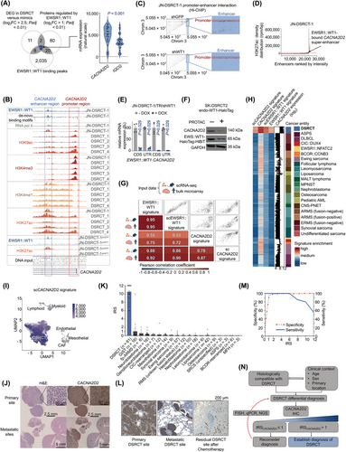
    Here we first focused on identifying promising candidates for validation as single, fast, and reliable diagnostic DSRCT markers. For this, we performed differential gene expression (DEG) analysis on datasets comprising patient samples from 32 DSRCT and 20 morphological mimics, identifying 23 genes overexpressed in DSRCT (log2 fold change (log2FC) > 2.5; adjusted P-value (Padj) < 0.01; Figure 1A, Supplementary Figure S1A). Secondly, we analyzed EWSR1::WT1 binding sites derived from chromatin immunoprecipitation followed by sequencing (ChIP-seq) data [5] obtained from the JN-DSRCT-1 cell line, identifying 2,065 genomic loci likely regulated by EWSR1::WT1 (Figure 1A). Third, we established JN-DSRCT-1 and SK-DSRCT2 cell lines expressing doxycycline (DOX)-inducible short hairpin RNA (shRNA)-mediated EWSR1::WT1 knockdown (KD) (Supplementary Figure S1B). Differential protein expression (DEP) analysis of these cells identified 104 proteins consistently regulated across both cell lines (log2FC > 1.0 and Padj < 0.01; Figure 1A, Supplementary Table S1). The intersection of these analyses revealed calcium voltage-gated channel auxiliary subunit alpha2delta 2 (CACNA2D2) and IQ motif containing G (IQCG) as potential DSRCT biomarkers (Figure 1A). CACNA2D2 was selected for validation due to its significantly higher expression in DSRCTs compared to IQCG (P < 0.001; Figure 1A). Indeed, DSRCT exhibited the highest expression of CACNA2D2 among all studied morphological mimics and normal tissues (P < 0.001; Supplementary Figures S1C-D). Further ChIP-seq data and motif analyses of EWSR1::WT1 binding coordinates and histone marks in JN-DSRCT-1 and four DSRCT patient samples [5, 6] suggested a direct regulatory role of EWSR1::WT1 through an enhancer interaction at the CACNA2D2 locus (Figure 1B). Notably, KD of EWSR1::WT1 in JN-DSRCT-1 resulted in a loss of the EWSR1::WT1 signal and Histone H3 lysine 27 acetylation (H3K27ac) enhancer marks at the CACNA2D2 locus (Figure 1B). Additionally, chromatin interaction data [6] revealed 19 loops connecting the EWSR1::WT1 binding site to the transcription start site of CACNA2D2, which were depleted upon KD of EWSR1::WT1 (Figure 1C). Super enhancer (SE) analysis further demonstrated that the EWSR1::WT1-bound enhancer exhibited a characteristic SE H3K27ac profile in JN-DSRCT-1, which was lost upon EWSR1::WT1 KD (Figure 1D, Supplementary Table S2).
    Details are in the caption following the image
    CACNA2D2 is a diagnostic hallmark and super-enhancer-driven EWSR1::WT1 signature gene. (A) Left: Venn diagram showing the overlap of genes significantly overexpressed (log2FC > 2.5; Padj < 0.01) in DSRCT compared to all other tumor entities (as in Supplementary Figure S1A) with genes or proteins potentially regulated by EWSR1::WT1, as determined by ChIP-seq and Mass spectrometry analyses. Right: Violin plot depicting CACNA2D2 and IQCG mRNA expression in 32 DSRCT patient samples. The black dotted line indicates the median, while blue dotted lines indicate quartiles. Statistical analysis: unpaired two-sided Mann-Whitney test. (B) EWSR1::WT1 de novo binding motifs and the epigenetic profile of the CACNA2D2 locus (chromosome 3:50,506,517-50,548,707, hg19) from four DSRCT tumor samples (DSRCT_1-4), JN-DSRCT-1 wild-type, and JN-DSRCT-1 cells carrying an shRNA against GFP (control) or EWSR1::WT1. ChIP-seq profiles from GSE156277 and GSE212977 are depicted for EWSR1::WT1 (blue), RNA polymerase II (green), H3K4me3 (red), H3K4me1 (yellow), H3K27ac (orange), and H3K9ac (salmon). Rectangles highlight the CACNA2D2 promoter and enhancer regions. (C) Scatter plot showing spatial chromatin interactions between genomic loci in chromosome 3, captured by Hi-ChIP (GSE212978). Dot density reflects the frequency of interactions. Highlighted dots represent interactions between the CACNA2D2 promoter and enhancer loci. The CACNA2D2 promoter region is marked in red, and the enhancer region is marked in blue. (D) Graph depicting H3K27ac signal density at active enhancer sites in the JN-DSRCT-1 cell line (GSE212977), ranked by normalized intensity. Red lines indicate the cutoff for super-enhancer identification (n = 859). (E) Bar plot showing relative mRNA expression levels of EWSR1::WT1 and CACNA2D2, quantified by qRT-PCR in JN-DSRCT-1 cell line expressing a DOX-inducible shRNA-mediated KD of EWSR1::WT1 for 96 hours. n = 4 biologically independent experiments. Horizontal bars represent mean expression levels, and whiskers indicate SEM. The number of analyzed samples is given in parentheses. Statistical analysis: unpaired two-sided Mann-Whitney test. (F) Western blot using antibodies against CACNA2D2, (EWSR1::)WT1, and GAPDH (loading control) in the SK-DSRCT2-endo-WT1-HaloTag cell line, which carries a knock-in of the HaloTag-HiBiT tag at the endogenous locus of EWSR1::WT1 C-terminus. Cells were cultured with 1 µmol/L PROTAC or DMSO as control for 24 hours. (G) x-y scatter plots illustrating the correlation of ssGSEA enrichment scores between (sc)EWSR1::WT1 and (sc)CACNA2D2 signatures of orthotopically xenografted single DSRCT cells. The numbers and colors in the correlation matrix represent Pearson correlation values for each signature enrichment in single cells from orthotopically xenografted DSRCT (top) or 32 bulk DSRCT patient samples (bottom). (H) Heatmap depicting ssGSEA signature enrichment scores for (sc)EWSR1::WT1 and (sc)CACNA2D2 signatures, along with log2 expression values for CACNA2D2 across 32 DSRCT patient samples and 20 additional cancer entities relevant for DSRCT differential diagnosis. (I) UMAP plot of scRNA-seq analysis of merged and integrated data from eleven samples, comprising tumor-derived and normal cells from four DSRCT patients (GSE263523), using the scCACNA2D2 signature. The color gradient indicates the ssGSEA score for scCACNA2D2 signature enrichment. (J) Representative histological images of CACNA2D2 IHC in a DSRCT murine orthotopic xenograft from a primary site and micrometastases in inner organs (muscle, liver, and kidney). DAB (brown chromogen) was used. (K) Bar plot showing individual IRS scores for CACNA2D2 staining in DSRCT and other morphological mimics. The number of analyzed samples is given in parentheses. Bars represent mean IRS values, and whiskers indicate SEM. DSRCT samples are highlighted in blue. Statistical analysis: unpaired two-sided Mann-Whitney test. (L) Left: Representative histological image of CACNA2D2 IHC in DSRCT primary tumor tissue. Middle: Representative histological image of CACNA2D2 IHC in a lung metastasis of DSRCT. Right: Representative histological image of CACNA2D2 IHC in a tumor sample with residual DSRCT cells after treatment. DAB (brown chromogen) was used. (M) Graph showing sensitivity (blue solid line) and specificity (red dashed line) of CACNA2D2 IHC, both expressed as percentages. (N) Diagram illustrating the proposed workflow for establishing a robust diagnosis of DSRCT. Abbreviations: AML, acute myeloid leukaemia; ARMS, alveolar rhabdomyosarcoma; ASPS, alveolar soft part sarcoma; BCOR::CCNB3, BCOR::CCNB3 rearranged sarcomas; CACNA2D2, Calcium voltage-gated channel auxiliary subunit alpha2delta 2 gene; CAFs, cancer-associated fibroblasts; CDS, coding sequence; ChIP-seq, chromatin immunoprecipitation followed by sequencing; Chrom, chromosome; CIC::DUX4, CIC::DUX4 rearranged sarcomas; CNS-PNET, primitive neuroectodermal tumor arising in the central nervous system; DAB, 3,3'-diaminobenzidine; DEG, differential gene expression; DLBCL, diffuse large B-cell lymphoma; DMSO, dimethyl sulfoxide; DOX, doxycycline; DSRCT, Desmoplastic small round cell tumor; DSRCT_1 – 4, DSRCT patient sample data; ERMS, embryonal rhabdomyosarcoma; EWS::WT1-HaloTag-HiBiT, EWSR1::WT1 protein fused to HaloTag-HiBiT protein; EWSR1::NFATC2, EWSR1::NFATC2 rearranged sarcomas; EWSR1::WT1, fusion protein of EWSR1 and WT1 proteins; FISH, Fluorescent in situ hybridization; GAPDH, Glyceraldehyde-3-phosphate dehydrogenase; GFP, Green fluorescent protein; GIST, gastrointestinal stromal tumor; H&E, hematoxylin and Eosin; H3K27ac, histone H3 lysine 27 acetylation; H3K4me1, histone H3 lysine 4 aminomethylation; H3K4me3, histone H3 lysine 4 trimethylation; H3K9ac, histone H3 lysine 9 acetylation; hg19, human genome version 19; Hi-ChIP, high-throughput chromatin immunoprecipitation followed by sequencing; IHC, immunohistochemistry; IQCG, IQ motif containing G gene; IRS, immune reactive score; JN-DSRCT-1/TR/shWT1, JN-DSRCT-1 cell line stably expressing a doxycycline inducible shRNA expression cassette targeting EWSR1::WT1 mRNA; kDa, kilodalton; log2FC, log2 fold change; MALT, mucosa-associated lymphoid tissue lymphoma; MFH, malignant fibrous histiocytoma; MPNST, malignant peripheral nerve sheath tumor; NGS, next generation sequencing; Padj, adjusted P-value; PROTAC, proteolysis targeting chimera; qRT-PCR, quantitative real-time polymerase chain reaction; RMS, rhabdomyosarcoma; RNA pol II, RNA polymerase II; rpm/bp, reads er million per base pair; scCACNA2D2, single-cell data derived CACNA2D2 signature; scEWSR1::WT1, single-cell data derived EWSR1::WT1 signature; scRNA-seq, single-cell RNA-sequencing; SEM, standard error of the mean; shGFP, shRNA targeting GFP mRNA; shRNA, short hairpin RNA; shWT1, shRNA targeting EWSR1::WT1 mRNA; SK-DSRCT2 endo-WT1-HaloTag, SK-DSRCT2 cell line expressing endogeneous CACNA2D2 protein fused to HaloTag protein; SRCSs, small round cell sarcomas; ssGSEA, single sample gene set enrichment analysis; UMAP, uniform manifold approximation and projection; UTR, untranslated region.

    Post-transcriptional and post-translational KD of EWSR1::WT1 in three DSRCT cell line models expressing different EWSR1::WT1 isoforms (Supplementary Figure S2A) resulted in a significant reduction in CACNA2D2 expression (Figures 1E–F, Supplementary Figure S1B, Supplementary Figures S2B–F). Additionally, ChIP-seq data derived from MeT-5A mesothelial cells [6] – the potential cell of origin of DSRCT [7, 8] – ectopically expressing different EWSR1::WT1 isoforms (-KTS, +KTS, or -KTS/+KTS) suggested direct regulation, as evidenced by the co-occurrence of H3K27ac signals and signals for V5- or HA-tagged EWSR1::WT1 isoforms at the CACNA2D2 enhancer region (Supplementary Figure S2G). Notably, MeT-5A cells transfected with a control vector showed no substantial signal at this locus (Supplementary Figure S2G). Publicly available RNA-sequencing (RNA-seq) data from MeT-5A cells [6] expressing different EWSR1::WT1 isoforms showed that CACNA2D2 was differentially expressed in the presence of EWSR1::WT1 (4.1 ≤ log2FC ≤ 5.9, Padj < 0.001) (Supplementary Figure S2H). Finally, quantitative polymerase chain reaction (qPCR) analysis of MeT-5A cells stably expressing a DOX-inducible ectopic EWSR1::WT1 expression cassette confirmed that upon EWSR1::WT1 induction, CACNA2D2 was significantly and highly overexpressed (Supplementary Figure S2I). Taken together, these results emphasize that EWSR1::WT1 is sufficient to drive CACNA2D2 expression. SE analysis of MeT-5A-derived data strikingly showed that the CACNA2D2 enhancer bound by EWSR1::WT1 became a SE upon ectopic expression of EWSR1::WT1− KTS + KTS (Supplementary Figure S2J).

    To explore whether CACNA2D2 could serve as a surrogate indicator of oncogenic EWSR1::WT1 transformation, we defined a CACNA2D2 gene set and gene signature by performing a correlation analysis of gene expression data from 32 DSRCT patient samples (Supplementary Figure S3A, Supplementary Tables S3-S4). Next, an EWSR1::WT1 signature was computed by performing a combined DEG analysis of newly generated in vivo and in vitro [4] material derived from three DSRCT cell lines upon EWSR1::WT1 KD (Supplementary Figure S3A, Supplementary Table S4). Notably, fast gene set enrichment analysis (fGSEA) of the CACNA2D2 gene set demonstrated a highly significant (Padj < 0.001) and strong positive enrichment for the EWSR1::WT1 signature (normalized enrichment score, NESEWSR1::WT1 = 3.6). Moreover, single sample gene set enrichment analysis (ssGSEA) of expression data from 32 DSRCT patient samples confirmed that the EWSR1::WT1 signature significantly correlated with that of CACNA2D2 (r = 0.75), highlighting a transcriptional interconnection between CACNA2D2 and EWSR1::WT1 in situ (Figure 1G). These observations were further supported by single-cell (sc)-derived signatures from orthotopically-generated tumors using two DSRCT cell lines with DOX-inducible KD of EWSR1::WT1 at primary (n = 221) and metastatic (n = 221) locations (Figure 1G, Supplementary Table S4). Indeed, ssGSEA of our single-cell data showed highly significant correlation between the NES of our generated EWSR1::WT1 and CACNA2D2 signatures (Figure 1G), regardless of tumor location, implying that CACNA2D2-associated genes are also characteristic features of metastasized DSRCT cells (Supplementary Figure S3B).

    To delineate the specificity of the interaction between CACNA2D2 and EWSR1::WT1 in DSRCT, we performed ssGSEA using our EWSR1::WT1 and CACNA2D2 signatures on expression data from 20 DSRCT morphological mimics (Figure 1H). Here, non-DSRCT cancer entities showed significantly lower NES and correlation strength for all signatures compared to DSRCT (Supplementary Figures S3C-D). These results further emphasized the high specificity of the CACNA2D2 and EWSR1::WT1 interplay in DSRCT. Moreover, both bulk- and sc-derived CACNA2D2 signatures precisely distinguished DSRCT cell clusters from non-tumor cells in single-cell RNA-sequencing (scRNA-seq) data from four DSRCT patients (n = 11 samples) [9] (Figure 1I, Supplementary Figure S3E). Concordantly, all predicted normal cell types within these tumors exhibited low enrichment of both CACNA2D2 signatures (Supplementary Figures S3F-G).

    Further, dimensional reduction of CACNA2D2-associated CpG sites in 24 DSRCT patient samples, compared with 192 samples from 13 morphological mimics [10] revealed distinct clustering of all DSRCT samples, which was unique to CACNA2D2 compared to other described EWSR1::WT1-regulated genes or IQCG (Figure 1A, Supplementary Figures S4A-B). Additionally, these CACNA2D2-associated CpG sites exhibited significant (P < 0.001) and specific hypomethylation in DSRCT patient samples, collectively suggesting that the CACNA2D2-associated methylation signature is a distinct and specific feature of DSRCT (Supplementary Figure S4C).

    To assess the diagnostic utility of CACNA2D2, we optimized a staining protocol for DSRCT cell line xenografts, achieving consistent and robust membranous or cytoplasmatic staining, even uncovering micrometastases (Figure 1J, Supplementary Figure S4D).

    Finally, we assembled the largest collection of fresh-frozen and paraffin-embedded DSRCT patient samples analyzed to date (n = 61), comprising primary, metastatic, and post-treatment samples, and supplemented it with 249 patient samples from 18 different DSRCT morphological mimics (Supplementary Table S5). CACNA2D2 immunoreactivity was evaluated using a modified Immune Reactive Score (IRS) (Supplementary Material and Methods). Excitingly, DSRCT tumor sections exhibited the highest IRS for CACNA2D2 (IRSmean = 10.5, 6 ≤ IRSDSRCT ≤ 12, P < 0.001) (Supplementary Figure S4E-F), with specificity reaching 98% when applying a cutoff of IRS > 1 (Figure 1K-M, Supplementary Figure S4E). Indeed, even samples derived from CIC- and BCOR-rearranged sarcomas, as well as fusion-positive alveolar rhabdomyosarcomas, showed negligible mean protein expression compared to DSRCT (IRSCIC = 0.21, IRSBCOR = 0, IRSfp-ARMS = 0.56). Furthermore, 100% sensitivity was achieved when applying an IRS cutoff of ≤ 6, implying that DSRCT samples consistently displayed strong staining for CACNA2D2 (Figure 1M). Thus, we recommend a single CACNA2D2 staining for clinically and histologically compatible DSRCT differential diagnosis. If IRSCACNA2D2 ≤ 1, the diagnosis should be reconsidered or re-evaluated using molecular diagnostic procedures (such as fluorescence in situ hybridization, qRT-PCR, or next-generation sequencing), if available (Figure 1N). Conversely, if IRSCACNA2D2 > 1, a diagnosis of DSRCT may be established. Also, CACNA2D2 staining may be used to rule out DSRCT within the broad spectrum of small-round-blue-cell tumors, potentially offering extensive diagnostic utility.

    Finally, the high, specific, and homogenous membranous expression of CACNA2D2 in DSRCT, combined with the highly specific antibody described here, makes CACNA2D2 an ideal candidate for targeted therapeutic approaches, including drug delivery using antibody-drug conjugates or CAR-T cell therapy. Future studies should investigate the precise role of CACNA2D2 in DSRCT biology, with a focus on its potential contributions in tumor cell fitness, differentiation, and tumorigenic potential.

    In conclusion, here we developed an extensive toolset for DSRCT research (Supplementary Figure S4G), a validated blueprint for how such resources could be harnessed in other cancer entities, and identified CACNA2D2 as a singular, powerful DSRCT biomarker.

    AUTHOR CONTRIBUTIONS

    Florian Henning Geyer, Florencia Cidre-Aranaz, and Thomas Georg Phillip Grünewald conceived the study. Florian Henning Geyer and Florencia Cidre-Aranaz wrote the paper and drafted all figures and tables. Florian Henning Geyer carried out all in vitro and in vivo experiments and performed all bioinformatic and statistical analyses. Florian Henning Geyer, Alina Ritter, and Thomas Georg Phillip Grünewald performed immunohistochemical evaluation and scoring of tumor samples and TMAs. Florencia Cidre-Aranaz, Roland Imle, and Ana Banito performed and/or coordinated in vivo experiments. Olivier Delattre provided microarray expression data. Seneca Kinn-Gurzo performed in vitro experiments on BER cell lines. Tobias Faehling and Clémence Henon performed single-cell bioinformatic analyses. Karim Aljakouch and Azhar Orynbek performed MassSpec and analyzed MassSpec data. Alina Ritter, Jing Li, Endrit Vinca, Laura Romero-Perez, Martin Sill, and Shunya Ohmura contributed to experimental procedures. Wolfgang Hartmann and Benjamin Friedrich Berthold Mayer provided clinical and/or histological guidance. Enrique De Álava, Juan Díaz-Martín, Stefanie Bertram, Sophie Postel-Vilnay, Martin Ebinger, Monika Sparber-Sauer, Daniel Baumhoer, Carine Ngo, David Horst, Yvonne Versleijen-Jonkers, Armin Jarosch, Sabine Stegmaier, and Thomas Kirchner provided clinical samples. Patrick Joseph Grohar, Thomas Georg Phillip Grünewald, and Jeroen Krijgsveld provided laboratory infrastructure. Florencia Cidre-Aranaz and Thomas Georg Phillip Grünewald supervised the study and data analysis. All authors read and approved the final manuscript.

    ACKNOWLEDGEMENTS

    We would like to thank Nadine Gmelin, Stefanie Kutschmann, and Felina Zahnow for their expert technical assistance, and Claudia Schmidt from the Light Microscopy Facility (German Cancer Research Center (DKFZ), Heidelberg, Germany) for her meticulous work in conducting immunohistochemical stainings. We thank the Microarray Core Facility (German Cancer Research Center (DKFZ)) for providing the Gene Expression Arrays and related services. We thank Katharina Bauer, Denise Keitel and Jan-Philipp Mallm from the Single-cell Open Lab (German Cancer Research Center (DKFZ)) for expert support in the preparation of single-cell libraries. We thank the Flow Cytometry Facility team (German Cancer Research Centre (DKFZ)) for their support with cell sorting. We thank Dr. Marc Ladanyi for sharing the SK-DSRCT2 cell line.

      CONFLICT OF INTEREST STATEMENT

      The authors declare no competing interests.

      FUNDING INFORMATION

      The laboratory of Thomas Georg Phillip Grünewald is supported by grants from the Matthias-Lackas Foundation, the Dr. Leopold und Carmen Ellinger Foundation, the European Research Council (ERC CoG 2023 #101122595), the Deutsche Forschungsgemeinschaft (DFG 458891500), the German Cancer Aid (DKH-70112257, DKH-7011411, DKH-70114278, DKH-70115315), the Dr. Rolf M. Schwiete foundation, the SMARCB1 association, the Ministry of Education and Research (BMBF; SMART-CARE and HEROES-AYA), and the Barbara and Wilfried Mohr foundation. The research team of Florencia Cidre-Aranaz was supported by the German Cancer Aid (DHK-70114111), and the Dr. Rolf M. Schwiete Stiftung (2020-028 and 2022-31). In addition, this work was delivered as part of the PROTECT team supported by the Cancer Grand Challenges partnership funded by Cancer Research UK, the National Cancer Institute, the Scientific Foundation of the Spanish Association Against Cancer And KiKa (Children Cancer Free Foundation). Florian Henning Geyer, Tobias Faehling, Endrit Vinca, and Alina Ritter were supported by the German Academic Scholarship Foundation. In addition, Endrit Vinca was supported by scholarships from the Heinrich F.C. Behr foundation and the Rudolf and Brigitte Zenner foundation, Tobias Faehling by the Heinrich F.C. Behr foundation, and Florian Henning Geyer and Alina Ritter are supported by the German Cancer Aid through the ‘Mildred-Scheel-Doctoral Program’ (DKH-70114866). This project is co-funded by the European Union (ERC, CANCER-HARAKIRI, 101122595). All views and opinions expressed are however those of the authors only and do not necessarily reflect those of the European Union or the European Research Council. Neither the European Union nor the granting authority can be held responsible for them.

      ETHICS APPROVAL AND CONSENT TO PARTICIPATE

      In vivo experiments were approved by the government of North Baden and conducted in accordance with ARRIVE guidelines and recommendations of the European Community (86/609/EEC) and UKCCCR (guidelines for the welfare and use of animals in cancer research). Open slides or tissue-microarrays from human formalin-fixed, paraffin-embedded or cryopreserved tissue samples were retrieved from the archives of the Institute of Pathology of the LMU Munich, the Charité Berlin, The Biobank of the Hospital Universitario Virgen del Rocío of Seville, the Hospital Gustave Roussy (Villejuif), the Bone Tumor Reference Center at the University of Basel, the University of Essen, the Cooperative Weichteilsarkom Studiengruppe (CWS) study center, the Klinikum Stuttgart (ethics committee from the Medical Faculty of the Eberhard-Karls University and University Hospital of Tübingen, approval no. 207/2022BO2), the Radboud University Medical Center, the Pathology Institute of the LMU Munich (approval no. 550-16 UE), and the University of Heidelberg (approval no. S-211/2021).

      DATA AVAILABILITY STATEMENT

      The microarray data are deposited at the National Center for Biotechnology Information (NCBI) GEO database with accession codes GSE273438 and GSE273441. All proteomic data is deposited at the PRoteomics IDEntifications database with accession code PXD053786. All other data supporting the findings of this study are available within the article and its supplementary information files, or from the corresponding author upon reasonable request.

        The full text of this article hosted at iucr.org is unavailable due to technical difficulties.