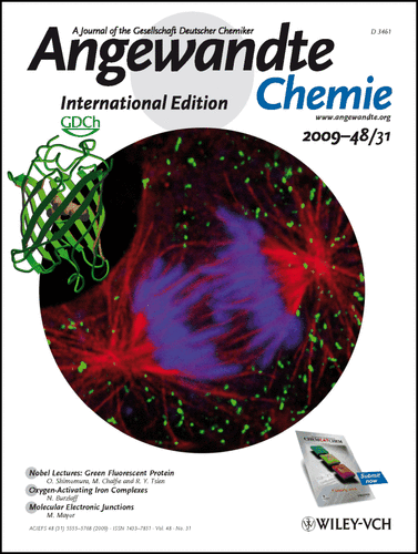
Cover Picture: Discovery of Green Fluorescent Protein (GFP) (Nobel Lecture) / GFP: Lighting Up Life (Nobel Lecture) / Constructing and Exploiting the Fluorescent Protein Paintbox (Nobel Lecture) (Angew. Chem. Int. Ed. 31/2009)
Graphical Abstract
The green fluorescent protein is an invaluable tool in molecular and cellular biology. The cover picture shows the confocal fluorescence image of a cell with GFP-labeled peroxisomes (blue: DNA, red: microtubules of the spindle fibers). At mitosis, most peroxisomes are randomly distributed in the cytoplasm and the majority are not associated with the microtubules in the mitotic spindle. As the cell divides, the peroxisomes are distributed randomly, together with the cytoplasm, to the daughter cells, which suggests that the inheritance of peroxisomes in cells is stochastic rather than ordered. All that can be achieved with the green fluorescent protein can be found in the Nobel Lectures by O. Shimomura, M. Chalfie, and R. Tsien on page 5590 ff. Picture courtesy of Thomas Deerinck, Mark Ellisman, and Roger Tsien, University of California San Diego.


