11q terminal deletion and combined immunodeficiency (Jacobsen syndrome): Case report and literature review on immunodeficiency in Jacobsen syndrome
Abstract
Antibody deficiency is common finding in patients with Jacobsen syndrome (JS). In addition, there have been few reports of T-cell defects in this condition, possibly because most of the reported patients have not been specifically evaluated for T-cell function. In this article, we present a child with an 11q deletion and combined immunodeficiency and we perform a literature overview on immunodeficiency in JS. Our patient presented with recurrent bacterial and prolonged viral infections involving the respiratory system, as well as other classic features of the syndrome. In addition to low IgM, IgG4, and B-cells, also low recent thymic emigrants, helper and naïve T-cells were found. We propose that patients with Jacobsen syndrome need thorough immunological evaluations as T-cell dysfunction might be more prevalent than previously reported. Patients with infections consistent with T-cell defects should be classified as having combined immunodeficiency. © 2016 Wiley Periodicals, Inc.
INTRODUCTION
Terminal 11q deletion syndrome, also known as Jacobsen syndrome (JS; OMIM#147791), is a rare genetic disorder that affects different body systems. Besides dysmorphic stigmata and intellectual disability; brain, skin, heart, hematological, gastrointestinal, and urogenital systems are often affected [Mattina et al., 2009].
Antibody deficiency was the first reported immune defect in patients with JS [Sirvent et al., 1998; Puglisi et al., 2009]. Severe infections are not characteristic for these patients, therefore thorough immunologic evaluation has been rarely performed. In a large prospective study of 110 patients with 11q terminal deletions, infections of the upper respiratory system were common, but with no life-threatening or opportunistic infections reported. Recurrent episodes of otitis media and/or sinusitis were most prevalent, occurring in one half of subjects. Only serum IgA levels were measured in a minority of these patients [Grossfeld et al., 2004]. Thorough immunologic evaluation, including T-cell concentration and proliferation response to mitogens was performed only in a few patients and showed combined immunodeficiency in both children and adults [von Bubnoff et al., 2004; Fernández-San José et al., 2011; Seppänen et al., 2014].
Lack of thorough immunologic evaluation in patients with 11q terminal deletions and reports of combined immunodeficiency in a few of these patients may result in underestimation of immunodeficiency in patients with JS.
We present a patient with confirmed JS in whom T-cell deficiency was observed in addition to antibody deficiency and other features of the syndrome. T-cell deficiency was not present during the initial evaluation, which highlights the importance of performing a thorough immunological follow up in patients with 11q terminal deletion syndrome. Review of the literature is focused on revealing the current lack of knowledge regarding T-cell deficiency in these patients.
Inclusion of JS in the category of primary immunodeficiencies [Picard et al., 2015] is important for appropriate evaluation of the immune system and subsequent clinical care of patients with JS.
MATERIALS AND METHODS
Cytogenetic Studies
Cytogenetic studies were carried out on peripheral blood lymphocytes from the patient and her parents. Chromosome analyses on metaphase spread preparations were performed according to standard procedures using GTG banding at a 500 band resolution level. A total of 30 metaphase cells were analyzed and karyotypes were described according to the International System for Human Cytogenetic Nomenclature. Additional FISH experiments were undertaken using Multi-Color FISH probe—mixture 11 (TelVysion Mix) for 11p, 11q, 18p, and CEP18. (Vysis, Abbott Molecular) on metaphase and interphase chromosome spreads. Hybridization and washing was done according to the manufacturer's protocol and a minimum of 100 interphase cells were analyzed. Chromosomes were counterstained with 4′,6-diamidino-2-phenylindole (DAPI) and images were captured using the CytoVysion Imaging System (Leica Microsystems, Wetzlar, DE).
Microarray Analysis
Array comparative genomic hybridization (aCGH) analysis was performed on genomic DNA extracted from peripheral blood sample using a commercial oligonucleotide array (Agilent 60K ISCA Oligo, Agilent Technologies, Santa Clara, CA) and sex-matched human reference DNA sample (Agilent Technologies). Data were analyzed with the Cytogenomics 3.0 Software (Agilent Technologies).
Lymphocyte Studies
B-cell analysis was done using washed whole blood to remove free antibodies.
Samples were stained with CD27-FITC or CD21-FITC, anti IgD-PE, CD19-PC7, and antiIgMCy5 all from Becton Dickinson. T-cells were stained with monoclonal antibodies for CD45RO APC, CD27 FITC, and CD4 PerCP or CD8 PerCP), all from Becton Dickinson. For cell proliferation assays, cells were stimulated with phytohemagglutinin (PHA; Sigma, St Louis, MO) or anti-CD3 and anti-CD28. Proliferating cells were identified by using a BrdU Flow Kit (Pharmingen, San Diego, CA), that uses anti-BrdUmAb to label BrdU that has been incorporated into newly synthesized DNA within dividing cells.
RESULTS
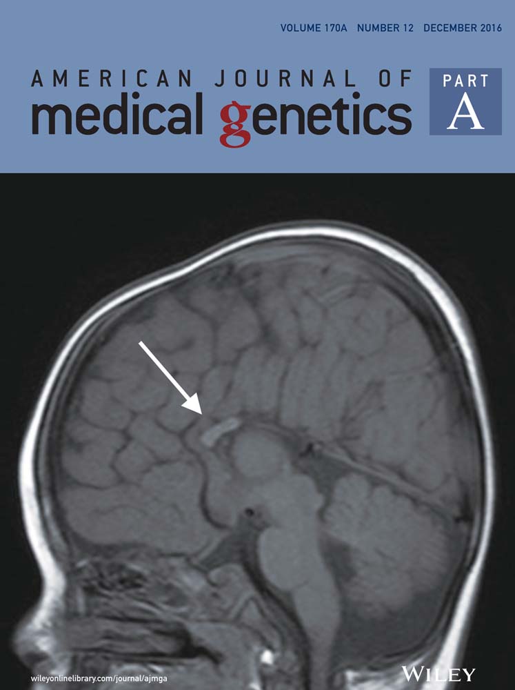
A 16-year-old female that was diagnosed with JS, presented with recurrent viral and bacterial infections of respiratory system and other features including: short stature, anteverted nostrils, long philtrum, high arched palate, malformed ears, retrognathia, genu valgum, short toes, and clinodactyily of fifth fingers. She was born after an unremarkable pregnancy and was the first child of non-consanguineous parents. The patient has suffered from recurrent purulent otitis media and mostly upper respiratory tract infections after inclusion in kindergarten. Immunologic workup performed was consistent with antibody deficiency: reduced IgM, IgG4, IgE, and B-cells. Total IgG, IgA, and specific antibody responses to conjugated pneumococcal, diphtheria and tetanus vaccine were all within normal limits. No other abnormalities of the immune system were detected at that time: T-cell, NK-cell concentrations, and complement activation were normal. She also had mild cognitive impairment with attention deficit hyperactivity disorder and short stature (normal IGF-1) among additional clinical features. Hypertension and left ventricular hypertrophy with diastolic dysfunction both resolved after antihypertensive treatment. Significant difference in the size of kidneys was observed without other structural abnormalities of genitourinary tract. Metabolic workup and brain MRI did not show any abnormalities. Keratoderma of palms and fingers was observed and histologically confirmed. Skin prick tests with inhalant allergens were negative.
The patient continued to have recurrent viral and bacterial upper respiratory tract infections, and lobar pneumonia at the age of 12 years. Thorough evaluation was repeated at the age of 16 years. A karyotype done initially was reported as normal but a repeat study at 16 years of age showed a terminal deletion of chromosome 11 with breakpoint approximately at 11q23 region, which is described as a cause of JS. The deletion was confirmed using specific FISH analysis and the combined karyotype was 46,XX,del(11)(q23.3).ish del(11)(q23.3)(D11S1037-)dn. (Supplementary material: Fig. S1).
Genomic microarray analysis revealed an 11.9 ± 0.1 Mb deletion at chromosome region 11q24.1q25 (arr[hg19] 11q24.1q25(122,984,714–134,868,407) × 1). This deletion results in the loss of one copy of 90 RefSeq genes. (Supplementary material: Fig. S2). The parental cytogenetic analyses did not demonstrate any chromosomal rearrangement, which is consistent with de novo origin of the deletion.
Repeated evaluation of the immune system (see Table I) showed reduced B-cells: 40 cells/mm3 (100–400) and reduced T-cell subtypes: T helper cells: 339 cells/mm3 (400–1300), naïve T-cells: 95 cells/mm3 (230–770), and recent thymic emigrants (RTE): 41 cells/mm3 (50–926). Mitogen response to PHA was reduced to 25% (42–63%) and low normal when repeated: 43% (42–63%). Mitogen response after stimulation with anti-CD3 and anti-CD28 was within normal limits: 80% (50–85%).
| Age (years) | 3 | Normal values (3 year) | 14 | 16 | 17 | Normal values |
|---|---|---|---|---|---|---|
| T-cell (CD3+;×106/mm3) | 3555 | 1400–3600 | 746 | 761 | 742 | 700–1900 |
| helper T-cell (CD3 + CD4+;×106/mm3) | 1712 | 700–2000 | 364 | 312 | 339 | 400–1300 |
| cytotoxic T-cell (CD3 + CD8+;×106/mm3) | 1756 | 500–1400 | 345 | 402 | 347 | 200–700 |
| naive T-cell (CD3 + CD4 + CD45 + RA+;×106/mm3) | 430–1500 | 146 | 106 | 95 | 230–770 | |
| memory T-cell (CD3 + CD4 + CD45 + RA-;×106/mm3) | 220–660 | 218 | 206 | 244 | 240–700 | |
| RTE (CD3 + CD4 + CD45 + RA + CD31+;×106/mm3) | 50–926 | 47 | 41 | 50–926 | ||
| Regulatory T-cell (CD3 + CD4 + CD25 + +; %) | 1–5 | 7 | 1–5 | |||
| NK-cell (CD16 + CD56+;×106/mm3) | 527 | 100–700 | 149 | 192 | 178 | 100–400 |
| B-cell (CD19+;×106/mm3) | 351 | 400–1500 | 43 | 34 | 40 | 100–400 |
| CD3 stimulation with PHA (%) | 42–63 | 25 | 43 | 42–63 | ||
| CD3 stimulation with anti-CD3 and anti-CD28 (%) | 50–85 | 80 | 50–85 | |||
| IgG (g/L) | 6.4 | 4.70–12.30 | 6.24 | 8.91 | 7.38 | 6.90–14.00 |
| IgA (g/L) | 0.4 | 0.21–1.45 | 0.32 | 0.53 | 0.41 | 0.7–4.10 |
| IgM (g/L) | 0.4 | 0.41–1.56 | <0.16 | <0.17 | <0.17 | 0.30–2.40 |
| Platelets (×1012/L) | 266 | 194–345 | 184 | 245 | 204 | 194–345 |
- RTE, recent thymic emigrants.
DISCUSSION
Jacobsen syndrome is a contiguous gene deletion syndrome caused by partial 11q deletion of different sizes, ranging from 5 to 20 Mb. The 11q23 chromosomal region is gene rich with 342 functional genes and several possible breakpoints [Mattina et al., 2009]. In a minority of patients with JS, the breakpoints were assigned to FRA11B fragile site with several (CGG) n repeats at 11q23.3 region. Previous reports have hypothesized that hypermethylation of this expanded CGG repeats is involved in breakage and impaired DNA replication [Michaelis et al., 1998; Jones et al., 2000]. Smaller deletions usually present with milder phenotypic expression and occur by other mechanism, most often they are mediated by low copy repeats (LCR) or palindromic AT-rich repeats [Giglio et al., 2001].
Patients with JS have typical dysmorphic features, short stature, and cognitive impairment. Thrombocytopenia is the most common laboratory finding [Grossfeld et al., 2004; Mattina et al., 2009]. Typical dysmorphic features, short stature, recurrent infections, borderline thrombocytopenia, and mild cognitive impairment suggestive of JS were all observed in our patient. The result of the first karyotype was misleading, as it did not show any abnormalities. Repeated karyotype showed a deletion consistent with the terminal 11q deletion syndrome. Using array CGH, deletion breakpoint was shown to be at the band 11q24.1, therefore not including 11q23 band, but encompassing most of the crucial genes responsible for typical phenotype of JS [Favier et al., 2015]. With more than 200 patients described worldwide so far, some genotype–phenotype correlations have been proposed. Special emphasis has been given to deciphering gene candidates for specific phenotypic features, mostly cognitive and behavioral characteristics, immune system disturbances, and congenital heart anomalies. Six genes from common deleted region are related to immune system regulation and response—TIRAP, ETS1, FLI-1, NFRKB, THYN1, SNX19, and all are deleted in our patient as well [Seppänen et al., 2014; Favier et al., 2015]. At the same time, our patient has a bigger deletion, extending proximally for additional 3,2 Mb compared to the case, reported by Seppänen et al. [2014]. In this region, there is another gene, SIAE, known to be involved in enhanced B cell receptor activation and defects in peripheral B cell development [Cariappa et al., 2009].
Propensity to recurrent and prolonged viral infections in our patient was consistent with a T-cell defect. B-cell defect manifested as recurrent purulent otitis media and lobar pneumonia. Recurrent purulent otitis is common in young children without primary immunodeficiency as well, but is usually outgrown, which was not the case in our patient. Poor drainage via narrow ear canals, which are common in patients with JS, could also cause recurrent ear infections, but would not explain lobar pneumonia. Even with these potential non-immune contributing factors for recurrent infections it seems that immune deficiency in presented patient is an important factor for morbidity.
Previous reports focused on antibody deficiency in patients with JS [Sirvent et al., 1998; Puglisi et al., 2009]. Absence of evident T-cell defect and lacking immunological evaluation in most reported patients with JS has led to the perception that JS is predominantly antibody deficiency.
Not all features of a particular syndrome are known until more patients are evaluated. In addition, it is known that some primary immunodeficiencies (PID) can have different presentations with important impact on treatment decisions and prognosis and are sometimes classified in more than one PID category in the International classification of PID [Picard et al., 2015]. One of the examples is DiGeorge syndrome, which is usually a mild immunodeficiency, except in rare cases, when it presents as severe combined immunodeficiency.
Combined immunodeficiency has already been described in both children and adults with 11q deletion syndrome. A child with JS and reduced IgG, IgM, T helper, T cytotoxic, and B-cells was described by Fernández-San José et al. [2011]. T-cell proliferation to PHA was normal but reduced when stimulated with anti-CD3. Late onset combined immunodeficiency was described as well in a 45-year-old woman with JS. This patient was found to have reduced IgG, specific antibody response, B-cells, switched memory B-cells, marginal zone B-cells, T helper, T naïve, and NK-cells. After TCR stimulation, her T helper and cytotoxic cells responded with pronounced IFNγ and TNFα production. Response to pokeweed mitogen was low but normal to concavalin A [Seppänen et al., 2014]. Reduced number of T helper cells and reduced T-cell response to PHA was also described in a 34-year-old patient with mosaic 46,XY,del(11)(q24.1)/46,XY karyotype [von Bubnoff et al., 2004]. In the largest cohort of 110 patients with JS, immunologic workup unfortunately did not include evaluation of IgG, IgM, and lymphocyte populations [Grossfeld et al., 2004]. Also recent review on JS by Mattina et al. [2009] recommends only immunoglobulin screen in these patients. However, low numbers of T lymphocytes and NK cells in addition to a significant decrease in the absolute number of memory B cells were recently described in a case series of patients with JS [Dalm et al., 2015].
We believe that findings in our patient and other reported cases show that T-cell deficiency can be present in patients with JS, therefore T-cell number and function should be evaluated thoroughly, especially in patients with recurrent or severe viral infections or infection with Pneumocystis jiroveci. JS has not yet been included in the International classification of PID [Picard et al., 2015]. Inclusion of immunodeficiency in JS in the PID classification can have important impact on future evaluation of patients. For example, patients with combined immunodeficiency might not have been evaluated for possible JS if JS would be classified as predominantly antibody immunodeficiency.
Although some patients with JS have been identified through screening for severe combined imunodeficiency (SCID) in the United States, those patients who develop T cell deficiency later in life would be missed in the screening [Puck, 2011].
Current lack of evidence for T-cell defect in patients with JS precludes entering a patient with JS into the European Society for Immunodeficiencies PID registry database as combined immune deficiency even if T-cell deficiency is present.
CONCLUSION
The prevalence of combined immunodeficiency in patients with JS is not known, as most patients have not yet been evaluated for possible T-cell deficiency. Development of T-cell defect with clinical relevance in presented patient and other reported patients with JS from literature demonstrate that T-cell deficiency could be part of JS. Evaluation of T-cell numbers and function in other patients with JS and report in the literature would elucidate the prevalence of combined immunodeficiency in this syndrome.
ACKNOWLEDGMENTS
Our thanks to Avčin T, Bertok S, Glavnik V, Levart Kersnik T, Kopač L, Kopač M, Mazić U, Tomažič M, Vesel T, who participated in the evaluation of the patient and to the family for their kind co-operation.


