PDZD7 and hearing loss: More than just a modifier
Abstract
Deafness is the most frequent sensory disorder. With over 90 genes and 110 loci causally implicated in non-syndromic hearing loss, it is phenotypically and genetically heterogeneous. Here, we investigate the genetic etiology of deafness in four families of Iranian origin segregating autosomal recessive non-syndromic hearing loss (ARNSHL). We used a combination of linkage analysis, homozygosity mapping, and a targeted genomic enrichment platform to simultaneously screen 90 known deafness-causing genes for pathogenic variants. Variant segregation was confirmed by Sanger sequencing. Linkage analysis and homozygosity mapping showed segregation with the DFNB57 locus on chromosome 10 in two families. Targeted genomic enrichment with massively parallel sequencing identified causal variants in PDZD7: a homozygous missense variant (p.Gly103Arg) in one family and compound heterozygosity for missense (p.Met285Arg) and nonsense (p.Tyr500Ter) variants in the second family. Screening of two additional families identified two more variants: (p.Gly228Arg) and (p.Gln526Ter). Variant segregation with the hearing loss phenotype was confirmed in all families by Sanger sequencing. The missense variants are predicted to be deleterious, and the two nonsense mutations produce null alleles. This report is the first to show that mutations in PDZD7 cause ARNSHL, a finding that offers addition insight into the USH2 interactome. We also describe a novel likely disease-causing mutation in CIB2 and illustrate the complexity associated with gene identification in diseases that exhibit large genetic and phenotypic heterogeneity. © 2015 Wiley Periodicals, Inc.
INTRODUCTION
Many molecular factors are essential for the development and lifelong maintenance of the auditory and visual systems. Although these systems differ greatly in their anatomy and physiological processes, they share multiple molecular components. Defects in these shared proteins may impair hearing and vision.
The most common dual sensorineural disorder—Usher syndrome (USH)—is characterized by hearing loss and visual impairment. A spectrum of clinical severity exists and accordingly USH is classified into three types (USH1, USH2, and USH3) based on the degree of hearing loss, onset of retinitis pigmentosa (RP), and presence of vestibular dysfunction [Millan et al., 2011; Ahmed et al., 2013; Fettiplace and Kim, 2014]. Consistent with this phenotypic variability, 10 genes have been causally implicated in USH (http://hereditaryhearingloss.org/). The encoded proteins participate in dynamic complexes essential for functional normality of cochlear hair cells and retinal photoreceptors.
The interactions amongst USH proteins are broadly grouped into the USH1 and USH2 interactomes. Investigating these protein networks is a highly active area of research [Overlack et al., 2012; Avraham, 2013; Lentz et al., 2013; Blackburn et al., 2014; Nagel-Wolfrum et al., 2014]. In the hair cell of the inner ear, the USH2 interactome is found at the base of the stereocilia. Four proteins are vital for its formation—USH2A (OMIM 608400), VLGR1 (OMIM 602851), WHRN (OMIM 607928), and PDZD7 (OMIM 612971, NM_001195263.1). The first three are well described proteins, defects in which cause either moderate hearing loss with late onset RP and normal vestibular function (USH2), non-syndromic hearing loss (NSHL) or non-syndromic RP [Adato et al., 2005; Sandberg et al., 2008; McGee et al., 2010;Sadeghi et al., 2013]. To date, mutations in the fourth protein, PDZD7, have not been implicated alone as a cause of either USH or NSHL, however, they have been shown to contribute to a digenic form of USH2 and can modify the USH2 phenotype by decreasing the age of onset and increasing the severity of RP [Ebermann et al., 2010].
PDZD7 is a scaffolding protein highly expressed in the stereocilia of inner ear hair cells [Liu et al., 2014] and in the connecting cilia of photoreceptors. It interacts via PDZ-domain binding with WHRN, USH2A and VLGR1, and in mice, it is essential for normal hearing and proper function, development and morphology of stereocilia [Chen et al., 2014; Hu et al., 2014; Zou et al., 2014]. In a zebrafish model, knockdown of Pdzd7 results in a phenotype similar to knockdown of the other three members of the USH2 interactome [Ebermann et al., 2010]. In humans, variants in PDZD7 have been described solely as modifiers of the retinal phenotype in USH2 [Ebermann et al., 2010]. Here, we demonstrate that PDZD7 represents more than just a modifier as we directly implicate it in the physiopathology of autosomal recessive non-syndromic hearing loss (ARNSHL) in four Iranian families segregating this phenotype.
MATERIALS AND METHODS
Subjects
Four Iranian families (L-455, L-775, L-8900092, and L-8600482) segregating apparent ARNSHL were ascertained for this study. Clinical examination of the subjects was performed by an otolaryngologist, ophthalmologist, and clinical geneticist. Pure tone audiometry was performed to determine air conduction thresholds at 0.25, 0.5, 1, 2, 3, 4, and 8 kHz. Ophthalmological examination was completed in all affected persons in each family. After obtaining written informed consent to participate in this study, blood samples were obtained from all family members. All procedures were approved by the human research Institutional Review Boards at the Welfare Science and Rehabilitation University and the Iran University of Medical Sciences, Tehran, Iran, and the University of Iowa, Iowa City, Iowa, USA.
Homozygosity Mapping and Haplotype Segregation Analysis
Genome-wide scan was completed in two families, L-455 and L-775, using short tandem repeat (STR) microsatellite markers as previously described [Babanejad et al., 2012; Jaworek et al., 2013]. Genotypes were resolved on an ABI 3730s Sequencer (Perkin Elmer, Waltham, MA) and analyzed using GeneMapper Software (Life Technologies, Madison, WI). Additional STRs were used to refine the DFNB57 locus. Haplotypes were constructed manually and segregation with the deafness phenotype was confirmed in all families.
Targeted Genomic Enrichment, Massively Parallel Sequencing, and Data Analysis
Targeted genomic enrichment with massively parallel sequencing (TGE + MPS) using the OtoSCOPE® v5 platform was performed to screen all genes implicated in NSHL and USH (90 genes; Supplemental Table SI) for possible mutations in one affected person from each family [Shearer et al., 2010]. Enriched libraries were sequenced on the Illumina HiSeq 2000 (Illumina, Inc., San Diego, CA) using 100 bp paired-end reads. Data analysis was performed on a local installation of the open-source Galaxy software running on a high-performance computing cluster at the University of Iowa, as described [Shearer et al., 2010; Azaiez et al., 2014; Azaiez et al., 2015]. Briefly, sequence reads were aligned using the Burrows–Wheeler Alignment (BWA) to the reference genome (hg19, NCBI Build 37). ANNOVAR and a custom workflow for variant annotation were used to annotate variants. Variants were filtered by quality (QD > 10); minor allele frequency (MAF) <1% in the 1000 Genomes Project database, the National Heart, Lung, and Blood Institute (NHLBI) Exome Sequencing Project Exome Variant Server (EVS) and the Exome Aggregation Consortium (ExAC); function (exonic and splice-site); conservation (GERP and PhyloP); and pathogenicity (Polyphen2, MutationtTaster, LRT and SIFT) assuming an autosomal recessive mode of inheritance. Samples were also analyzed for copy number variations (CNVs) using a sliding-window method to assess read-depth ratios [Shearer et al., 2014]. Validation and segregation of candidate variants was completed by Sanger sequencing on an ABI 3730 Sequencer (Perkin Elmer, Waltham, MA). All sequencing chromatograms were compared to published cDNA sequence; nucleotide changes were detected using Sequencher v5 (Gene Code Corporation, Ann Arbor, MI).
Molecular Modeling
Homology models for PDZ1 and PDZ2 domains in the PDZD7 protein were acquired and refined using the AMOEBA polarizable force field as a part of the Force Field X (FFX) software package [Ren et al., 2011; Shi et al., 2013]. The model refinement consisted of local minimization followed by rotamer optimization around the mutation and then a second minimization step. The first minimization step eliminates obvious steric clashes in the protein; rotamer optimization allows side chain atoms of residues near the mutation to be altered into a specific set of discrete conformations (rotamers) with low energy [Shapovalov and Dunbrack, 2011]; and the final minimization step allows rigid conformations in side chains to relax. The original model was first refined using this protocol to remove model bias before modeling mutations; wild-type and mutant models were superimposed using the PyMOL molecular visualization program.
RESULTS
Subjects
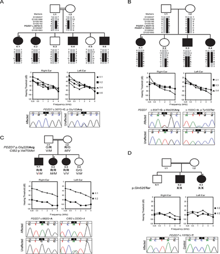
Ascertained families originated from different parts of Iran: North East (L-445 and L-8900092), Central (L-755) and North West (L-8600482) (Table I). Families L-8900092, L-8600482, and L-445 reported consanguinity (Fig. 1A, C, and D). Physical examination in affected persons was remarkable only for hearing loss. Audiological examination in affected individuals in families L-445 and L-755 revealed prelingual mild-moderate downsloping to severe hearing loss in high frequencies whereas the two patients in family L-8900092 reported prelingual severe-profound hearing loss across all frequencies (Fig. 1A, B, and D). In family L-8600482, two different phenotypes were observed. The proband (II.2) presented with severe-to-profound hearing loss whereas the sibling (II.1) has mild-moderate downsloping to severe hearing loss in high frequencies (Fig. 1C) similar to the phenotypes observed in families L-445 and L-775. Ophthalmological examination revealed no abnormalities on funduscopy. In family L-445, three patients had a refractory error, myopia (−3.75), corrected with contact lens and/or glasses.
| Family ID | L-755 | L-8600482 | L-445 | L-8900092 | ||||||||
|---|---|---|---|---|---|---|---|---|---|---|---|---|
| Ethnicity region | Iranian Central-Shiraz | Iranian North East-Birjand | Iranian North West-Tabriz | Iranian North West-Tabriz | ||||||||
| Patient ID | II.1 | II.3 | II.4 | II.1 | II.2 | II.3 | II.1 | II.2 | II.4 | II.6 | II.2 | II.3 |
| Age at examination | 38 | 33 | 25 | 40 | 33 | 20 | 36 | 30 | 25 | 21 | 31 | 23 |
| Ophthalmologic examination (years) | N | N | N | N | N | N | N* | N* | N* | N | N | N |
| Audiologic pattern | Down sloping | Down sloping/flat | Down sloping | Flat | ||||||||
| Severity of HL | Moderate-severe | Moderate-severe/profound | Moderate-severe | Profound | ||||||||
| Type of HL | Prelingual | Prelingual | Prelingual | Prelingual | ||||||||
- N represents a normal funduscopic report. An asterisk indicates a refractory error (myopia [−3.75]).

Homozygosity Mapping and Haplotype Segregation Analysis
Genome-wide scanning was completed on two families: L-445 and L-775. In family L-445, linkage analysis identified a single region of homozygosity-by-descent that segregated with the hearing loss phenotype. The region mapped to chr10q21.2–q26.13 between markers D10S1652 and D10S587 (60 Mb) with a maximum LOD score of 3.26. This interval was further refined to 34 Mb between markers D10S1686 and D10S1693, an interval that overlaps with the reported deafness locus, DFNB57 (10q23.1–q26.11) (Fig. 1A). In Family L-775, affected individuals shared haplotypes between markers D10S185 and D10S597 (Fig. 1B). Proximal and distal boundaries were defined by Individual II.3 (between markers D10S185 and D10S192) and his sister II.4 (between markers D10S192 and D10S597), respectively. DFNB57 lies distal to a known deafness gene, CDH23, and proximal to a candidate deafness gene, TECTB. Direct sequencing of both of these genes was normal.
Variant Identification
TGE + MPS on probands from each family generated an average of 6.9 million reads per person with an average coverage of 620 and greater than 99% coverage at 20X (Table II). Variant filtering applying the guidelines described in Materials and Methods Section, yielded an average of six variants per sample (Table II). In three of the families, only variants in PDZD7 (NM_001195263.1) represented plausible deafness-causing candidates because of their high conservation and pathogenicity prediction. As expected, probands from consanguineous families were homozygotes for these variants, while the proband from L-755 was a compound heterozygote. In family L-8600482, two plausible mutations were identified—one in PDZD7 and one in CIB2 (NM_006383). There were no other plausible deafness-causing variants. CNV analysis was negative (Table II).
| OtoSCOPE® results | |||||||||
|---|---|---|---|---|---|---|---|---|---|
| % Coverage | |||||||||
| Family ID | Patient | Average target coverage | Reads overlapping target | 1X | 10X | 20X | Variants after filtering | CNV | Candidate variant(s) |
| L-445 | II.1 | 622 | 7700415 | 99.88 | 99.59 | 99.35 | 3 | 0 | 1 |
| L-755 | II.1 | 513 | 5976821 | 99.86 | 99.52 | 99.18 | 6 | 0 | 2 |
| L-8600482 | II.2 | 648 | 7353056 | 99.93 | 99.68 | 99.44 | 7 | 0 | 2 |
| L-8900092 | II.3 | 697 | 9254099 | 99.90 | 99.63 | 99.47 | 8 | 0 | 1 |
| Average | 620 | 7571098 | 99.89 | 99.61 | 99.36 | 6 | 0 | 1 | |
PDZD7 Variant Analysis
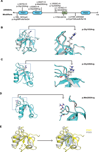
Segregation analysis was completed for all families in all individuals for whom DNA samples were available. In family L-455, all affected persons were homozygous for a single PDZD7 variant, c.307G>C (p.Gly103Arg), which lies in the PDZ1 domain of the protein (Fig. 2A) and has a MAF of 0.007% and 0.0008% in EVS and ExAC, respectively. Two novel PDZD7 variants were identified in family L-755: a missense mutation c.854T>G (p.Met285Arg) and a nonsense mutation c.1500C>A (p.Tyr500Ter): all affected individuals were compound heterozygotes, while the unaffected sibling carried only (p.Tyr500Ter). The (p.Met285Arg) mutation is located in the PDZ-2 domain (Fig. 2A). In consanguineous families L-8900092 and L-8600482, homozygous mutations c.1576C>T (p.Gln526Ter) and c.682G>A (p.Gly228Arg; also in PDZ-2), respectively, were identified in affected probands. In Family L-8600482, a novel mutation in CIB2 c.223G>A (p.Val75Met) was also identified in the proband but this variant did not segregate with the deafness phenotype in the family (Fig. 1C). All three missense mutations in PDZD7 and the single missense variant in CIB2 are in conserved amino acids and predicted to be deleterious by Polyphen2, SIFT, Mutation Taster and LRT (Table III). None of these mutations was seen in 300 ethnically matched controls.

| MAF (%) | ||||||||||||||||
|---|---|---|---|---|---|---|---|---|---|---|---|---|---|---|---|---|
| Phenotype | Family origin | ID | Genotype | Nucleotide change | Amino acid change | Domain | 1 kg | EVS | ExAC | GERP | PhyloP | Poly phen2 | SIFT | Mutation taster | LRT | Reference |
| ARNSHL | Iranian | L-445 | Homo | c.307G>C | p.Gly103Arg | PDZ1 | 0 | 0.012 | 0.0008 | C | C | D | D | D | D | This study |
| L-8600482 | Homo | c.682G>A | p.Gly228Arg | PDZ2 | 0 | 0 | 0.0008 | C | C | D | D | D | D | |||
| Homoa | c.94G>A | p.Val32Met | EF1 | 0 | 0 | 0.0008 | C | C | D | D | D | D | ||||
| L-8900090 | Homo | c.1576C>T | p.Gln526Ter | – | NA | NA | 0 | |||||||||
| L-755 | Comp | c.854T>G | p.Met285Arg | PDZ2 | 0 | 0 | 0 | C | C | D | D | D | D | |||
| Het | c.1500C>A | p.Tyr500Ter | NA | NA | 0 | |||||||||||
| USH2 modifier | Canadian | Fac | Het | c.166_167insC | p.Arg56fsTer24 | – | NA | NA | 0 | Ebermann [2010] | ||||||
| Homob | c.4338_4339del | p.Cys1447fsTer | FTIII | NA | NA | 0 | ||||||||||
| German | GER1 | Het | c.1750-2A>G | Splice site | HNL | NA | NA | 0 | ||||||||
| Hetb | c.4515_4518del | p.Arg1505fsTer | – | NA | NA | 0 | ||||||||||
| Hetb | c.13316C>T | p.Thr4439Ile | FTIII | 0 | 0 | 0 | C | C | P | T | D | U | ||||
| GER2 | Het | c.2194_2203del | p.Cys732fsTer18 | – | NA | NA | 0 | |||||||||
| Hetc | c.17137delG | p.Ala5713fsTer | – | NA | NA | 0 | ||||||||||
| Hearing loss | Korean | SB112-204 | Het | c.76_77del | p.Ser26fsTer53 | – | NA | NA | 0 | Kim [2015] | ||||||
| Hetc | c.3022 + 2T>G | Splice site | – | NA | NA | 0 | ||||||||||
- Nucleotide numbering: the A of the ATG translation initiation site is noted as +1 using isoform NM_001195263 of PDZD7, NM_006383 for CIB2, NM_206933 for USH2A, and NM_032119 for ADGRV1. NSHL, non-syndromic hearing loss; USH2, Usher syndrome Type 2; C, predicted conserved; D, predicted damaging or deleterious; P, probably damaging; T, tolerated; U, unknown; NA, data not available; PDZ, PDZ domain; EF, EF-hand; HNL, harmonin-N-like domain; FTIII, fibronectin type-III. Dashes denote mutations outside functional domains.
- a Mutation found in CIB2.
- b Mutations reported in USH2A.
- c Mutations reported in ADGRV1.
Molecular Modeling and Mutation Analysis
Two of the missense mutations involve identical amino acid changes although in different PDZ domains (p.Gly103Arg in PDZ1; p.Gly228Arg in PDZ2). For both of these mutants, the glycine residue present in the wild-type structure is in a backbone conformation that is not typically seen with arginine (ϕ = 74°, ψ = 175° for Gly103 in PDZ1; ϕ = 80°, ψ = 170° for Gly228 in PDZ2). In addition to a decrease in backbone conformational flexibility, mutation from glycine to positively charged arginine results in new interactions with backbone or side chains amino acids (Fig. 2B and C). The (p.Met285Arg) substitution in the PDZ2 domain replaces a polar methionine with the positively charged arginine, which results in different side chain interactions and backbone movement upstream in the sequence (Fig. 2D). In all three cases, the mutations occur on the surface of a PDZ domain, which suggests a disease mechanism based on alteration of intermolecular PDZ interactions, as opposed to destabilizing the fold of the PDZ domain in isolation.
DISCUSSION
In this study, we implicate pathogenic variants in PDZD7 as a novel cause of ARNSHL. PDZD7 encodes a 1,033 amino acid protein, PDZD7, which has three PDZ-domains and one harmonin-N-like (HNL) domain [Schneider et al., 2009; Chen et al., 2014] (Fig. 2A). Expressed in the sensory cells of the inner ear and retina, PDZD7 is highly homologous to two other proteins with similar expression patterns, harmonin and whirlin, both of which are well described scaffolding proteins that give rise to USH or ARNSHL when mutated [Ebermann et al., 2010; Grati et al., 2012; Hu et al., 2014]. This shared similarity suggests that PDZD7 is also a scaffolding protein and functions as such in the ear and the eye.
In the cochlea, PDZD7 localizes to the base of stereocilia along with other USH-related proteins: USH2A, VLGR1, and WHRN [Grati et al., 2012; Zou et al., 2014]. These proteins are an indispensable part of the USH2 interactome and form the ankle links, a narrow web-like network that runs parallel to the apical surface of hair cells just above the insertion of stereocilia, connecting each stereocilium to its nearest neighbors [Ahmed et al., 2013]. USH2A and VLGR1 are large proteins with extensive and dissimilar extracellular domains that form the network described above. In common to both, however, are cytoplasmic PDZ-binding motifs (PBMs), which facilitate cytoplasmic protein–protein interactions [Millan et al., 2011]. Although initially posited that WHRN, with its three PDZ-domains, effectively coupled VLGR1 and USH2A, and in turn anchored the entire complex cytoplasmically at the base of the stereocilia, recent evidence suggests a more complex picture [Chen et al., 2014; Zou et al., 2014].
Mouse mutants homozygous for the targeted deletion of Pdzd7 have congenital profound deafness as assessed by auditory brainstem response testing, although their electroretinogram responses from both rod and cone photoreceptors are normal at 1 month of age. At the microscopic level, stereocilia bundles are disorganized, and at the molecular level, localization of the USH2 interactome is disrupted, implying a direct role for PDZD7 in organizing the ankle links in the developing cochlea [Zou et al., 2014].
PDZD7 forms homodimers through its PDZ2 domains. These domains also interact with VLGR1 through the latter's cytoplasmic PBM, and heterodimerize with WHRN through its PDZ2 domain. While PDZD7 also has the ability to interact with USH2A, the PDZD7/USH2A interactions are weak and not preferential [Chen et al., 2014]. The functional role of the other domains of PDZD7 (PDZ1, PDZ3, and HNL) remains to be determined.
The five mutations we identified in PDZD7 are conserved and predicted to have a deleterious impact on protein function (Table III). Of the three missense mutations, one—(p.Gly103Arg)—is in the PDZ1 domain while the remaining two—(p.Gly228Arg) and (p.Met285Arg)—are in the PDZ2 domain (Fig. 2A). Interestingly, two of the missense changes are identical, changing a small polar glycine to a large bulky basic arginine, which by modeling alters the conformation of PDZ1 and PDZ2 (Fig. 2B and C, Supplement Figure S1), (p.Met285Arg) has a similar effect (Fig. 2D). These changes suggest that the decreased flexibility of PDZ2 impedes binding with VLGR1 and/or WHRN. The two nonsense mutations, (p.Tyr500Ter) and (p.Gln526Ter), lie outside functional domains (Fig. 2A), but are predicted to yield null alleles by nonsense-mediated mRNA decay (NMD) [Lu et al., 2009]. As such, affected persons in family L-8900092 have no functional copies of PDZD7; in contrast, affected L-755 family members carry both (p.Met285Arg) and (p.Tyr500Ter) and will have one fully translated allele with impaired binding to WHRN and/or VLGR1. In families L-455 and L-8600482, both copies of PDZD7 are translated but the segregating mutations are predicted to disrupt protein interactions with their respective PDZ domains (Fig. 2E). Although protein modeling is a robust prediction tool for structural impacts of missense mutations, its underlying assumption of protein production from the mutated allele may be inaccurate. Further functional studies are needed to fully assess and understand the consequences of the identified variants on gene expression and protein function and interactions.
Mutations in PDZD7 have been described to modify the retinal phenotype of USH2 or have been implicated in digenic USH. All patients with divergent phenotypes had moderate downsloping hearing loss with either more severe or milder RP than expected, as summarized in Table III [Ebermann et al., 2010; Kim et al., 2015]. While these patients carried USH2A mutations, they were also heterozygous for mutations in PDZD7. In one patient, the mutation, p.Arg56ProfsX24 was identified. This patient had a more severe retinal phenotype. In another patient, a splice mutation c.1750-2A>G was identified; this change causes an in-frame insertion that disrupts the HNL domain while probably preserving the PDZ3 domain. This patient had less severe RP. Finally, two patients have been described with mutations in ADGRV1 and PDZD7. The first patient had an USH2 phenotype and one mutation in ADGRV1. After screening the coding sequence of all USH related genes, the authors identified a frameshift mutation in PDZD7 (p.Cys732LeufsX18), raising the possibility that this mutation in combination with the ADGRV1 mutation might be USH causing. The second patient had mild NSHL with one mutation in ADGRV1 and one mutation in PDZD7 [Kim et al., 2015]. USH2 was not excluded as the patient was too young to exhibit RP and so it remains unclear whether this case represents digenic USH or digenic NSHL. Phenotypic ambiguity of NSHL mimics versus true NSHL was also raised by Schneider and colleagues in an 8-year-old patient with a homozygous reciprocal translocation that disrupted the open reading frame of PDZD7 [Schneider et al., 2009].
The absence of RP is noteworthy in the families we studied. This finding suggests that the functional roles of PDZD7 in the retina are compensated for by other scaffolding proteins. With respect to the hearing loss, three of the families we report have a downsloping audiogram, which matches the audiometric profiles of other USH2 genes [Sadeghi et al., 2013; Steele-Stallard et al., 2013]. Persons in family L-8900092, however, have profound hearing loss across all frequencies that may reflect the fact that these patients segregate with a homozygous stop mutation. Family L-8600482 segregates with two different audio profiles; profound across all frequencies in one person and a mild-moderate downsloping to severe loss in the sibling. Interestingly, the person with the more severe phenotype also carries a homozygous mutation in the CIB2 gene, mutation of which is responsible for DFNB48 and Usher Syndrome type 1J (USH1J) [Riazuddin et al., 2012] (Fig. 1D). This variant is novel and is predicted to be pathogenic by multiple pathogenicity prediction algorithms. It is located in the first EF-hand domain of the CIB2 protein, between two previously described pathogenic mutations that alter protein conformation and negatively affect calcium binding. We hypothesize that this novel variant has a similar effect [Schneider et al., 2009; Jan, 2013]. The phenotype of this person matches the deafness phenotype associated with mutations in CIB2, making it possible that the CIB2 mutation is causally related to the more severe phenotype. It is interesting that this person segregates two mutations in two USH-related genes and yet at the age of 33 years old does not have any vision problems.
In diseases that exhibit extensive genetic and phenotypic variability, such as deafness, identifying the genetic cause of disease can be difficult. With over 100 genes causally related to hearing loss (http://hereditaryhearingloss.org/), providing an accurate interpretation of patients' results can be difficult [Rehman et al., 2014; Richards et al., 2015]. Each misdiagnosed case is a missed opportunity for clinical intervention and appropriate genetic counseling. Until now, patients segregating possibly pathogenic alleles in PDZD7 have been left undiagnosed largely due to the restriction of PDZD7's role as only a modifier of the USH2 phenotype. Here, we correct this misinterpretation and expand the phenotypic spectrum related to PDZD7.
In summary, we have identified PDZD7 as the gene responsible for ARNSHL at the DFNB57 locus. Our finding adds to the phenotypic spectrum associated with mutation of PDZD7 and offers additional insight into the USH2 interactome. We also describe a novel disease-causing mutation in CIB2 and illustrate the complexity associated with gene identification in diseases that exhibit large genetic and phenotypic heterogeneity. Further studies aimed at clarifying the complex interactions of the USH2 interactome are needed to define what drives phenotypic differences, as this knowledge may offer novel strategies to alter expected phenotypes in subtle but clinically important ways.
ACKNOWLEDGMENTS
We thank the families reported here for their collaboration in this study. This research was supported in part by NIDCD RO1s DC003544, DC002842, and DC012049 to RJHS.


