Near-Infrared Manipulation of Membrane Ion Channels via Upconversion Optogenetics
Abstract
Membrane ion channels are ultimately responsible for the propagation and integration of electrical signals in the nervous, muscular, and other systems. Their activation or malfunctioning plays a significant role in physiological and pathophysiological processes. Using optogenetics to dynamically and spatiotemporally control ion channels has recently attracted considerable attention. However, most of the established optogenetic tools (e.g., channelrhodopsins, ChRs) for optical manipulations, are mainly stimulated by UV or visible light, which raises the concerns of potential photodamage, limited tissue penetration, and high-invasive implantation of optical fiber devices. Near-infrared (NIR) upconversion nanoparticle (UCNP)-mediated optogenetic systems provide great opportunities for overcoming the problems encountered in the manipulation of ion channels in deep tissues. Hence, this review focuses on the recent advances in NIR regulation of membrane ion channels via upconversion optogenetics in biomedical research. The engineering and applications of upconversion optogenetic systems by the incorporation multiple emissive UCNPs into various light-gated ChRs/ligands are first elaborated, followed by a detailed discussion of the technical improvements for more precise and efficient control of membrane channels. Finally, the future perspectives for refining and advancing NIR-mediated upconversion optogenetics into in vivo even in clinical applications are proposed.
1 Introduction
As essential cell surface components, membrane ion channels are primarily responsible for the propagation and integration of electrical signals in the nervous, muscular, and other systems, their activation or malfunctioning plays significant roles in physiological and pathophysiological processes, such as brain thinking, muscle contraction, and channelopathies.1 Precisely regulating various membrane channels and spatiotemporally balancing the relevant dynamic processes are crucial to understand the biological implications of cellular ion channels, as well as provide insight into the future therapy of neurological and other life-threatening diseases.2 Conventional strategies through chemical, genetic or electrical approaches have been well developed for modulation of ion channel activity and conductance in biochemical research.3 Despite the initial success in practice, these commonly used methods are faced with challenges of dissecting the role of dynamic nature. For instance, nonspecific accumulation and action of chemical drugs with the blood circulation would be inevitable, which may compromise the spatial accuracy of the control.4 In addition, the irreversibility of chemical or genetic perturbations would be another inherent concern that impedes the patterned stimulation with a high temporal resolution.[3] Furthermore, although the electrical patterns show great promise for spatiotemporal mapping and modulation of voltage-gated ion channels, even for deep brain's stimulation (DBS), the highly invasive implantation of electrodes or chips, and associated adverse events are considerable concerns for the clinical application.5 Therefore, the development of unique and reliable techniques, which exhibit high spatiotemporal precision, minimal invasion as well as broad applicability for membrane ion channels modulation and future translational studies are highly desirable.
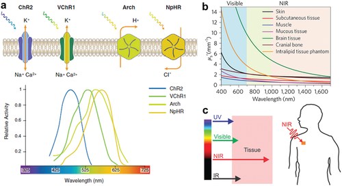
In recent years, using light to control biomolecules and biological processes has gained much attention based on its unsurpassable flexibility and spatiotemporal precision.6 One typical technology, termed as “optogenetics,” has been extensively utilized for optical manipulation neural activity in neuroscience with high specificity and temporal resolution (millisecond-timescale).7 Most importantly, the rapid evolution of light-sensitive microbial opsins also opens up opportunities for driving optogenetic ion channels manipulation into more complex systems, ranging from single cell level in vitro to brain circuitries-mediated behavioral analyses in freely moving animals.8 However, despite the remarkable achievements, currently established opsins or other optogenetic tools for membrane channels modulation are mainly sensitized in visible window (Scheme 1a), which significantly limits their uses for in vivo applications due to the unsatisfactory light penetration and less controlling efficacy caused by biological tissues absorption and scattering (Scheme 1b).[7, 9] Although the approach by optical fiber or LED implantation could partly achieve deep tissue stimulations, the highly invasive procedure raises tremendous safety concerns.10 Hence, the engineering of novel optogenetic technique that allows minimized invasion in deep tissues for controlling of membrane channels is of great importance.
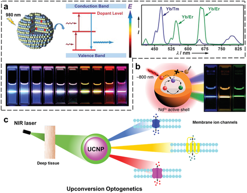
Notably, extensive efforts have been devoted to achieve the goal for in vivo optogenetic stimulation, in which shifting the excitation wavelength to the near infrared (NIR) window is considered to be favorable for deeper tissue penetration.[9, 13] Although significant progress has been acquired in genetically engineering of red-shifted ChRs,14 current optogenetics is still constrained within the visible wavelength window (e.g., <650 nm). Alternatively, by employing a combination of NIR light responsive nanomaterials and optogenetic tools encourages the possibility of enhanced penetration, where the nanomaterials mainly serve as photon transformer in optogenetic stimulation.15 To this end, lanthanide-doped upconversion nanoparticles (UCNPs) as emerging photonic nanomaterials with their fascinating capability to convert NIR light (e.g., at 980 or 808 nm, Scheme 2a,b) into multiple emissions ranging from UV–vis to NIR region, have attracted numerous interests in bioimaging and nanomedicine due to the less scattering and deeper penetration depth into tissues.16 It is noted that the development of NIR light-triggered optogenetics though incorporating multiple emissive UCNPs into various light-sensitive opsins, terms as upconversion optogenetcis (Scheme 2c), have been witnessed in the past few years, relevant progress and results in membrane ion channels control have already been widely reported.[15, 17]
Therefore, in this review, we focus on the recent advances of upconversion optogenetics for NIR manipulation of membrane ion channels in biomedical research. The engineering of upconversion optogenetic systems by incorporation specific UCNPs into desired ChRs or other light-sensitive proteins will be first elaborated, followed by detail discussion of their broad applications as well as technique improvements, such as enhancing photon-converting efficacy of UCNPs, minimizing heating effect and localized regulation for more precise and efficient optogenetic manipulation of membrane channels. Finally, the conclusion and future perspectives for refining and advancing NIR-mediated upconversion optogenetic manipulations of ion channels into in vivo applications, even in clinical studies are proposed.
2 Advances of Upconversion Optogenetics in NIR Membrane Ion Channel Manipulations
The combination of upconversion nanotechnology and optogenetics provides great opportunities for NIR manipulation of membrane ion channels in either neuronal or nonneuronal research. In particular, significant efforts in neuroscience have been made by utilization of upconversion optogenetics, which lifts such flexible optical interrogation tools to a level of broader applicability.[15, 21] Hence, the recent advances of diverse applications are discussed from the following perspectives: upconversion optogenetic systems, stimulation platforms, and applied models.
2.1 Upconversion Optogenetics
As summarized in Table 1, by incorporation appropriate UCNPs into light-sensitive opsins, flexible and multiplexed activation/inhibition of specific ion channels can be achieved in response to NIR stimulation. The idea proposed first by Deisseroth and Anikeeva in a patent application in 2011,22 where compositions and methods for noninvasively delivering light to light-responsive opsins-expressed neurons on the plasma membrane via the use of various UCNPs are provided. In 2013, Han et al. also proposed in a grant to develop the wireless upconversion optogentics for in vivo neuronal manipulations.23
| Opsin | λmaxa) | Threshold limit valueb) | Functions | UCNP | Xc) | Ex. | Em.d) |
|---|---|---|---|---|---|---|---|
| ChR2 | 480 nm |
P: ≈1.1 mW mm−2 S: ≈1.05 mW mm−2 |
Excitatory cation channel; native from Chlamydomonas reinhardtii | NaYF4: Yb/X@NaYF4 | Tm | 980 nm | 450, 475, 800 nm |
| VChR1 | 545 nm | Not tested | Red-shifted, excitatory cation channel; native from Volvox carteri | Er | 545, 665 nm | ||
| ChIEF | 450 nm |
P: ≈1.65 mW mm−2 S: ≈1.38 mW mm−2 |
Fast kinetics, excitatory cation channel; chimera of ChR1 and VChR1 | Pr | 480 606 nm | ||
| ChETA | 470 nm |
P: ≈5.02 mW mm−2 S: ≈0.62 mW mm−2 |
Fast kinetics, excitatory cation channel; substitutions in ChR2 | Tb | 480–650 nm | ||
| Step function opsins |
470 nm on 590 nm off |
Not tested | Excitatory cation channel; single mutations in ChR2 | Eu | 570–720 nm | ||
| Chronos | 500 nm | ≈0.3 mW mm−2 | Fast kinetics, excitatory cation channel; native from Stigeoclonium helveticum | Sm | 601 nm | ||
| Chrimson | 590 nm | ≈4.0 mW mm−2 | Red-shifted, excitatory cation channel; Native from Chlamydomonas noctigama | Ho | 540 nm | ||
| C1V1 | 540 nm | ≈7.2 mW mm−2 | Red-shifted, excitatory cation channel; chimera of ChR1 and VChR1 | Dy | 486, 575 nm | ||
| NpHR | 590 nm | ≈21.7 mW mm−2 | Inhibitory chloride pump; native from Natronomonas pharaonis | NaGdF4: Yb/X@NaGdF4 | As above | As above | |
| ChloC | 480 nm | Not tested | Inhibitory anion channel; mutagenesis of ChR2 | ||||
| GtACR2 | 520 nm | Not tested | Inhibitory anion pump; native from Guillardia theta | NaYF4: Nd/Yb/X@NaYF4: Nd | As above |
980 nm or 808 nm |
As above |
| ArCh | 570 nm | Not tested | Inhibitory proton pump; native from Halobacterium sodomense |
- a) Peak λ indicates the wavelength of light that maximally activates the opsin
- b) P and S indicate peak response and steady-state response, respectively
- c) X indicates the doped lanthanide ions within UCNPs
- d) Upconverting emissions with corresponding lanthanide ion-doped UCNPs. The table of both the optogenetic tools and UCNPs is reproduced with permission.[9, 24] Copyright 2010, The Authors, Journal compilation copyright, 2011, The Physiological Society. Copyright 2016, John Wiley & Sons. Copyright 2013, Royal Society of Chemistry.
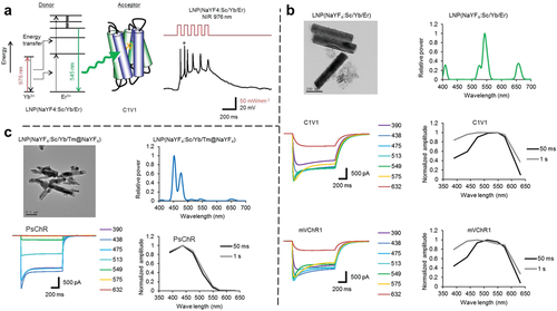
Hereafter, successful implementation of upconversion optogenetics was reported in 2015 by different research groups independently.17, 25 For example, Yawo and co-workers used rare-earth elements doped UCNPs as luminous donors to activate ChRs, in which different types of upconversion optogenetic systems (termed “LNP-ChRs,” Figure 1) were established for membrane ion channels and neural activities manipulation.17 Upon NIR (976 nm) irradiation, the LNP(NaYF4: Sc/Yb/Er) emitted green light (550 nm) that in turn activated C1V1 or mVChR1 to generate a photocurrent in the expressing cells (Figure 1b). Similarly, the blue emissive (peaks at 450 and 480 nm) LNP(NaYF4:Sc/Yb/Tm@NaYF4) could effectively activate PsChR ion channel with significant photocurrent generation (Figure 1c). Moreover, the results of NIR stimulations also demonstrated time-dependent and power-dependent manners. However, there remains room for refinements of this study, such as the light-upconverting efficiency of UCNP-ChR systems, the biosafety of UCNPs and NIR optics, to further boost upconversion optogenetics into in vivo applications.
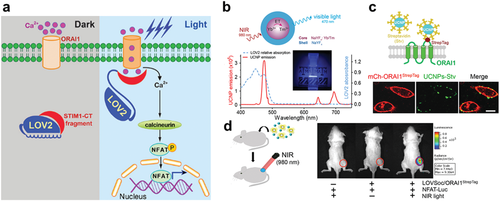
In addition to the superior UCNP-ChR systems for NIR manipulation ion channels, Zhou and co-workers demonstrated another type of UCNP-based optogenetic platform, termed “Opto-CRAC” (Figure 2a).[25] Such unique optogenetic system was capable of remote and selective control Ca2+ influx, subsequent Ca2+-dependent gene expression and further photomodulate of immune-inflammatory responses both in vitro and in vivo. In which, blue emissive NaYF4:Yb/Tm@NaYF4 UCNPs upon 980 nm excitation was applied to manipulate Ca2+ gate-controlling protein that was genetically engineered to enable blue light-activation (Figure 2b–d). Moreover, the optogenetic module-LOVSoc27 can reversibly generate both sustained and oscillatory Ca2+ signals through the light input adjustment (e.g., laser pulse and intensity). Most importantly, Opto-CRAC mediated light-generated Ca2+ signaling pathway can lead to specific physiological responses in cells of the immune system and further enable its potential application for (patho)physiological studies in animal models.
The lanthanide-doped UCNPs with tunable emission can be applied to activate/inhibit a wide range of genetically photosensitive ion channel proteins (e.g., ChRs and CRAC). In combination with the deep light delivery in vivo, such promising and multiplexed upconversion optogenetics will provide great opportunities to specifically and precisely control ion-dependent signaling pathways, biological activities and even the ultimate goal of disease therapy.
2.2 Stimulation Platforms
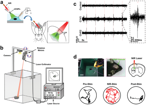
Despite the flexible adaptability of upconversion optogenetic systems, great efforts have addressed that the development of preferable stimulation platforms could intensively facilitate efficient NIR stimulations of light-sensitive ion channels. As shown in Figure 3a, Lee and co-workers reported hybrid upconversion nanomaterials, UCNPs (NaYF4: Yb/Tm@NaYF4) embedded poly(lactic-co-glycolic acid) (PLGA) 0.5 mm films, which were used as underlying scaffolds for neurons culture and optogenetic stimulation.[25] Such hybrid polymeric scaffolds converted NIR light to blue luminescence as the internal excitation light source, and subsequently activated blue light-sensitive channelrhodopsin-2 (ChR2)-expressed neurons. The neurons generated time-locked, sustained naturalistic impulses with millisecond resolution in response to 980 nm laser pulses at 1, 5, and 10 Hz. Most critically, this upconversion platform could be further optimized for NIR-mediated optogenetic control by balancing multiple physicochemical features of the nanomaterial (e.g., morphology, size, emission spectra, surface modification, concentration), thus enabling an early demonstration of rationally designing nanomaterial-based strategies for advanced neural applications.

Additionally, relevant studies regarding the effective optogenetic light delivery by different stimulation platforms and methods were also demonstrated in Yawo and co-workers.17 The UCNPs were set close to the recording cell by either Method 1 or Method 2 (Figure 3b). Both two methods showed inward photocurrent response in a manner dependent on the laser power, which indicated that the upconverting luminescence (green emission) emitted from UCNPs effectively activated the ChR-based membrane channel (C1V1). However, Method 1 showed higher efficiency than Method 2 upon the same power of NIR excitation, it is suggested that UCNPs should be positioned as close as possible to the ChR protein since the power density is proportional to the reciprocal square of the distance. This result provides valuable information to clarify optimized conditions for further applications of upconversion optogenetic systems.
Other than the progress of developing upconversion platforms for in vitro optogenetic stimulation, Shi et al. first packaged UCNPs into glass micro-optrodes to form implantable transducers that can convert NIR energy to visible light and subsequently stimulate neurons with different ChRs expression (Figure 3c).28 These microdevices showed superb long-term biocompatibility and allowed remotely NIR modulation of brain function, even to control complex animal behaviors. Most significant, such fully implantable microdevices not only represented the feasibility of NIR optogenetic practical applications in living animals but also opens up new possibilities for remote and multiplexed control of neural activities in the brains.
Very recently, Sheng et al. reported another type of infrared-to-visible upconversion concept-based microscale optoelectronic devices for highly efficient optogenetic neuromodulation (Figure 3d).29 The thin-film, ultraminiaturized devices realize NIR to visible red or yellow upconverting emission that was linearly dependent on incoherent, low-power excitation, with a quantum yield of about 1.5%. Moreover, these microscale devises showed promising biocompatibilities in the heterogeneous biological environment. Most notably, in vitro and in vivo studies demonstrated the possible utility of such injectable microdevices for optogenetic neuromodulation upon implanted in behaving animals. Compare to UCNP-based systems, this type of optoelectronic platform provides another versatile route to achieve photon-upconversion throughout the entire visible spectral range with higher efficiency. It is also feasible for optogenetic stimulation various ChRs in living animals via upconversion spectral tuning.
2.3 Applied Models
Upconversion optogenetic techniques have been successfully applied into broad biomedical research in a highly controlled manner and spatiotemporal precision. Such NIR manipulating system via incorporating UCNPs with genetically expressed photosensitive ion channels that in principle overcome the limitations, including low penetration of excitation light and the invasiveness of light source implantation, encountered in commonly used optogenetics in vivo.[15] Considering the inherent complexity of living conditions, so far, it has remained extremely difficult to experimentally clarify the fundamental physiological/pathophysiological implications based on current NIR optogenetics and other technical approaches. Evolutionary studies on upconversion optogenetics for NIR manipulation membrane ion channels and relevant biological activities in different animal models and varying purposes are discussed further below.
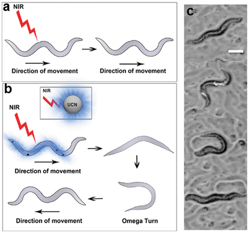
Other than the successful upconversion optogenetic manipulation of membrane channels in vitro, Zhang and co-workers utilized quasi-continuous wave NIR excitation approach to improve the efficacy and applicability of optogenetic neuromodulation in Caenorhabditis elegans model expressing ChR2 (Figure 4).30 This work offers possibility to enhance multiphoton emissions from UCNPs, furthermore, it achieves NIR light-triggered upconversion optogenetic activation of the membrane channel in vitro and manipulation of touch-akin reversal behavior of C. elegans in vivo. Moreover, such noninvasive NIR optogenetic control strategy also exhibits flexibility for advancing optogenetic operations in other animal models and living system studies.
Zebrafish have proved to be a suitable in vivo model for biomedical studies in bioimaging and theranostics due to their physiological and genetic homology with mammals, high fecundity and easy operability.31 Therefore, the utilization of zebrafish in optogenetic manipulation would be highly convenient for spatiotemporally regulating and monitoring biological events. Xing and co-workers creatively applied upconversion optogenetics in living zebrafish for remotely activating the ion-channel (ChR2) and Ca2+ mediated biological functions under NIR (808 nm) light stimulation (Figure 5).32 Furthermore, site-specific covalent localization of UCNPs on the cell membrane by means of glycan metabolic labeling enabled more precise and efficient optogenetic regulation. It was revealed that the feasibility of NIR-light-mediated activation of ion channels to evoke apoptosis in vitro and in vivo. Very recently, another optogenetic study in living zebrafish from Xing et al. was reported to precise manipulate membrane ion channel and Ca2+ influx triggered signaling pathway.33 As mentioned above, the practicality and reliability of zebrafish model in upconversion optogenetic studies would greatly promote further applications on the manipulation of biological events.

Over the past few decades, the mouse has emerged as a prevalent model system in biomedical research considering its high genomic and physiological homology with humans as well as enormous technical advances in genetic engineering and characterization techniques.34 Although the application of mice in deep-tissue optical stimulation is substantially dependent on special techniques and complex manipulations, recent progress in optogenetics and upconversion nanotechnology have addressed the possibility of NIR optogenetic control mouse (or rat) brain activity.
Shi et al. reported an all-optical method by utilizing UCNPs based microdevices to remote control neurons expressing different opsin proteins (ChR2 or C1V1) in rat brain (Figure 6). Upon NIR illumination with the robotic laser projection system, this neural stimulation technique was able to reliably trigger spiking activity and spatiotemporally modulate brain activity in various regions, including the striatum, ventral tegmental area, and visual cortex. Moreover, this study provided an innovative tetherless upconversion optogenetic strategy for brain stimulation in behaving rats. Notably, another work by Shi et al. applied the similar upconversion based strategy for multiplexed optogenetic neural stimulation both in vitro and in vivo by fabricating spectrum-selective UCNPs.35 Such unique technique would significantly benefit both fundamental physiological regulation and translational neuroscience research.
Furthermore, several other upconversion optogenetic studies are also significantly contributing to this field. For instance, McHugh and Liu recently explored feasibility of using UCNPs as optogenetic actuators to miniinvasively stimulate deep neurons in the mouse brain (Figure 7).36 Moreover, the NIR-mediated upconversion optogenetics could evoke dopamine release from genetically tagged neurons in the ventral tegmental area (VTA), and induce brain oscillations through activation of inhibitory neurons in the medial septum (MS). Most importantly, such upconversion optogenetics can also silence seizure by inhibition of hippocampal excitatory cells, and thus trigger memory recall. Such a promising technology will enable NIR manipulation of neuronal activity with the great potential for remote therapy of the neurological disease.

3 Strategies to Improve Upconversion Optogenetics
Despite the great promise, upconversion optogenetics is still facing challenges of low photon-conversion efficacy and heating effect caused by NIR illumination.[15, 37] To address these issues, multitudinous strategies have been established to increase the upconversion luminescence, including surface passivation, material design, engineering of the excitation source, or a combination of multiple parameters.38 Moreover, site-specific optogenetic manipulation by internalizing or localizing UCNPs inside cell or on cell surface has proved to be an alternative method for overcoming above limitations.32 These improvement approaches provide exciting opportunities to promote NIR-mediated upconversion optogenetics for more efficient and precise biomedical modulations, and enabling future translational and clinical research.
3.1 Enhancing Photon-Converting Efficacy of UCNPs
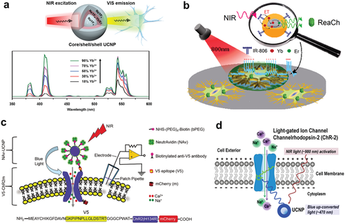
As summarized in Table 2, the recent advances to enhance photon-converting efficacy of UCNPs provide flexible solutions for further improving upconversion optogenetics. Among numerous strategies, surface passivation by preparing core–shell nanostructured UCNPs was an effective and commonly used approach in majority of NIR optogenetic studies. Besides, very recent efforts from Wang and co-workers have been made to enhance the luminescence of UCNPs through fabricating core-shell-shell structure and optimizing the doping contents of ytterbium ions (Yb3+) (Figure 8a),39 which achieved nearly threefold luminescent enhancement over traditional core−shell UCNPs and were further utilized as optical transducers to develop a fully implantable upconversion-based device for in vivo optogenetic inhibition in eNpHR-expressed behaving mice. Meanwhile, another previous work by Prasad and co-workers applied a new type of core-shell UCNPs (NaYbF4: Tm@NaYF4) in NIR optogenetics, such photon-transformers presented exceptionally high, about 6-times higher luminescent intensity than conventional NaYF4: Yb/Tm@NaYF4 UCNPs.40 Other than the aspect of UCNPs optimization, Zhang and co-workers enhanced the blue emission intensity of UCNPs by employing quasi-continuous wave as excitation source to achieve efficient optogenetic neuroregulations.30 These progresses regarding upconversion luminescence enhancement provide great opportunities for the feasibility of using UCNPs in NIR optogenetics.
3.2 Controlling Heating Effects
The utilization of UCNPs can effectively activate light-sensitive membrane ion channels in deep tissues. However, it should be noted that conventional UCNPs were excited by 980 nm laser, such NIR illumination possibly leads to potential thermal damage to the local tissue due to the strong water absorption.[38] To this end, shifting the excitation wavelength to 800 nm (where water absorption was largely reduced) could be useful for minimizing laser-induced local overheating effect.55 Accordingly, Han et al. used 800 nm NIR light to activate ion channel protein (ReaChR) in hippocampal neurons, which were cultured on thin films of poly(methylmethacrylate) embedded with dye-sensitized core/shell UCNPs (Figure 8b).56 This strategy not only amplifies the upconversion luminescence efficiency but also significantly reduces the temperature increasing during NIR light stimulation in a mouse model. Another method by exploring Nd3+ sensitized UCNPs with 808 nm laser excitation could also overcome the heating effect.55 Such type of UCNPs was successfully applied in NIR optogenetic regulation of membrane cation channel (ChR2) in cells and in zebrafish by Xing and co-workers.32 On the other hand, high power density or long-time laser irradiation can apparently enhance upconversion optogenetic efficiency, but it will result in potential heat damage. As shown in Table 3, a moderate power density (<50 mW mm−2) of NIR optogenetic stimulation was applied for most of optogenetic studies in cells or in the zebrafish model. In addition, engineering UCNPs as implantable microdevices also demonstrated efficient transcranial neural stimulation at various depths in mouse or rat brains with relative low NIR laser power density (<10 mW mm−2). It should be noted that a very recent study for deep brain stimulation via upconversion optogenetic in mouse model was well performed with a power density of 1.4 W mm−2.36 As above, although the noninvasive deep-tissue optogenetic regulations without opening skin or skull show great promising in the future translational study, the potential temperature increase needs to be tightly controlled due to the relative high power density of 980 nm laser irradiation.37 Overall, the advances in rational refinements of UCNPs and stimulation platforms will greatly promote upconversion optogenetics in practical biomedical research and even clinical translations.
| Upconversion optogenetics | Power density of NIR stimulationa) In vitro In vivo | Ex./Em. wavelength | Refs. | |
|---|---|---|---|---|
| UCNP-ChR2 |
8.22 W mm−2 slice |
1.4 W mm−2 mouse |
980 nm/470 nm | 36 |
|
22.6 mW mm−2 cell |
– | 57 | ||
|
8 mW mm−2 cell |
8 mW mm−2 zebrafish |
808 nm/470 nm | 32 | |
|
7 mW mm−2 cell |
7 mW mm−2 rat |
980 nm/470 nm | 35 | |
| UCNP-C1V1 |
5.3 mW mm−2 cell |
4.4 mW mm−2 rat |
980 nm/540 nm | |
|
50 –100 W mm−2 cell |
– | 975 nm/550 nm | 17 | |
| UCNP-mVChR1 | ||||
| UCNP-PsChR | 975 nm/450 nm | |||
| UCNP-ReaChR |
20 mW mm−2 cell |
– | 800 nm/540 nm | 56 |
| UCNP-eNpHR3.0 | – |
6 mW mm−2 rat |
980 nm/540 nm | 39 |
| “Opto-CRAC” |
30 mW mm−2 cell |
50 mW mm−2 mouse |
980 nm/470 nm | [25] |
- a) The stimulation platform and the distance between the laser source and the subject vary from different studies.
3.3 Localized Optogenetic Manipulation
Other than the improvements of UNCP-based photon-transducers, localized stimulations could be additional approaches that contribute to more efficient and remote optogenetic manipulations. Recent study from Chen and co-workers demonstrated that reducing the distance between UCNPs and light-sensitive ion channel proteins (ChRs) to the molecular level could minimize the NIR energy and achieve highly specific optogenetic stimulation (Figure 8c).57 The demonstrated study provides a generic approach on the basis of specific binding between NAv-UCNPs and V5-ChR2m on the cell membrane, which can be further duplicated for various optogenetic systems. Apart from this, other different strategies have been reported to realize the cell surface localized optogenetic regulations. Of which Zhou and co-workers performed the membrane-localized optogenetic manipulation by specifically anchoring streptavidin-conjugated UCNPs to engineered ORAI1 channels (Figure 2b).[25] Xing et al. innovatively applied glycan metabolic strategy and click chemistry for covalently localizing UCNPs on the cell surface (Figure 5a).32 Moreover, Prasad et al. demonstrated that subcellular optogenetics also exhibits high precision of ion channel activation, owing to upconverted light generated in situ by intracellular UCNPs in close vicinity to the optogenetic proteins (Figure 8d).40
These efforts from different aspects definitely promote the development of NIR optogenetics. However, current studies in this field are still at their infant stage. Therefore, more extensive research needs to be fully engaged to identify the significances or potential disadvantages, and to further clarify the feasibilities of these evolutions within upconversion optogenetics. For example, the horizontal comparison and analysis of various labeling strategies as well as different localized stimulations in the same conditions would be of great interest and importance.
4 Summary and Perspectives
Currently, the rapid development of upconversion optogenetics for NIR manipulation of membrane ion channels has been witnessed over the past few years. Such unique hybrid techniques have advanced a broad range of applications in neuroscience research and beyond, including neural/brain activity control, intracellular signaling regulation, pathophysiological profiling and therapeutic purposes.[15] However, despite the significant achievements, there are still some challenges remain in current biomedical study and future clinical translation.
First of all, the efficiency of upconversion optogenetic manipulations should be further improved due to the low quantum yields and poor targeting capability of UCNPs. So far, the highest NIR-to-visible upconverting efficiencies in UCNPs are approximately 5% under the excitation source power density below 100 W cm−2, while the quantum yields of UCNPs are typically <1% in practical bio-applications considering the maximum permissible exposure of skin (e.g., 980 nm, MPE <1 W cm−2).[38, 58] Recent research efforts demonstrated the possibility for enhancing the photon-upconverting efficacy by manipulating the local environment or improving the energy transfer in UCNPs, including surface passivation by synthesizing core-shell structures, surface modifications by sensitizers/activators and the excitation source engineering.[38, 59] Although these findings have disclosed that the low upconverting luminescence efficiency of UCNPs is attributed to multiple factors, the detail investigation and innovative strategy are highly desired to fundamentally resolve this issue and achieve efficacious optogenetic manipulations.
Secondly, the targeting capability of UCNPs is an additional limitation for the remote regulation of cell-type/tissue-specific ion channels.60 For example, optogenetic manipulations in deep brain where the blood-brain barrier (BBB) is a huge obstacle for foreign substance delivery. Therefore, UCNPs should be ultimately designed and modified to cross the BBB. Recent studies revealed that several major parameters show clear correlations among nanoparticles (NPs) and BBB penetrability: 1) size (less than 100 nm); 2) shape (usually spherical); 3) zeta potential (preferably moderate to high negative potential); 4) ligand modification (ligands that mediate NPs' pharmacokinetics, biodistribution and clearance pathway).61 Besides, the remote and sustained delivery of NPs could also be beneficial for effective and prolonged CNS targeting.62 Despite the prominent progress in nanomaterial field, so far, very few well-conjugated UCNPs have been applied in optogenetics for the noninvasive and selective photo-transformer delivery and optogenetic modulation of ion channel-mediated neural activity, demonstrating that the research in upconversion optogenetics is still at its early stage. Further research is necessary to develop brighter and targeted UCNPs for more precise and effective optogenetic manipulation of tissue-specific ion channels, which would significantly advance upconversion optogenetics into a broader range of applications and potential clinical translation.
The third key issue critical for facilitating upconversion optogenetics in biomedicine and even clinical applications is the potential toxicity or biocompatibility issues of the metallic nanomaterials' utilization in vivo.63 Extensive studies investigated the nanotoxicity of UCNPs with varying morphology, chemical composition, size distribution and surface modifications in vitro and in vivo.64 In spite of the feasibility for predicting and assessing nanotoxicity, these data are difficult to evaluate the long-term toxicological aspects of UCNPs, such as NPs induced immune response and mutagenic effects.65 In fact, there is no report to date addresses the long term (i.e., a few animal generations) toxicity of the UCNPs itself or relevant reagents and ligands used in the surface functionalization. Nevertheless, based on current research results, the possible strategies to circumvent the toxicity concern of UCNPs are 1) synthesis of ultrasmall size (<10 nm) NPs for enhancing the biological clearance;66 2) surface modification with suitable ligands or detoxifying agents, such as polyethylene glycol (PEG), polyethylene imine (PEI), SiO2 and other biofunctional materials for improving the biocompatibility64 3) development of high luminescence efficiency UCNPs to lower the dosage in practice. Furthermore, the high power density of lasers causes heating effects and leads to possible tissue damage,67 which may represent another potential threat for ion channels' manipulation by upconversion optogenetics. Notably, the shift of the UCNPs excitation wavelength to 808 nm could be a key step to reduce overheating effects caused by the relatively strong absorption of water at 980 nm.55 Besides, necessary efforts in revolutionizing and standardizing the stimulation methods including the excitation source, power density, irradiation time, and frequency are also highly required. Notwithstanding the fact that several studies claimed the negligible or low toxicity of UCNPs by carefully selecting the chemical composition (of NPs alone), optimizing surface state and utilizing in appropriate dosage.[64] It is expected that future advances in UCNPs nanotechnology will encourage the clinical research of upconversion optogenetics in the area of biomedicine and pharmacy.
In the fourth place, despite the initial success in neuronal or nonneuronal modulation via upconversion optogenetics, ion-selective (e.g., Ca2+, Na+ or K+) and cell-specific manipulations are considerable challenges in biomedical applications. There have been few reports that underpin the possible strategies to address the issues: one way is to genetically engineer novel ChRs or other optogenetic toolboxes for selective ion channel modulation, in which Ca2+ release-activated Ca2+ (CRAC) channel-based optogenetic platform (Opto-CRAC) represented promising capability to achieve selective Ca2+ regulation in vitro and in vivo;[25, 68] another approach is the employment of targeted optogene delivery system,69 recent progress in cell/tissue-specific lentiviral infection technique has greatly boosted the optogenetic manipulation in clinical research.70 In terms of the evolution of ChR-based optogenetic tools is still on the journey, which would significantly benefit remote ion channels manipulation with higher precision and specificity.
Finally, other than perspectives of improving UCNPs and optogenetic tools for NIR ion channels manipulation, the reliable instrumentation is of great importance in practical upconversion optogenetic applications. To date, most of the instruments used in optical characterizations of UCNPs are custom built. In addition, specialized instruments toward NIR laser-mediated optogenetic stimulation/monitor in animal models are also very limited.71 Considering the unsatisfied reproducibility and robustness of upconversion optogenetic manipulations vary from different research groups, the development of commercial instrumentation, such as spectrometers, microscopes and stimulators, is a critical step for standardizing the protocols and enabling comparative results. Overall, as upconversion optogenetic research is a highly multidisciplinary field, the cross-continental constructive cooperation and integration from academia and industry would also be great helpful to translate this technology into real-world applications.
In summary, the emerging upconversion optogenetics by incorporating multiple emissive UCNPs into various light-sensitive optogenetic toolboxes allow more precise NIR manipulation of membrane ion channels with deeper penetration, higher spatiotemporal resolution, and less invasiveness, which provides great opportunities for overcoming the problems encountered in the classical optogenetic approaches. Along with remarkable efforts in improving the efficacy, minimizing the potential toxicity and developing the dependable instrumentation of upconversion optogenetics, we envision that such powerful techniques will deepen our understanding toward physiological and pathophysiological implications of ion channels, as well as provide insight into clinical translations for the future therapy of neurological and other life-threatening disorders.
Acknowledgements
This work was partially supported by NTU-AIT-MUV NAM/16001, RG110/16 (S), NTU-JSPS JRP grant (M4082175.110) and Merlion 2017 program (M408110000) awarded in Nanyang Technological University (NTU) and National Natural Science Foundation of China (NSFC) (No. 51628201).
Conflict of Interest
The authors declare no conflict of interest.
Biographies

Zhimin Wang received his bachelor's degree and master's degree from China Pharmaceutical University in 2013 and 2016. He is currently pursuing his Ph.D. under the supervision of Prof. Bengang Xing in the Division of Chemistry and Biological Chemistry, School of Physical and Mathematical Sciences, Nanyang Technological University. His research focuses on the development of near infrared photon-converting nanoplatforms for bioimaging and biofunctional regulation.

Bengang Xing received his Ph.D. degree from Nanjing University in 2000, after which he did his postdoctoral studies in HKUST, UCLA, and Stanford University. He joined Nanyang Technological University in 2006. Currently he is an associate professor in the School of Physical & Mathematical Sciences. His research interest is at the interface of bioimaging, nanomedicines, and chemical biology, focusing on the development of “smart” small molecules, peptides, and nanomaterials for biological process monitoring and diagnosis.


