Extraosseous Cemento-Ossifying Fibroma of the Cheek
Abstract
Background. Cemento-ossifying fibroma (COF) is a relatively rare tumor of the maxillary bones, classified among the fibro-osseous lesions Feller et al. (2004). The lesion that develops appears within the bone, although in some cases, it involves the soft tissues Kaufmann et al. (1999), Jung et al. (1999). In literature there is not report of COF in the thickness of the cheek. Methods. A 24-year-old Caucasian woman presented a hard mass of 1.5 cm in the thickness of the right cheek; no signs of damaged tissues were present. Radiographically all the mass appeared radiopaque as bone, and well demarcated with an evident capsule, without invasing the adjacent structures. The lesion was resected en bloc. Result. Pathological examination of the excised mass revealed an encapsulated cemento-ossifying fibroma that did not invade the adjacent tissues. The case was resolved with no complicance and with restitutio ad integrum. Conclusion. Typically, the unusual characterisitcs of a pathology get difficult as for its diagnosis and therapy. This is a case report of a rare cemento-ossifying fibroma of the cheek. Clinical and instrumental examinations exclude a malignant pathology and lead to an appropriate conservative surgical therapy. Only the histological examination confirmed the clinical diagnosis of extraosseous COF.
1. Introduction
The Ossifying Fibroma (OF) is a benign tumor of the maxillary bones [1].
It is a relatively rare lesion, considered as an osteogenic tumor (nonodontogenic) with variable expressiveness. Clinically it is defined as a well-demarcated and occasionally encapsulated lesion.
The histopathology consists in a fibrous tissue that may vary in cellularity from areas with closely packed cells to nearly acellular parts within the same lesion. The mineralized component may consist of woven bone, lamellar bone, and acellular to poorly cellular basophilic, and smoothly contoured deposits thought to be cementum [2].
Because of these characteristics, the WHO (2005) defines the OF and cemento-ossifying fibroma (COF) synonyms [3].
There are two histologic variants of OF that are the Juvenile trabecular ossifying fibroma (JTOF) and the juvenile psammomatoid ossifying fibroma (JPOF). The juvenile ossifying fibroma instead represents an active/aggressive form.
To start explaining the nature of COF and its mineralized component like cementum, it was supposed to have origin from the periodontal membrane, therefore, with double embryonic origin (ectodermic and mesodermic). In fact connective tissue of the periodontal membrane can contemporarily elaborate both bone and cementum. But the localization of COF out of the maxillary bones, like skull and extracranial sites, has given rise to discussions regarding the real nature of cement nature in the calcification.
Bernier hypothesized that the ethiopathogenesis of the COF in the bone might be caused by an irritant stimulus (such as tooth extraction) which may activate the production of new tissue from the remaining periodontal membranes [4].
The ethiopathogenesis of the COF in the extraosseous forms where there is no periodontal tissue is discussed. Çakir and Karadayi [5] suggested that extraosseous COF originated from embryologic nests while Brademann et al. explained that ectopic periodontal membrane was the origin of the COF [6].
In the end the WHO (2005) defined cementum as a mineralized material covering the rooths surface of the teeth and outside this location, its distinction from bone is equivocal and out of clinical relevance [3].
Usually this lesion occurs in the mandibular molar or premolar regions although in some occasions it involves extra jaws bones and the soft tissues [5, 7–10].
In the current literature, there have been reports of some cases with cemento-ossifying fibroma (COF) involving the nose, the orbit, the ethmoid, maxillar and sphenoid sinuses, the occipital and the temporal bones, and nasopharynx [7, 8, 11–14].
In the present work, the authors present a rare case of COF located in the thickness of the cheek, which represented a difficult initial diagnosis because of its unusual localization.
2. Case History
A 24-year-old Caucasian woman was referred to have a mass growing in the thickness of her right cheek, located in the region of the angle of the mandible (Gonion), five months before coming to the authors’ clinic.
During the clinical observation, the face appeared asymmetric for the presence of a painless mass. The mucous and the external skin did not show signs of lesions. The dimensions were around 2 cm in diameter.
The palpation performed in rest and in clenching of the teeth by the patient showed a hard consistent lesion close to the massetere muscle but not adherent to it, and on the tissues around it; these appeared soft and easily palpable. No latero-cervical lynphoadenopathy was present. No clinical pathology was observed in the anamnesis; there were no traumatic events or allergies. The patient performed an X-ray of the chest, an arch dental radiography, and a TAC of the face.
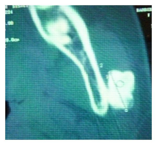
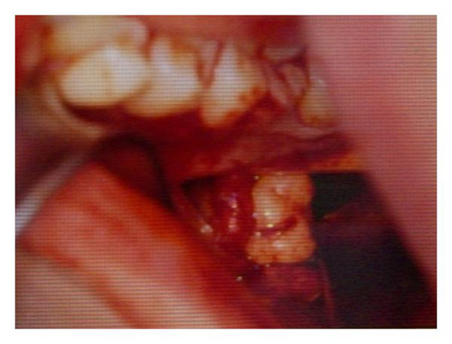
The TAC report carried a 1.5-cm oval formation-like bone-radio density with clean and regular borders (Figures 1, 2). The formation is independent of the bone of the mandible.


The X-Ray of the chest was negative.
The patient was submitted to surgical therapy under local anesthesia by intra oral access.
The lesion was easily dissected from the surrounding tissue. It was a bony-like tissue covered by a thin layer of soft tissue.
There were no complications after surgery.
3. Histopathology
3.1. Macroscopic Findings
The specimen consisted of a large, hard solid mass measuring 1.5 × 1.7 × 1.4 cm.
This tissue had a spherical-ellipsoidal form; its colour was white-gray and was not completely covered by a soft tissue capsule.
3.2. Microscopic Findings
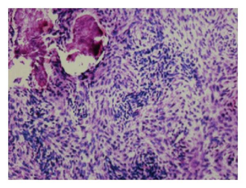
A well-circumscribed and half-encapsulated fibro-osseous lesion has an abundant cellular fibrous tissue with scattered trabeculae of lamellar bone. No cellular atipies and dental tissues were present.
The morphological pattern is compatible with the ossifying fibroma (Figure 3).

4. Discussion and Conclusions
The clinical comparison of the extraosseous forms of COF is rare.
In 1999, Kaufmann et al. described a case of COF in a 22-year-old man, situated in the right ear auricle dx [7], while in the same year Jung introduced a case of COF in the parapharyngeal and masticatory spaces [8].
Subsequently, Keefe et al. (2000) and Galdeano Arenas et al. (2004) reported two different cases of COF in the gingival region [9, 10].
More cases were observed in the skull and other bones out of the jaws such as frontal bone, nasal cavity, ethmoid, sphenoid orbit, and mastoidea region [6, 11, 13, 15].
In the light of literature reports, the unusual localization of COF in the case reported does not let us imagine about its nature.
Even if clinic and rx exams were comfortable to exclude a malignant lesion, we had doubts about the real nature of the lesion.
- (1)
Myositis ossificans: this is a benign process characterized histologically by a central fibroblastic tissue with an intermediate zone with trabeculae and a peripheral zone of calcification and mature lamellar bone.
- (2)
Well-differentiated osteosarcoma: this is characterized by lack of atypical nuclear in the spindle cells.
- (3)
Fibrous dysplaisa: this is characterized by a woven bone arising directly from the fibrous tissue, without osteoblastic activity.
- (4)
Odontogenic lesion (odontogenic fibroma, ameloblastic fibroma, ameloblastic fibro-sarcoma, ameloblastic fibro-odonto-sarcoma): this is characterized by the presence of dental epithelial structures as an integral tumor component.
We could obtain a definitive diagnosis only through the histological exam.
Prognosis was excellent, and the patient got the restitution ad integrum of the lesion.
This experience allows us to consider less in doubt this kind of lesions.


