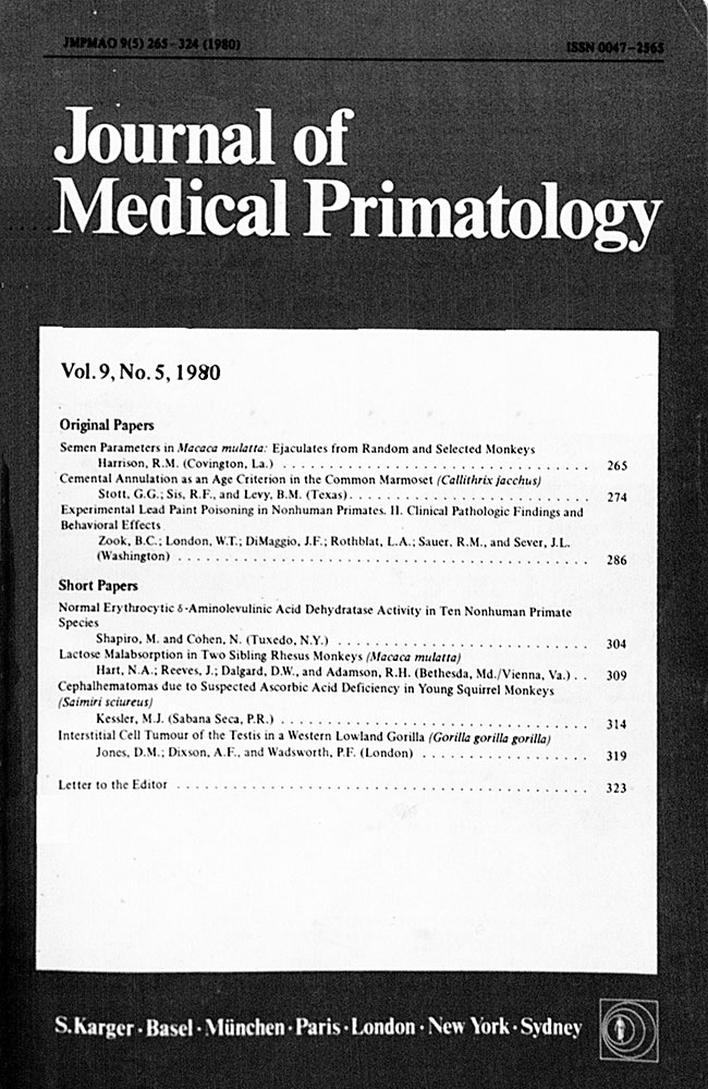
Experimental Lead Paint Poisoning in Nonhuman Primates
II. Clinical Pathologic Findings and Behavioral Effects1
Corresponding Author
B.C. Zook
Department of Pathology, School of Medicine and Health Sciences, and Department of Psychology, The George Washington University, Washington, D.C.
Infectious Disease Branch, Collaborative and Field Research, National Institute of Neurological Diseases and Stroke, NIH, Bethesda, Md.
Pathology Division, National Zoological Park, Smithsonian Institution, Washington, D.C.
Bernard C. Zook, DVM, The George Washington University, Ross Hall, B-12, 2300 I Street, N.W., Washington, DC 20037 (USA)Search for more papers by this authorW.T. London
Department of Pathology, School of Medicine and Health Sciences, and Department of Psychology, The George Washington University, Washington, D.C.
Infectious Disease Branch, Collaborative and Field Research, National Institute of Neurological Diseases and Stroke, NIH, Bethesda, Md.
Pathology Division, National Zoological Park, Smithsonian Institution, Washington, D.C.
Search for more papers by this authorJ.F. DiMaggio
Department of Pathology, School of Medicine and Health Sciences, and Department of Psychology, The George Washington University, Washington, D.C.
Infectious Disease Branch, Collaborative and Field Research, National Institute of Neurological Diseases and Stroke, NIH, Bethesda, Md.
Pathology Division, National Zoological Park, Smithsonian Institution, Washington, D.C.
Search for more papers by this authorL.A. Rothblat
Department of Pathology, School of Medicine and Health Sciences, and Department of Psychology, The George Washington University, Washington, D.C.
Infectious Disease Branch, Collaborative and Field Research, National Institute of Neurological Diseases and Stroke, NIH, Bethesda, Md.
Pathology Division, National Zoological Park, Smithsonian Institution, Washington, D.C.
Search for more papers by this authorRM. Sauer
Department of Pathology, School of Medicine and Health Sciences, and Department of Psychology, The George Washington University, Washington, D.C.
Infectious Disease Branch, Collaborative and Field Research, National Institute of Neurological Diseases and Stroke, NIH, Bethesda, Md.
Pathology Division, National Zoological Park, Smithsonian Institution, Washington, D.C.
Search for more papers by this authorJ.L. Sever
Department of Pathology, School of Medicine and Health Sciences, and Department of Psychology, The George Washington University, Washington, D.C.
Infectious Disease Branch, Collaborative and Field Research, National Institute of Neurological Diseases and Stroke, NIH, Bethesda, Md.
Pathology Division, National Zoological Park, Smithsonian Institution, Washington, D.C.
The authors thank Margaret R. Ashworth, Robert L. Brown, and Sherry L. Thomas for their technical assistance. Dr. Sauer is presently with the Gillette Medical Evaluation Laboratories in Rockville, Md. A portion of this work by J.F. DiMaggio has been accepted by The George Washington University in partial fulfillment for a Master of Arts degree.Search for more papers by this authorCorresponding Author
B.C. Zook
Department of Pathology, School of Medicine and Health Sciences, and Department of Psychology, The George Washington University, Washington, D.C.
Infectious Disease Branch, Collaborative and Field Research, National Institute of Neurological Diseases and Stroke, NIH, Bethesda, Md.
Pathology Division, National Zoological Park, Smithsonian Institution, Washington, D.C.
Bernard C. Zook, DVM, The George Washington University, Ross Hall, B-12, 2300 I Street, N.W., Washington, DC 20037 (USA)Search for more papers by this authorW.T. London
Department of Pathology, School of Medicine and Health Sciences, and Department of Psychology, The George Washington University, Washington, D.C.
Infectious Disease Branch, Collaborative and Field Research, National Institute of Neurological Diseases and Stroke, NIH, Bethesda, Md.
Pathology Division, National Zoological Park, Smithsonian Institution, Washington, D.C.
Search for more papers by this authorJ.F. DiMaggio
Department of Pathology, School of Medicine and Health Sciences, and Department of Psychology, The George Washington University, Washington, D.C.
Infectious Disease Branch, Collaborative and Field Research, National Institute of Neurological Diseases and Stroke, NIH, Bethesda, Md.
Pathology Division, National Zoological Park, Smithsonian Institution, Washington, D.C.
Search for more papers by this authorL.A. Rothblat
Department of Pathology, School of Medicine and Health Sciences, and Department of Psychology, The George Washington University, Washington, D.C.
Infectious Disease Branch, Collaborative and Field Research, National Institute of Neurological Diseases and Stroke, NIH, Bethesda, Md.
Pathology Division, National Zoological Park, Smithsonian Institution, Washington, D.C.
Search for more papers by this authorRM. Sauer
Department of Pathology, School of Medicine and Health Sciences, and Department of Psychology, The George Washington University, Washington, D.C.
Infectious Disease Branch, Collaborative and Field Research, National Institute of Neurological Diseases and Stroke, NIH, Bethesda, Md.
Pathology Division, National Zoological Park, Smithsonian Institution, Washington, D.C.
Search for more papers by this authorJ.L. Sever
Department of Pathology, School of Medicine and Health Sciences, and Department of Psychology, The George Washington University, Washington, D.C.
Infectious Disease Branch, Collaborative and Field Research, National Institute of Neurological Diseases and Stroke, NIH, Bethesda, Md.
Pathology Division, National Zoological Park, Smithsonian Institution, Washington, D.C.
The authors thank Margaret R. Ashworth, Robert L. Brown, and Sherry L. Thomas for their technical assistance. Dr. Sauer is presently with the Gillette Medical Evaluation Laboratories in Rockville, Md. A portion of this work by J.F. DiMaggio has been accepted by The George Washington University in partial fulfillment for a Master of Arts degree.Search for more papers by this authorAbstract
Oral administration of lead-containing paint to rhesus monkeys induced anemia, more profound in older primates. Erythrocytes were microcytic and hypochromic, but tended to become macrocytic terminally. Stippled erythrocytes were increased in all poisoned monkeys, especially in those with high blood lead levels and anemia. Proteinuria, glycosuria, casts and sloughed tubular cells containing acid-fast inclusion bodies were found on urinalysis. Terminal elevations of blood urea nitrogen were associated with profound anemia and renal tubular damage. Repeated blood lead values over 200 μg/dl were associated with a moribund termination while monkeys which had levels under 100 μg/dl remained apparently healthy. Behavioral studies in a small number of subclinically poisoned juveniles and neonates failed to reveal deficiencies of visual acuity or cognitive ability, nor was there evidence of alterations in levels of activity.
References
- 1Albert, R.E.; Shore, R. E.; Sayers, A. J.; Strehlow, C.; Kneip, T. J.; Pasternack, B. S.; Friedhoff, A. J.; Covan, F., and Cimino, J.A.: Follow-up of children overexposed to lead. Environ. Health Perspect. 7: 33—39 (1974).
- 2Allen, J.R.; McWey, P. J., and Suomi, S.J.: Pathological and behavioral effects of lead intoxication in the infant rhesus monkey. Environ. Health Perspect. 7: 239—246 (1974).
- 3Bederka, J.P. and McLellan, J.S.: Developmental effects of lead; in Khan and Bederka, Survival in toxic environments, pp. 275—279 (Academic Press, New York 1974).
10.1016/B978-0-12-406050-0.50030-7 Google Scholar
- 4Brown, D.R.: Neonatal lead exporsure in rats: decreased learning as a function of age and blood-lead concentrations. Toxicol appl. Pharmacol 32: 628—637 (1975).
- 5Burde, de la B. and Choate, M.S.: Does asymptomatic lead exposure in children have latent sequelae? J. Pediat. 81: 1088—1091 (1972).
- 6Bushnell, P.J.: Behavioral toxicology of lead in the infant rhesus monkey; PhD thesis, University of Wisconsin, Madison (1978).
- 7Bushnell, P.J.; Bowman, R. E.; Allen, J. R., and Marlar, R.J.: Scotopic vision deficits in young monkeys exposed to lead. Science 196: 333—335 (1977).
- 8Bushnell, P.J. and Bowman, R.E.: Reversal learning deficits in young monkeys exposed to lead. Pharmacol. Biochem. Behav. 10: 733—742 (1979).
- 9Carson, T.L.; Van Gelder, G.A.; Karas, G. G., and Buck, W.B.: Development of behavioral tests by the assessment of neurologic effects of lead in sheep. Environ. Health Perspect. 7: 233—237 (1974).
- 10Clasen, R.A.; Hartmann, J. F.; Coogan, P. S.; Pandolfi, S.; Laing, I. and Becker, R.A.: Experimental acute lead encephalopathy in the juvenile rhesus monkey. Environ. Health Perspect. 7: 175—185 (1974).
- 11Cohen, N.; Kneip, T. J.; Goldstein, D. H., and Muchmore, E. A. S.: The juvenile baboon as a model for studies of lead poisoning in children. J. med. Primatol. 1: 142—155 (1972).
- 12Cohen, N.; Kneip, T. J.; Rulon, V., and Goldstein, D.H.: Biochemical and toxicological response of infant baboons to lead driers in paint. Environ. Health Perspect. 7: 161—173 (1974).
- 13David, O.; Clark, J., and Voeller, K.: Lead and hyperactivity. Lancet ii: 900—903 (1972).
- 14Fisher, L.E.: Lead poisoning in a gorilla. J. Am. vet. med. Ass. 125: 478—479 (1954).
- 15Goode, J.W.; Johnson, S., and Calandra, J.C.: Evaluation of chronic oral administration of lead to rhesus monkeys. Toxicol. appl. Pharmacol. 26: Abstr. 70, pp. 465—466 (1973).
- 16Hausman, R.; Sturtevant, R. A., and Wilson, W.J.: Lead intoxication in primates. J. forens. Sci. 6: 180—196 (1961).
- 17Hopkins, A.: Experimental lead poisoning in the baboon. Br. J. ind. Med. 27: 130—140 (1970).
- 18Houser, W.D. and Frank, N.: Accidental lead poisoning in a rhesus monkey (Macaca mulatta). J. Am. vet. med. Ass. 157: 1919—1922 (1970).
- 19Krehbicl, D.; Davis, G. A.; LeRoy, L. M., and Bowman, R.E.: Absence of hyperactivity in lead-exposed developing rats. Environ. Health Perspect. 18: 147—157 (1976).
- 20Lamport, C. et Desmedt, J.E.: La production de nevrites expérimentales chez le singe: premier essais d'intoxication chronique au plomb. C.r. Séanc. Soc. Biol. 160: 2504—2507 (1966).
- 21Landrigan, P.J.; Whitworth, R. H.; Baloh, R. W.; Staehling, N. W.; Barthel, W. F., and Rosenblum, B.F.: Neuropsychological dysfunction in children with chronic low-level lead absorption. Lancet ii 708—712 (1975).
10.1016/S0140-6736(75)91627-X Google Scholar
- 22Millar, J.A.; Battistini, V.; Cumming, R.L.C.; Carswell, F., and Goldberg, A.: Lead and γ-aminolaevulinic acid dehydratase levels in mentally retarded children and in lead-poisoned suckling rats. Lancet ii: 695—698 (1970).
10.1016/S0140-6736(70)91962-8 Google Scholar
- 23Moore, M.R.; Meredith, P. A., and Goldberg, A.: A retrospective analysis of blood-lead in mentally retarded children. Lancet i: 717—719 (1977).
- 24 National Academy of Sciences: Committee on Biological Effects of Atmospheric Pollutants. Lead: airborne lead in perspective (National Academy of Sciences, Washington 1972).
- 25Ogilvie, D.M.: Sublethal effects of lead acetate on the Y-mase performance of albino mice (Mus musculus L.). Can. J. Zool. 55: 771—775 (1977).
- 26Overmann, S.R.: Behavioral effects of asymptomatic lead exposure during neonatal development in rats. Toxicol, appl. Pharmacol. 41: 459—471 (1977).
- 27Pueschel, S.M.; Kopito, L., and Schwachman, H.: Children with an increased lead burden: a screening and follow-up study. J. Am. med. Ass. 222: 462—466 (1972).
- 28Silbergeld, E.K. and Goldberg, A.M.: A lead-induced behavioral disorder. Life Sci. 13: 1275—1283 (1973).
- 29Snowdon, C.T.: Learning deficits in lead-injected rats. Pharmacol. Biochem. Behav. 1: 599—603 (1973).
- 30Sobotka, T.J. and Cook, M.P.: Post-natal lead acetate exposure in rats: possible relationship to minimal brain dysfunction. Am. J. ment. Defic. 79: 5—9 (1974).
- 31Vermande-van Eck, G.J. and Meigs, J.W.: Changes in the ovary of the rhesus monkey after chronic lead intoxication. Fert. Steril. 11: 223—224 (1960).
- 32Waldron, H.A.: The anemia of lead poisoning: a review. Br. J. ind. Med. 23: 83—100 (1966).
- 33Whitworth, R.H.; Rosenblum, B. F.; Dickerson, M. S., and Baloh, R.W.: Follow-up on human lead absorption. Morbid. Mortality 23: 157—159 (1974).
- 34Wilpizeski, C.R.: Effects of lead on the vestibular system: preliminary findings. Laryngoscope, St Louis 84: 821—832 (1974).
- 35Winneke, G.; Brockhaus, A., and Baltissen, R.: Neurobehavioral and systemic effects of long-term blood lead elevation in rats. Arch. Tox. 37: 247—263 (1977).
- 36Zook, B.C.; Eisenberg, J. F., and McLanahan, E.: Some factors affecting the occurrence of lead poisoning in captive primates. J. med. Primatol. 2: 206—217 (1973).
- 37Zook, B.C.; London, W. T.; Sever, J. L., and Sauer, R.M.: Experimental lead paint poisoning in nonhuman primates. I. Clinical signs and course. J. med. Primatol. 5: 23—40 (1976).
- 38Zook, B.C.; Sauer, R. M.; Bush, M., and Gray, C.W.: Lead poisoning in zoo-dwelling primates. Am. J. phys. Anthrop. 38: 415—424 (1973).
- 39Zook, B.C.; McConnell, G., and Gilmore, C.E.: Basophilic stippling of erythrocytes in dogs with special reference to lead poisoning. J. Am. vet. med. Ass. 157: 2092—2099 (1971).
- 40Zook, B.C.; London, W. T.; Wilpizeski, C. R., and Sever, J.L.: Experimental lead paint poisoning in nonhuman primates. III. Pathologic findings. J. med. Primatol. (in press).


