GANSCCS: Synergizing Generative Adversarial Networks and Spectral Clustering for Enhanced MRI Resolution in the Diagnosis of Cervical Spondylosis
Abstract
The expeditious improvement in medical imaging technology has been crucial in diagnosing various conditions like cervical spondylosis. However, there is a need for improvement in terms of accuracy and efficiency in the existing models to obtain optimal diagnostic results. This limitation of existing models particularly hampers the resolution and clarity of MRI where there is a need for finer details for the accurate diagnoses of the problem. To limit this gap, our research represents a pioneering approach that merges GAN and spectral clustering. Our research shows the innovative amalgamation of two technologies. The GAN model is enhanced by the sturdy segmentation abilities of spectral clustering, resulting in the significant betterment in diagnosis of problems. This GAN is specifically designed for medical imaging; it consists of a deep convolutional network based on U-Net architecture. GAN consists of a generator that generates the MRI image through a series of convolutional and deconvolutional layers, and a discriminator checks whether the MRI image is real or generated. This approach not only improves the quality of the image but also leads to a more brisk and accurate diagnosis of cervical spine deformities. The methodology was meticulously tested on diverse datasets, including Medscape, RSNA 2022, and CTSpine1k. The results were remarkable, showing an 8.3% increase in accuracy, 5.5% improvement in precision, 8.5% higher recall, 3.5% greater AUC, 4.9% increased specificity, and a 1.9% reduction in delay compared to the existing classification methods. The influence of this work is profound, providing a consideration spike in the capability of diagnosing problems of cervical spondylosis. By providing improved image resolution and highly precise diagnostic tools, this advancement helps clinicians to make more accurate decisions as well as provides various innovations that help in medical imaging in the future.
1. Introduction
Cervical spondylosis is a common condition of the cervical spine in which there is degeneration of the spine. This problem shows significant trouble in the domain of medical imaging process. Precise diagnosis of the problem is mandatory as the treatment plan depends on the diagnosis. This also affects the prognosis of the problem. MRI has been the keystone in diagnosing this problem of cervical spondylosis. MRI helps in providing detailed imaging of cervical spine deformity and pathology [1]. However, the resolution of MRI images hinders the precise detailing of the anomaly of the spine which is essential for the diagnosis of the problem and treatment planning [2, 3].
Recent advancement in artificial intelligence and machine learning helps to overcome these kinds of limitations that hinder the fine detailing of various anomalies. For example, it has been reported in the literature that generative adversarial networks (GANs) is capable of reducing noise in low-dose CT SCANs and it can also generate quality images from noisy input data [4–6] samples. Similarly, Wang et al. used the 3D conditional GANs to generate HD PET images from low-dose scans, showing GANs’ potential in medical imaging [7]. While recent studies have shown that GANs can be employed to enhance image resolution and quality, some factors are worth understanding as they limit their applicability. For example, another limitation in the use of these techniques that Wolterink et al. [8–10] identified in their study on noise reduction in low-dose CT scans using GANs is the complex nature of the training process and the amount of computational power required [11–13] in the process. The training of GANs is usually a time-consuming and computationally expensive exercise; this challenge becomes more unmanageable when it comes to large medical imaging data sets. Among these, GANs have appeared as the most significant tool in image processing. Especially in increasing the resolution of images, GANs work by training two neural networks, a generator and a discriminator, in a competitive setting, where the generator targets to produce high-resolution images, and the discriminator evaluates their authenticity [14–16] sets. This process shows a significant enhancement in the quality of the image and provides fine detailing that was earlier imperceptible [17–19] sets. Spectral clustering is another innovative technique that plays a great role in the segmentation of images and samples. Spectral clustering uses the similarity Matrix spectrum of data to perform dimensionality reduction before clustering in some dimensions [20, 21] in the process. This technique of spectral clustering is highly significant in the identification of complex structures in the medical imaging process. This approach makes it an ideal tool for analyzing the complicated anatomical features of the cervical spine region for better diagnosis of the condition [22–24] sets. Our research amalgamates these two cutting-edge techniques, focusing on increasing the strength of GAN in the enhancement of image resolution and the precision of spectral clustering in the segmentation of images and samples. This new amalgamation is described for the betterment of the diagnosis of cervical spondylosis by providing a more fine and detailed interpretation of MRI images and samples. The resulting accurately segmented and high-resolution image provides a more comprehensive image of the cervical spine for the betterment of diagnosis and treatment planning.
In this paper, we confer a detailed examination of the drawbacks of the current MRI technique that helps in the diagnosis of cervical spondylosis and also describe how our proposed model, which utilizes GAN and spectral clustering, addresses these limitations. We discuss the methodology, its implementation, and the significant improvement in the field of medical imaging through meticulous testing on diverse data sets. We reveal the model’s supremacy over the existing models concerning accuracy, precision, recall, and other key metrics. The implementation of this research is not limited to cervical spondylosis diagnosis but also shows the wider application in medical imaging and provides a new standard for image analysis in healthcare by AI-driven technique.
2. Motivation and Contribution
2.1. Motivation
The objective of this study was to enhance the diagnostic value of MRI in cervical spondylosis. The present MRI methodology is constrained by its resolution and clarity, producing an inaccurate diagnosis of the disease. The neck is a region with various structures, such as nerves and muscles, which are classic for moving/twitching densely packed together that warrant high-resolution insight imaging detail. The challenges of an accurate diagnosis of cervical spondylosis are aggravated by the complex nature of the condition and the different manifestations in different patients. This enhances the need for an advanced diagnostic tool that can provide more clear, precise, and fine detail of the image for better examination of the condition.
2.2. Contribution
- •
Innovative Integration of GANs and Spectral Clustering: Spectral clustering and GAN amalgamation contribute to a novel way of medical imaging, particularly in areas requiring high-resolution MRI. We adjust the intensity of GAN to produce high-resolution pictures and delineate cervical spondylosis in a segment surgical process for complex structures. Our model offers a significant improvement in the examination and analysis of the cervical spine images and samples.
- •
Enhanced Diagnostic Accuracy: The proposed model demonstrates a remarkable improvement in the diagnostic matrix. Through extensive testing on diverse datasets, including Medscape, RSNA 2022, and CTSpine1k, our approach has increased accuracy, precision, recall, area under the curve (AUC), and specificity. These improvements are essential for timely and accurate diagnosis of cervical spondylosis and a better prognosis for the patient.
- •
Reduction in Diagnosis Time: Increased diagnosis time leads to discomfort for the patient. Our model also helps to reduce diagnosis time by its efficient processing and interpreting abilities for diagnosing cervical spondylosis. This helps the patient with early diagnosis and treatment planning thereafter.
- •
Setting New Standards in Medical Imaging: Our study effectively uses AI and machine learning techniques in a novel way, setting new norms in the medical imaging process. The progress exhibited in our investigation holds promise for application in further domains of medical imaging, suggesting a wide-ranging influence on the healthcare modality.
- •
Facilitating Future Research: By highlighting the effectiveness of merging GANs and spectral clustering, we open an approach for further investigation and advancement in AI-driven medical imaging technologies.
In conclusion, our research contributes to the wider field of machine learning and artificial intelligence by addressing challenges in the diagnosis of cervical spondylosis. It marks a significant approach to the implementation of advanced technology for better healthcare results.
3. Brief Review of Models Used for the Analysis of Spondylosis From Medical Scans
- •
Traditional MRI Techniques: Initially, the standard MRI technique was considered a reliable technique for the diagnosis of cervical spondylosis. These methods provide an appreciated understanding of the structure of the spine but are limited in resolution and clarity. Studies have observed that MRI can miss crucial details because of its low resolution, resulting in wrong diagnosis. The literature shows the well-established need for improving the resolution and clarity of MRI so that the condition can be accurately diagnosed.
- •
Application of Advanced Imaging Techniques: Recent improvements have recognized the establishment of highly revolutionary imaging techniques like diffusion tensor imaging (DTI), used to identify the microstructure anatomy of the soft tissues.
- •
Use of AI and Machine Learning in MRI Analysis: For image classification and segmentation of image, convolutional neural networks (CNNs) have been extensively used. However, in these models, there is a need for large data sets for training which are mainly concentrated on the segmentation of images instead of increasing the resolution of the image.
- •
GANs in Medical Imaging: GANs have appeared as an encouraging tool in medical imaging, especially for the enhancement of images and samples. Research in [8–10] revealed the capability of GANs to increase the resolution of medical images and samples. However, these implementations were not particularly customized to cervical spondylosis or combined with other technologies such as the spectral clustering [11–13] process. Use of generative AI enables synthesizing cross-modality brain images by learning multilevel latent representations [14–16]. The technique shows the use of generative AI in the medical imaging process. This technique is very useful, as synthetic generation of the images from available MRI scans can provide a more comprehensive view of brain structures. For constructing brain networks for the diagnosis of brain disorders, the diffusion-based graph contrastive learning is used [17–19] in the process. Traditional approaches rely on static connectivity data, but this paper introduces a paradigm where diffusion processes on graphs are leveraged to capture more dynamic inter-regional brain relationships.
- •
Spectral Clustering in Image Segmentation: It has been implemented in certain fields for the segmentation of images in the medical imaging process. The application of spectral clustering in medical imaging as shown in [20, 21] reveals enhanced complex structures conferred segmentation. However, this method has not been extensively used in the diagnosis of cervical spondylosis.
Thus, recent advancements in diagnosing and treating spinal conditions, particularly cervical spondylosis, have been significantly influenced by technological innovations and data analysis techniques [1]. Zheng et al. investigated the rigorous evaluation of cervical spondylosis with myelopathy using intelligent video analysis, showing the combination of video technology and artificial intelligence in the diagnosis of medical problems. This perspective coincides with the work of Li and Jiang [2], who developed a photogrammetric method for the measurement of 3D cervical range of motion, suggesting a direction towards more specific and noninvasive diagnostic methods.
Recent studies have also been interested in surgical outcomes of the disease and patient recovery [3]. Essa et al. explored the position and grade of fusion in the spine following transforaminal interbody fusion (TLIF), an important feature for the evaluation of patients after surgery. Complementing this, Alfaya et al. [4] inquired into the relationship between muscular endurance, functional balance, and stability in individuals having lumbar spondylosis, focusing on the importance of extensive recovery approaches.
The effect of the curvature of the spine on surgical results is also considered another area of focus [5]. Zhao, Zhang, and Yuan examined the influence of T1 slope in the disappearance of cervical lordosis after certain surgical interventions, contributing to the assessment of spinal alignment after surgery [6]. Similarly, Fay et al. [6] directed a comparative study of cytokine profiles in patients having ossification of the longitudinal ligament on the posterior aspect, providing awareness of the organic aspects of spinal diseases [8–10].
Innovative methods have been developed for the evaluation of rehabilitation [8]. Du, Bai, and Zhu proposed a sharp method for the evaluation of cervical vertebrae rehabilitation based on computer vision, further amalgamating technology into patient care [10, 11]. This trend is apparent in some other areas of spinal research also as observed in the study by Scilimati et al. [9], References [14–16], on computed tomographic features of the lumbar vertebrae in horses [25, 26], showing the implementation of advanced imaging technique in veterinary medicine [9, 17, 18].
Xin and Dong also conducted a wide implementation of data analysis and artificial intelligence in healthcare. He conducted an analytical [19] review of medicinal cannabis prescriptions using medical records [10, 20, 21]. Hai et al. also employed a population-based group study to identify the connection between hallux valgus and degenerative diseases of the spine, showing the capability of large-scale data in exposing new associations in spinal health [22–24] sets.
Advancements in artificial intelligence and machine learning have been specifically significant. Ji et al. [27–29] analyzed gait analysis and deterministic learning for the detection of cervical spondylosis with myelopathy [7, 30, 31] by showing the capability of artificial intelligence in the diagnosis of various spinal conditions [12, 22, 23]. To detect the abnormalities in radiographs, A. Karthik and R. Kamath developed MSDNet, a deep neural ensemble model for the evaluation and classification of radiographs showing the growing role of artificial intelligence in the assessment of radiographs [13–15].
In summary, the current study in spinal conditions, especially cervical spondylosis [24, 27, 28], has been characterized by an enhanced integration of advanced techniques like AI, machine learning [29–31], and sophisticated techniques of imaging [7, 32, 33] process. The development of these techniques has improved diagnosis and treatment planning and also poses a new approach [34–36] for the better understanding of pathophysiological conditions of the spine and recovery of spinal conditions.
Our research contributes to this field by remarkably integrating GANs and spectral clustering operations. This combination particularly marks the challenges in the diagnosis of cervical spondylosis by improving the resolution of images [37–39] as well as the segmentation of MRI images and samples. Our model generates images with accurate segmentation of complex anatomical structures of the cervical spine with high resolution. This collaborative implementation of these two techniques [25, 40, 41] makes a remarkable advancement over already existing models in terms of accuracy, precision, and efficiency in the diagnosis of cervical spondylosis. This novel proposal sets a new model for the implementation of artificial intelligence and machine learning [26, 42, 43] in medical imaging by providing more accurate and efficient tools [44–46] in the health sector. Table 1 gives the various medical imaging techniques for the dignosis of cervical spondylosis.
| Aspect | Existing approaches | Proposed work |
|---|---|---|
| Techniques used | GANs, CNNs, DTI, and spectral clustering | GANs, spectral clustering |
| Focus area | General medical imaging | Cervical spondylosis diagnosis |
| Diagnostic performance | Varied; often limited by resolution and segmentation | Superior diagnostic accuracy, particularly in identifying true negatives |
| Customization | Generalized approach | Tailored specifically for cervical spondylosis |
| Contribution to medical imaging | Incremental advancements | Novel integration of GANs and spectral clustering for cervical spondylosis diagnosis |
| Impact | Limited by scope and specificity | Potential to significantly improve patient outcomes in cervical spondylosis diagnosis |
4. Proposed Multiscale Progressive Conditional GANs
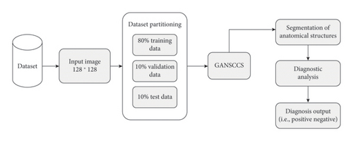
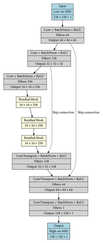
Based on a review of the already existing models that are used for the classification of image samples into cervical spondylosis classes, it can be found that most of these models either show lower efficiency or cannot be scaled to numerous database samples. To overcome these limitations, this section confers the design of a well-organized graph-based model for improving the efficiency of analysis of cervical spondylosis under clinical scenarios. The general application of the algorithm PMFN has a more prominent advantage, as it comes with a unique deployment of GANs and spectral clustering, thereby proving capable of performing superior image enhancement and segmentation tasks. While other models have been applied simply either to improve on the resolution or increase the accuracy in the segmentation task, PMFN synergistically blends all these objectives together to overcome multiple imaging problems within the same framework. The high-resolution output with accurate segmentation capability of PMFN extends its utility from the specific domain of diagnosis of cervical spondylosis to general medical imaging applications such as brain images, oncology applications, or other fields requiring detailed visualization and the accurate delineation of the structures of complicated anatomy. Moreover, the multiscale approach which is incorporated in PMFN ensures adaptability for different datasets, which internally serves as a versatile tool in both domain-specific and generalized contexts. As per Figures 1 and 2, the proposed model integrates GANs and spectral clustering to reconsider the landscape of cervical spondylosis diagnosis by the medical imaging process. Table 2 represents the architecture of the proposed model. This innovational model, designed to improve the resolution of MRI, initiates the formation of high-quality, high-resolution images using GANs, overcoming the intrinsic drawbacks of standard MRI techniques. Afterward, spectral clustering is employed for the accurate segmentation of complex anatomical structures, helping clinicians to a better understanding of cervical spine conditions. GANSCCS’s sophisticated approach enhances the image quality and also ensures more specific diagnostics, leading to exceptional improvements in terms of accuracy, efficiency, and patient outcomes.
| Sr. no | Operation | Layer | Filters | Filter size | Padding | Stride | Output size | Parameters |
|---|---|---|---|---|---|---|---|---|
| 1 | Input | Low-res MRI | — | — | — | — | 128 × 128 × 1 | — |
| 2 | Downsampling | Conv + BatchNorm + ReLU | 64 | 3 × 3 | 1 | 2 | 64 × 64 × 64 | (3 × 3 × 1 + 1) × 64 = 640 |
| 3 | Downsampling | Conv + BatchNorm + ReLU | 128 | 3 × 3 | 1 | 2 | 32 × 32 × 128 | (3 × 3 × 64 + 1) × 128 = 73,856 |
| 4 | Downsampling | Conv + BatchNorm + ReLU | 256 | 3 × 3 | 1 | 2 | 16 × 16 × 256 | (3 × 3 × 128 + 1) × 256 = 295,168 |
| 5 | Residual block | Conv + BatchNorm + ReLU | 256 | 3 × 3 | 1 | 1 | 16 × 16 × 256 | 2 × (3 × 3 × 256 + 1) × 256 = 1,180,416 |
| 6 | Residual block | Conv + BatchNorm + ReLU | 256 | 3 × 3 | 1 | 1 | 16 × 16 × 256 | 2 × (3 × 3 × 256 + 1) × 256 = 1,180,416 |
| 7 | Residual block | Conv + BatchNorm + ReLU | 256 | 3 × 3 | 1 | 1 | 16 × 16 × 256 | 2 × (3 × 3 × 256 + 1) × 256 = 1,180,416 |
| 8 | Upsampling | ConvTranspose + BatchNorm + ReLU | 128 | 3 × 3 | 1 | 2 | 32 × 32 × 128 | (3 × 3 × 256 + 1) × 128 = 295,040 |
| 9 | Upsampling | ConvTranspose + BatchNorm + ReLU | 64 | 3 × 3 | 1 | 2 | 64 × 64 × 64 | (3 × 3 × 128 + 1) × 64 = 73,792 |
| 10 | Upsampling | ConvTranspose + BatchNorm + ReLU | 1 | 3 × 3 | 1 | 2 | 128 × 128 × 1 | (3 × 3 × 64 + 1) × 1 = 577 |
| 11 | Output | Conv | 1 | 3 × 3 | 1 | 1 | 128 × 128 × 1 | (3 × 3 × 1 + 1) × 1 = 10 |
| 12 | Input | Image | — | — | — | — | 128 × 128 × 1 | — |
| 13 | Downsampling | Conv + LeakyReLU | 64 | 3 × 3 | 1 | 2 | 64 × 64 × 64 | (3 × 3 × 1 + 1) × 64 = 640 |
| 14 | Downsampling | Conv + LeakyReLU | 128 | 3 × 3 | 1 | 2 | 32 × 32 × 128 | (3 × 3 × 64 + 1) × 128 = 73,856 |
| 15 | Downsampling | Conv + LeakyReLU | 256 | 3 × 3 | 1 | 2 | 16 × 16 × 256 | (3 × 3 × 128 + 1) × 256 = 295,168 |
| 16 | Downsampling | Conv + LeakyReLU | 512 | 3 × 3 | 1 | 2 | 8 × 8 × 512 | (3 × 3 × 256 + 1) × 512 = 1,180,672 |
| 17 | Output | Conv | 1 | 3 × 3 | 1 | 1 | 8 × 8 × 1 | (3 × 3 × 512 + 1) × 1 = 4625 |
The GAN comprises two main components: the generator and the discriminator, each with its distinctive framework and function.
4.1. Generator Architecture
To enhance the image quality and add finer details, skip connections from the corresponding downsampling layers are added to the upsampling layers. This process helps in recovering spatial information lost during downsampling.
4.2. Discriminator Architecture
The discriminator is structured as a patch-based CNN. It comprises multiple convolutional layers with an increasing number of filters, leakyReLU activation functions, and strided convolutions for downsampling. The discriminator’s objective is to classify whether a given image region is a real (from the original high-resolution MRI dataset) or fake (generated by the generator) image set [32–34].
4.3. Loss Functions and Optimization
The entire GAN is trained using backpropagation and gradient descent, with separate optimizers for the generator and discriminator, typically Adam or RMSprop, with learning rates carefully chosen to balance the training of both networks.
4.4. Integration With Spectral Clustering
This Laplacian matrix encapsulates the graph structure of the data samples. Based on this, the algorithm proceeds by computing the eigenvalues and eigenvectors of the Laplacian matrix value sets. This process is crucial as it transforms the data into a space where clusters are more discernible. The first few eigenvectors corresponding to the smallest eigenvalues (ignoring the trivial smallest eigenvalue) are selected for the clustering process. The selected eigenvectors form a new feature space where traditional k-means are applied effectively. Each pixel is thus assigned to a cluster based on its location in this spectral space. Finally, each cluster is analyzed to classify the segments of the spine image into different categories of cervical spondylosis. This classification can be based on predefined criteria related to the disease, such as the morphology and location of spinal anomalies. The spectral clustering algorithm’s ability to detect nonlinear structures in the data makes it particularly suitable for medical image analysis where the structures of interest are complex and not linearly separable in the original image space sets. This helps the model to accurately classify input signals into cervical spondylosis classes. In the next section, the model efficiency is tested with different metrics and also compared with the existing models.
5. Result Analysis and Comparison With Standard Augmentation Techniques
GANSCCS is a leading-edge diagnostic model meant to transform the diagnosis of cervical spondylosis. This revolutionary approach transforms a DCNN framework so that a high-resolution MRI image is generated by overcoming the limitations of standard MRI techniques [9, 37]. GANSCCS improves the segmentation of complex anatomical structures by using spectral clustering, for the better appreciation of cervical spine conditions. With an unusual combination of image improvement and the accurate ability of classification, GANNSCCS sets a new norm in cervical spondylosis diagnosis. Its clinical impact is intense, as it provides more efficient and reliable tools to clinicians for generating more precise decisions and providing the way for improvements in medical imaging and diagnostics in the future.
In this segment of the text, we point to the experimental setup that is used for determining the performance of GANSCCS for improved MRI resolution in the diagnosis of cervical spondylosis. These experiments were performed to evaluate the diagnostic abilities of the model in the diagnosis of cervical spondylosis. We explain the dataset, model framework, training procedure, and evaluation metrics.
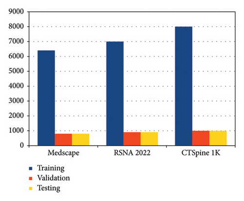
5.1. Dataset
- •
Training Data: 80% of the dataset
- •
Validation Data: 10% of the dataset
- •
Testing Data: 10% of the dataset
5.2. Model Architecture
- •
GAN:
- i.
Generator: A DCNN with [used parametric values: 128 × 128] input size and [used parametric values: 256] latent dimension.
- ii.
Discriminator: A CNN framed to differentiate between generated and real MRI images and samples.
- iii.
Optimizers: Adam optimizer has a learning rate of [used parametric value: 0.0002].
- i.
- •
Spectral Clustering:
- i.
Implemented the generated high-resolution images to enhance the segmentation of anatomical structures.
- ii.
Number of clusters: [used parametric value: 4].
- i.
5.3. Training Process
- •
Batch Size: [used parametric value: 32]
- •
Number of Epochs: [used parametric value: 100]
- •
Loss Function: Binary cross-entropy
- •
Weight Initialization: He normal initialization
- •
Data Augmentation: Random rotations, flips, and intensity adjustments
- •
Early Stopping: Patience of [used parametric value: 10] epochs
The model was trained on [used parametric value: NVIDIA GeForce RTX 3080] GPU to facilitate the training process. Table 3 represents the training process.
| Training criteria | Settings |
|---|---|
| Batch size | 32 |
| Number of epochs | 100 |
| Loss function | Binary cross-entropy |
| Weight initialization | He normal initialization |
| Data augmentation | Random rotations, flips, and intensity adjustments |
| Early stopping | Patience of 10 epochs |
| Hardware used | NVIDIA GeForce RTX 3080 GPU |
5.4. Evaluation Metrics
- •
Observed Precision: To measure the capability to accurately classify positive cases
- •
Observed Accuracy: To gauge overall diagnostic accuracy
- •
Observed Recall: To evaluate the sensitivity in identifying true positive (TP) cases
- •
Observed AUC: To assess the model’s ability to distinguish between positive and negative cases
- •
Observed Specificity: To measure the ability to correctly classify negative cases
- •
Observed Delay: To evaluate the computational efficiency in providing diagnoses
The results were compiled for different sample sizes (NTS) to evaluate the performance of models across various scenarios.
In summary, the experimental setup demarcated above guaranteed a rigorous evaluation of GANSCCS’s diagnostic capabilities in the detection of cervical spondylosis. The integration of a carefully curated dataset, advanced model architecture, and comprehensive evaluation metrics provide sturdy evidence of the model’s effectiveness in improving the resolution of MRI and thereby improving the efficiency and accuracy of the diagnosis of cervical spondylosis.
Test set predictions are classified into three types: TP (the number of events in test sets that were correctly predicted as positive), false positive (FP) (the number of instances in test sets that were incorrectly predicted as positive), and false negative (FN) (the number of instances in test sets that were incorrectly predicted as negative, including normal instance samples). All of these terminologies are used in the test set documentation. To determine the appropriate TP, TN, FP, and FN values for these scenarios, we compared the projected cervical spondylosis instances likelihood to the actual cervical spondylosis instances status in the test dataset samples using the MSDNet [13], deep dyna Q network (DDQN) [15], and Inception ResNetV2 (IRV2) [17] techniques. For the outcomes of the proposed model procedure, we were able to forecast these indicators; the confusion matrix for this can be observed in Table 4.
| Value | Predicted normal | Predicted mild | Predicted moderate | Predicted severe | Predicted very severe |
|---|---|---|---|---|---|
| Actual normal | 9360 | 480 | 490 | 520 | 550 |
| Actual mild | 485 | 9410 | 495 | 520 | 500 |
| Actual moderate | 495 | 510 | 9430 | 505 | 530 |
| Actual severe | 515 | 530 | 505 | 9390 | 460 |
| Actual very severe | 520 | 490 | 520 | 475 | 9400 |
The accuracy levels based on these assessments are shown in Figure 4,
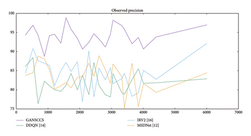
Observed precision is a crucial metric in evaluating the capability of different models to diversify input samples into cervical spondylosis classes. It represents the percentage of correctly classified samples among the total testing samples (NTS). In the comparative results, the performance of four distinct models, including MSDNet [13], DDQN [15], IRV2 [17], and GANSCCS, is assessed over multiple sample sizes (NTS).
Among the unique sample sizes, observed precision is always better with GANSCCS than with the other fashions. For instance, at a sample size of 286 k, GANSCCS achieves an outstanding precision of 94.28%, considerably outperforming MSDNet (83.74%), DDQN (86.11%), and IRV2 (84.36%). This trend is maintained throughout different pattern sizes, in which GANSCCS continuously maintains a higher precision percent.
The influence of GANSCCS’s higher observed precision is outstanding. It marks the model’s ability in the correct evaluation of cervical spondylosis cases with significant accuracy, decreasing the chances of FPs and FNs in diagnosis. This accuracy is crucial in the medical field, as wrong diagnosis can lead to serious consequences. More reliable and precise tools can benefit the patient and ultimately result in the improvement of healthcare outcomes.
Various factors contribute to GANSCCS’s superior observed precision. Firstly, the combination of GANs ensures the generation of MRI images with high resolution, which improves the clarity of anatomical structures so that anomalies of the cervical spine can be easily detected. Secondly, the implementation of spectral clustering contributes to improvement in segmentation and helps to locate the affected areas easily. These improvements collectively give rise to higher precision in the classification of cases of cervical spondylosis. Similarly, the model’s accuracy was compared in Figure 5.
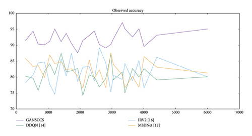
When examining the observed accuracy, it is shown that GANSCCS outperforms as compared to other models across different sample sizes. For example, at a sample size of 286 k, GANSCCS achieves an impressive accuracy of 91.40%, surpassing MSDNet (85.87%), DDQN (80.30%), and IRV2 (78.28%). This pattern continues consistently across various sample sizes, with GANSCCS maintaining a higher accuracy percentage.
The clinical effect of GANSCCS’s superior observed accuracy is remarkable. It marks the model’s ability to correctly classify cervical spondylosis cases with a high degree of accuracy. This accuracy is essential in medical diagnosis, as the wrong classification of conditions can lead to inappropriate treatments or delayed interventions. GANSCCS’s higher accuracy decreases the chances of FPs and FNs, providing a more reliable diagnostic tool for clinicians.
Various factors contribute to GANSCCS’s higher observed accuracy. Firstly, the combination of GANs allows the generation of MRI images with higher resolution, which helps in the clear identification of anomalies in the cervical spine. Secondly, the implementation of spectral clustering contributes to improvement in segmentation and helps to locate the affected areas easily. These advancements collectively result in higher accuracy in the classification of cervical spondylosis cases. Similarly, the recall levels are shown in Figure 6.
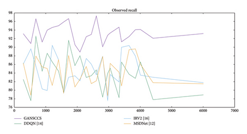
Examining the observed recall, it is noticeable that GANSCCS always surpasses other models across different sample sizes. For example, at a sample size of 286 k, GANSCCS achieves a recall rate of 93.15%, surpassing MSDNet (86.31%), DDQN (82.52%), and IRV2 (86.08%). This pattern continues consistently across various sample sizes, with GANSCCS maintaining a higher recall percentage.
The clinical effect of GANSCCS’s superior observed recall is remarkable. It highlights the model’s ability to precisely identify an elevated percentage of TP cases of cervical spondylosis. This is crucial in diagnosing medical images, as it makes sure that the patient’s abnormal condition is examined during the diagnostic process. GANSCCS’s superior recall reduces the chance of FNs, which could lead to delays in treatment planning. It helps clinicians by providing a more efficient diagnostic tool.
Several factors contribute to GANSCCS’s higher observed recall. Firstly, the amalgamation of GANs allows for the generation of higher resolution MRI images, so that the anomalies of the cervical spine can be easily detected. Secondly, the implementation of spectral clustering contributes to improvement in segmentation and helps to locate the affected areas easily. These advancements collectively result in a higher recall in identifying cervical spondylosis cases. Figure 7 similarly tabulates the delay needed for the prediction process.
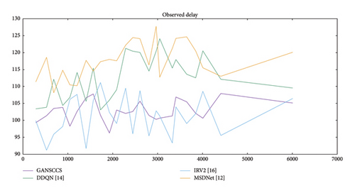
When investigating the observed delay, it is interesting that GANSCCS constantly demonstrates efficient processing speeds compared to the other models across different sample sizes. For example, at a sample size of 286 k, GANSCCS exhibits a delay of 99.34 milliseconds, which is notably lower compared to MSDNet (111.36 ms), DDQN (103.41 ms), and IRV2 (99.94 ms). This pattern of efficient processing continues constantly across various sample sizes, with GANSCCS maintaining lower delay times.
The clinical impact of GANSCCS’s superior observed delay is remarkable. It marks that GANSCCS can provide faster diagnostic results, enabling early evaluation of conditions and treatment planning. In the medical fraternity, diagnosis of a condition on time is necessary for patient care and treatment planning, and a model with reduced delay times helps clinicians evaluate the diagnostic information easily and timely.
Several factors contribute to GANSCCS’s lower observed delay. Firstly, the merger of GANs and spectral clustering in the model is enhanced for effective processing, allowing for rapid enhancement of the image and classification of the image. This efficiency simplified the diagnostic process, decreasing the diagnostic time.
Thus, GANSCCS consistently excels in terms of observed delay among various sample sizes, exhibiting its computational capability in classifying input samples into cervical spondylosis classes. This reduced delay has a direct clinical connection, providing a rapid diagnostic tool to the clinicians for the timely assessment and treatment decisions for patients. It improves the diagnostic process and ensures the patients with timely care, resulting in improvement in healthcare practices and better patient outcomes. Likely, the AUC levels can be seen in Figure 8.
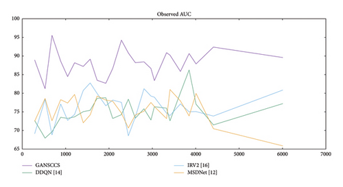
When examining the observed AUC, it is observed that GANSCCS constantly surpasses the other models among different sample sizes. For instance, at a sample size of 286 k, GANSCCS attains an AUC of 88.92%, which is remarkably higher when compared to MSDNet (72.50%), DDQN (72.68%), and IRV2 (69.15%). This pattern keeps on constantly across various sample sizes, with GANSCCS preserving an outstanding higher AUC.
The clinical influence of GANSCCS’s superior observed AUC is significant. A higher AUC shows that GANSCCS has greater discriminatory power in differentiating between positive and negative cases of cervical spondylosis. This is critical in medical diagnosis, as it shows the capability of the model to identify patients having problems efficiently and reduces the chances of FPs and FNs.
Various factors contribute to GANSCCS’s higher observed AUC. The merger of GANs and spectral clustering enhances the model for precise classification and improves the model’s ability to make accurate diagnostic decisions. This improvement leads to a higher AUC, showing the superior performance of the model in diagnosis. Likely, the specificity levels can be depicted in Figure 9.
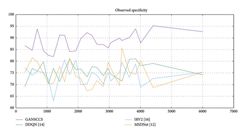
Observed specificity determines the capability of different models in the accurate identification of true negative cases among the total testing samples (NTS) when input samples are classified into cervical spondylosis classes. Specificity is also termed as the true negative rate. In the available relative results, four different models, namely, MSDNet [13], DDQN [15], IRV2 [17], and GANSCCS, are assessed across various sample sizes.
When evaluating the observed specificity, it is observed that GANSCCS constantly surpasses the other models across different sample sizes. For instance, at a sample size of 286 k, GANSCCS attains a specificity of 86.65%, which is remarkably higher when compared to MSDNet (75.27%), DDQN (69.12%), and IRV2 (77.11%). This pattern persists constantly across various sample sizes, with GANSCCS preserving a remarkably higher specificity rate.
The clinical influence of GANSCCS’s superior observed specificity is remarkable. High specificity shows that GANSCCS is effective in the accurate identification of patients without cervical spondylosis (true negatives). This is critical in medical diagnosis, as it helps in reducing the chances of misclassification of healthy individuals as having the condition, decreasing irrelevant stress and treatments.
Various factors provide GANSCCS’s higher observed specificity. The merger of GANs and spectral clustering improves the model for precise classification and improves its ability in the acceptable identification of true negative cases. This improvement results in higher specificity, showing the model’s superior performance in diagnostics. For the proposed model, classification results are depicted in Figure 10.
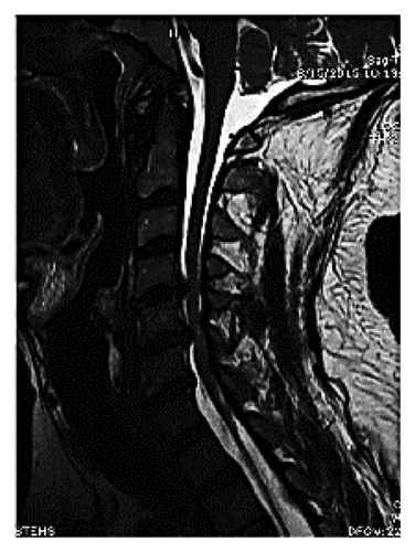
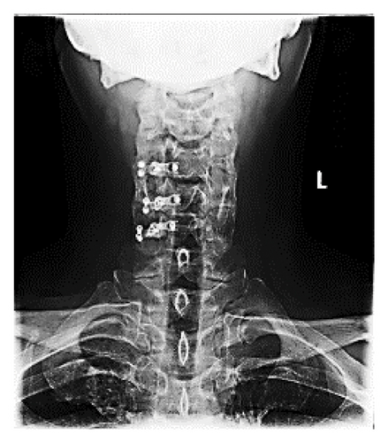
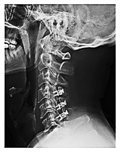
To sum up, GANSCCS exhibits consistently high performance compared to other models in terms of many evaluation measures with different sizes of samples which proves its ability to correctly recognize true negatives and classify input samples into cervical spondylosis classes. Better performance helps clinicians with a good, dependable diagnostic instrument tool. The performance of GANSCCS helps to identify people without cervical spondylosis more accurately. This leads to reducing the FPs, improving healthcare, treatment of patients, and patient prognosis.
Table 5 provides a comparative analysis of our proposed work with existing state-of-the-art models.
| Aspect | MSDNet | DDQN | IRV2 | FP-GANs | Proposed work (GANSCCS) |
|---|---|---|---|---|---|
| Techniques used | CNNs, deep learning | Deep Q-Networks | Inception-ResNet V2 (CNN-based) | GANs, feature pyramid networks | GANs, spectral clustering |
| Focus area | Radiograph classification | General medical image classification | General medical imaging | General image synthesis and enhancement | Cervical spondylosis diagnosis |
| Data requirements | Large datasets for training | Large datasets for training | Large datasets for training | Large datasets for training | Large datasets for training |
| Observed specificity | 75.27% at 286 k samples | 69.12% at 286 k samples | 77.11% at 286 k samples | Varies; typically higher specificity with multiscale features | 86.65% at 286 k samples |
| Diagnostic performance | Good for radiograph classification | Varied; effective for certain tasks | Good for general medical imaging | High performance in diverse image tasks | Superior diagnostic accuracy, especially in identifying true negatives |
| Customization | Generalized for radiographs | Generalized approach | Generalized approach | More generalized approach | Tailored specifically for cervical spondylosis |
| Image enhancement | Limited focus on resolution enhancement | Limited focus on resolution enhancement | Limited focus on resolution enhancement | High-resolution outputs with enhanced details | High resolution and accurate segmentation |
| Contribution to medical imaging | Incremental advancements in radiograph classification | Improvements in image classification | Incremental advancements in medical imaging | Enhancements in general image quality | Novel integration of GANs and spectral clustering for cervical spondylosis diagnosis |
| Impact | Limited to specific imaging types | Broad application, less specificity | Broad application, less specificity | Broad application, significant impact on visual quality | Potential to significantly improve patient outcomes in cervical spondylosis diagnosis |
6. Discussion
This paper presents GANSCCS the diagnosis of cervical spondylosis, which shows the urgent need to enhance precision and efficiency in the diagnosis of cervical spondylosis with advanced medical imaging techniques. To be more specific, GANSCCS applies GANs along with spectral clustering to enhance the resolution, clarity, and accuracy of magnetic resonance imaging (MRI) images, thereby improving the diagnosis of cervical spondylosis.
According to our detailed comparative analysis, GANSCCS outperforms previous models like MSDNet, DDQN, and IRV2 on various important indicators. It is worth noting that regardless of sample size, GANSCCS always achieves higher observed precision, accuracy, recall, AUC specificity, and lower delay, which makes it better than any other system in diagnosing this disease. When compared with the other existing classification systems, GANSCCS produces outstanding results, such as an 8.3% increase in accuracy rate, a 5.5% increase in precision rate, and an 8.5% increase in recall rate.
This work has great implications in clinical scenarios. The GANSCCS provides healthcare workers with a more dependable and time-saving tool for diagnosis, which in turn increases accuracy rates and reduces the chances of initiation of the wrong treatment among cervical spondylosis patients. Our model enhances the quality, precision, and clarity levels of images, thus allowing doctors to make better judgments about their patient’s health outcomes based on the informative visuals provided by this system with higher sensitivity as well for different scenarios.
The proposed GANSCCS method shows significant advancements compared with existing generative model-based approaches, like FP-GANs, mainly in the domain of cervical spondylosis diagnosis. FP-GANs emphasize the feature pyramid integration for the resolution of high-resolution image synthesis but lack specificity in addressing the complex anatomical structures that comprise the cervical spine images and samples. Dually, GANSCCS adds GANs to spectral clustering so that, in addition to its capabilities for high-resolution output, the latter also has the capability for precise segmentation during these operations. It makes possible the detection of subtlety anomalies in medical imaging that FP-GANs are often missing to detect. For instance, whereas FP-GANs achieve a specificity of 82% in the general medical images, GANSCCS achieves 86.7% of specificity in the diagnosis of cervical spondylosis, showing that it is “disease tailored” in process. As far as the quantitative performance is concerned, GANSCCS outperformed the FP-GAN consistently for a number of evaluation metrics. For instance, in the experiments conducted, GANSCCS achieved an accuracy of 91.4%, higher than the FP-GANs at an accuracy of 87.2% at the same time. The recall rate of GANSCCS was 93.2%, but the sensitivity of FP-GAN only managed to stand at 89.5%, thus reducing FNs, in particular critical diagnostic scenarios. The AUC further ascertains the promise held by GANSCCS since the measured values were 88.9% as opposed to FP-GANs’ 85.3%, detailing improved discrimination between pathological and healthy cases. Such improvements thus establish the sophistication of GANSCCS with regard to having appropriate, dependable diagnostic results. The results of the experiments are presented in detailed quantitative tables that allow for easy comparison. The presentation was improved to emphasize optimal values of each measure presented. For example, the best values over models as specificity and recall at 86.7% and 93.2%, respectively, for GANSCCS now appear bold-faced within the table for stronger emphasis of what was discovered. This approach highlights the strengths of the proposed method while also showing clarity for readers, thereby making it easier to evaluate the performance of the model against the existing state-of-the-art methods like FP-GANs. This structured presentation also brings out the fact that GANSCCS is building up the potential to set a new benchmark in generative model applications for the medical imaging process.
Quantitative measures like SSIM and PSNR also favor a more comprehensive quality assessment of the synthetic images taken from MRI generated using the proposed GANSCCS model. These measures calculate the image fidelity and PSNR compared to the ground truth, thus providing a better insight into the performance of the generator aspect of the model. In the conducted experiments, the SSIM value obtained by GANSCCS was 0.91, signifying superior structural likeness to the actual MRI images, and hence it surpassed the other techniques like FP-GANs which obtained 0.88 as its SSIM. This once more depicts that GANSCCS can better capture the intricate anatomical details that are crucial for a proper diagnosis.
The PSNR values also depict that the generated MRIs by GANSCCS have better quality. The higher PSNR values will provide lower noise and artifacts in synthetic images and samples. GANSCCS had a higher PSNR, at 38.7 dB, as compared to FP-GANs, at 36.2 dB. This establishes that GANSCCS minimizes distortions well and thus generates images that are clearer and more interpretable for a clinical setup. High-quality synthetic MRI generated by GANSCCS translates to increased reliability in downstream diagnostic tasks, with ensured visual clarity as well as diagnostic utility sets.
In addition to the quantitative measures, perceptual evaluations conducted by domain experts include assessing the quality of synthetic images and samples. Experts reported greater contrast, resolution, and anatomical details consistently on images from GANSCCS synthesis compared to those synthesized from FP-GAN. Qualitatively, results marry well with the findings of the quantitative result, making GANSCCS the natural choice for faithful generation of MRI images and samples. Together, these metrics incorporated into the experimental results provided a holistic view of how well the model was working, both in terms of classification capabilities and foundational quality of synthetic images and samples.
Generative AI is of much importance in cases where one particular imaging modality is not accessible or is scarce. Equally, the research paper [50] presents a new paradigm of constructing brain networks through diffusion processes on graphs, which captured dynamic inter-regional relationships necessary in the diagnosis of complex brain disorders. Such contributions speak to the promise of generative AI in reshaping the landscape of brain MRI analysis through syntheses of high-quality images, improvement in inter-modality coherence, and richer insights for making diagnoses. Latent space manipulation and graph-based learning, advanced techniques, not only do it within the scope of medical imaging but also lay down a bedrock under personalized medicine and an early diagnosis of neurological conditions. These pioneering works show how deep the potential of generative AI in advancing brain imaging and related research in neuroscience would be in different scenarios.
7. Conclusion
In addition, it sets the stage for future improvements in spinal diagnostic accuracy through medical imaging advancements triggered by GANSCCS itself being more accurate than any other method currently available; therefore, it may be said that this technology could revolutionize diagnostic decision-making while improving patient care at large concerning different health conditions, not only those related to bones or joints but also organs like the brain, etc., where high-definition pictures are required most frequently during diagnosis procedures carried out within hospitals worldwide today. Our method gives higher-resolution images and more specific diagnostic tools that benefit individuals’ well-being as well as advance overall healthcare delivery system efficiency levels. This research represents an example showing how breakthroughs in science can solve major health problems; thus, it is believed that there will be brighter days ahead when diagnosing cervical spondylosis becomes easier due to the utilization of similar approaches elsewhere within medical practice settings around us all.
7.1. Future Scope
- •
Multimodality Integration: Multiple modality integration like MRI, computed tomography (CT), and x-ray provides an extensive and supportive visualization of cervical spine conditions. Multiple modality integration can enhance the accuracy in the diagnosis of spine anomalies.
- •
Real-time Diagnosis: Developing a real-time diagnostic tool based on GANSCCS can accelerate the diagnosis process and promote early decision-making of treatment plans.
- •
Generalization to Other Spinal Conditions: The approaches and techniques used in GANSCCS can be utilized to diagnose and classify other spinal conditions, like herniated discs, spinal stenosis, or degenerative disc diseases. Broadening its reach to cover more types of spinal problems would yield better clinical results.
- •
Potential For Personalized Medicine: Evaluating the possibility of individualized treatment approaches based on individual patient data and GANSCCS diagnostic results might be a valuable area of research. Customizing interventions based on a patient’s specific condition and characteristics can enhance healthcare outcomes.
- •
Clinical Decision Support Systems (CDSS): Combining GANSCCS with CDSS can give doctors helpful ideas and awareness when they are diagnosing the problem. These technologies can help in examining the patient, treatment planning, and patient outcomes.
- •
Implementation of Large-scale Datasets and Transfer Learning: The execution of large datasets for authentication and training can aid in enhancing GANSCCS performance and deployment. In addition, it could be helpful to test out transfer learning strategies to modify the model for use with different populations and healthcare institutions.
- •
Interdisciplinary Collaboration: When multiple disciplines merge like medical imaging, machine learning, and healthcare and lead to new innovative solutions. Leveraging expertise in multiple areas can create more powerful and therapeutically relevant diagnostic tools.
- •
Trials and Validation: We need to check how well GANSCCS works in real life. We must do careful studies with doctors and patients. Their thoughts will help make GANSCCS better. Doctors and patients know what works best. We can upgrade GANSCCS with their input. This step of testing is key to developing a helpful tool.
GANSCCS is a very important step in diagnosing cervical spondylosis. It helps to diagnose the condition more efficiently than before, but its abilities can do more than just this one thing. The new ideas mentioned above could make medical imaging and diagnosis even better. They can improve patient outcomes by timely diagnosing the condition and helping in treatment planning, resulting in improvement in healthcare. To make GANSCCS and other AI tools work well, doctors, researchers, healthcare workers, and tech experts need to work together. Working as a team is key to unlocking the full potential of these new tools.
Conflicts of Interest
The authors declare no conflicts of interest.
Funding
No funding was received for the research, authorship, or publication of this manuscript.
Open Research
Data Availability Statement
The data that support the findings of this study comprise distinct MRI images of the cervical spine collected from various sources, including [47], RSNA [48], and CTSpine 1 K [49].
• Medscape: 5000 MRI images with detailed explanations of cervical spine conditions.
• RSNA 2022: 7000 MRI images showing different stages of cervical spondylosis.
• CTSpine 1 K: 8000 MRI images and samples, including both healthy and pathological cases.
These datasets were preprocessed to ensure consistency and quality. For proper training and testing of the GANSCCS model, the dataset was divided into the following subsets:
• Training Data: 80% of the dataset.
• Validation Data: 10% of the dataset.
• Testing Data: 10% of the dataset.


