Radiation Hardness Analysis of Inorganic Scintillators Irradiated by a 6 MeV Electron Beam
Abstract
This study aims to establish evaluation criteria for selecting suitable scintillators by investigating how inorganic scintillators respond to particle radiation, particularly electron beams. We conducted a radiation tolerance study of inorganic scintillators irradiated with a 6 MeV electron beam, examining factors such as beam energy, radiation dose, crystallinity, and operating environment. The energy spectral performance revealed a dose-dependent decrease in the relative light outputs and energy resolutions of Lu3Al5O12:Pr (LuAG:Pr) and Gd2Si2O7:Ce (GPS:Ce) when irradiated with a 137Cs radioactive source, indicating a reduced radiation detection efficiency. After exposure to a 3 kGy dose of electron beam, the relative light outputs of these scintillators reduced to 66.2% and 56.9%, respectively. In contrast, Lu2−xYxSiO5:Pr and Gd3Al2Ga3O12:Ce maintained their spectral performances after electron beam irradiation. Changes in the surface microstructure due to surface defects and the formation of color centers in bulk sites, induced by electron beam irradiation, hinder scintillation light detection and transmission, highlighting the importance of defect mitigation strategies and selection of suitable scintillators for radiation detection systems. Transmittance measurements of LuAG:Pr and GPS:Ce demonstrated that with increasing radiation dose, their optical performances and photoluminescence intensities decreased, indicating the impact of defects on light collection efficiency. In addition, their reduced thermoluminescence intensity confirmed that the irradiated electron beam-induced changes in the defect density within the scintillators. These results suggest that scintillators for medical and aerospace applications should be selected based on their operating environmental conditions. Further studies on defect behavior and its effect on scintillation properties will guide the advancement of radiation detection technology.
1. Introduction
Inorganic scintillators have potential applications in radiation detection, medical imaging, homeland security, oil logging, and nondestructive inspection [1]. However, their effectiveness in different operating environments, such as high temperatures, radiation, and humidity, depends on their specific properties [2, 3]. Moreover, the scintillation and optical properties of inorganic scintillators are critical for efficient detection of various types of radiation. Therefore, selection of competitive scintillators is critical in radiation-hardened environments.
Advances in crystal growth technologies for inorganic scintillators have expanded their applications to high-energy particle and space radiation detection [4]. Exposure to extreme radiation or high temperatures can degrade the quantum detection efficiency of these detectors, leading to signal losses and inaccuracies in signal processing. High temperatures can induce quenching, causing loss of light output and performance degradation, thereby complicating the radiation detection process [5]. Furthermore, high-energy radiation can introduce defects such as color centers in crystals with poor radiation tolerance. Thus, radiation detectors require support from detection components, circuits, and signal-processing technologies to operate effectively in extreme environments [6].
Inorganic scintillation crystals are extensively used in various devices, such as positron emission tomography, computed tomography, and LINAC machines, to detect electromagnetic waves (X-rays and γ-rays) and particle beams (electrons, neutrons, and protons) in medical diagnostics and therapeutics. The radiation detection efficiency of these scintillators depends on their scintillation and optical properties, which are, in turn, affected by environmental factors, including high temperatures and radiation levels. Previous studies have demonstrated the performance of specific scintillators under high-temperature conditions and exposure to photons [2]. For example, at temperatures exceeding 150°C, Lu3Al5O12:Pr (LuAG:Pr) and Gd2Si2O7:Ce (GPS:Ce) scintillators exhibit competitive spectral performances without showing abrupt degradation of their light outputs and energy resolutions. In contrast, the scintillation performance of Lu2−xYxSiO5:Pr (LYSO:Ce) deteriorates under similar conditions. Conversely, LYSO:Ce shows promising results upon exposure to a high radiation dose of up to 4 MGy, maintaining 73.4% of its original relative light output. However, the spectral performances of LuAG:Pr and GPS:Ce deteriorate with increasing radiation doses. LuAG:Pr and GPS:Ce have garnered considerable attention owing to their performance in high-temperature environments [2]. Studies have been performed to assess the feasibility of employing Lu2SiO5:Ce (LSO:Ce), Lu2−xYxSiO5:Pr (LYSO:Ce), and other crystals in next-generation heavy-crystal scintillators owing to their resilience against γ-rays, X-rays, and hadrons [7]. With the extensive utilization of particle radiation in research and therapeutics, the focus has shifted toward the detection, utilization, and study of particle radiation. A scintillation crystal subjected to prolonged exposure to particle radiation exhibits safety issues and a reduced radiation detection efficiency. In addition, inorganic scintillation crystals are actively utilized in the development of instruments for space exploration, and studies on the development of radiation-tolerant detection devices are ongoing. Space radiation consists of energetic electrons, protons, heavy ions, and photons. For example, the radiation in the outer belt mainly comprises electrons with energies of up to 10 MeV, whereas the solar wind mainly consists of electrons, protons, and α-particles [8]. Although the effects of radiation on matter are known to vary according to the radiation type [9], the effects of electron beam irradiation on scintillation crystals have been rarely explored to date.
In this study, our aim was to establish evaluation criteria for selecting appropriate scintillators by investigating their response to particle radiation, specifically electron beams. We employed 6 MeV electron beams to assess the radiation tolerance of inorganic scintillation crystals. The degradation of the scintillation and optical properties following electron beam exposure depends on several factors, including environmental conditions such as temperature, atmosphere, and previous radiation exposure. We selected LuAG:Pr, GPS:Ce, LYSO:Ce, and Gd3Al2Ga3O12:Ce (GAGG:Ce) scintillators to address these factors and examine their performances under 6 MeV electron beam irradiation. We selected GAGG:Ce scintillators as one of the target detectors in this study owing to their excellent luminescent properties [10, 11] and proton radiation tolerance [12]. We conducted a comparative study on the deterioration of the scintillation performances of these inorganic scintillators at different radiation doses. Identification of competitive inorganic scintillators from among these materials will facilitate their application in particle radiation detection in medical and aerospace applications. Subsequent photoluminescence (PL) and thermoluminescence (TL) analyses provided evidence of defect formation due to electron beam irradiation. Additionally, we present a thermal annealing process to elucidate the origin of the radiation-induced defect formation and recovery [13].
2. Materials and Methods
2.1. Sample Preparation and Electron Beam Exposure
We prepared several inorganic scintillators using a diamond wire saw (South Bay Technology, Model-850) with a block shape: (a) LuAG:Pr (Kinheng, China) of 3 × 3 × 5 mm³, (b) GPS:Ce (Oxide crystals, Japan) of 3 × 3 × 5 mm³, (c) GAGG:Ce single crystal (TPS, Republic of Korea) of 5 × 5 × 2 mm³, (d) GAGG:Ce ceramic (Chosun Refractories Co. Ltd., Republic of Korea), and (e) LYSO:Ce (Kinheng, China) of 4 × 4 × 5 mm³. The surface finish was conducted using a polisher (Ted Pella, XP 8) with an alumina suspension (particle size: 1 μm). In addition, to remove unfavorable crystallographic defects, all the samples were subjected to thermal annealing for 10 h at 1000°C in air. All the polished faces were rinsed with deionized water and dried in air.
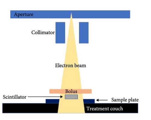
The samples were positioned on the treatment couch, and an electron beam was generated using a linear accelerator (Varian, Novalis Tx linear accelerator), as illustrated in Figure 1. An incident electron beam with an energy of 6 MeV penetrated the scintillator from top to bottom. The source-to-sample distance was set to 100 cm, and all the experiments were conducted at room temperature. Electron beam doses of 50, 100, 500, 1000, and 3000 Gy were calculated and irradiated on the scintillators.

2.2. Spectral Performances
γ-ray detection was performed by coupling each scintillator with an enhanced specular reflector (3M Enhanced Specular Reflector, 3M ESR) cap to a photomultiplier assembly (Hamamatsu Photonics, H11934-100). A bias voltage of −900 V was applied using a high-voltage power supply (Bertan, 225-30R). The output signal was then connected to a digitizer (CAEN, DT5730) for data acquisition and analysis on a PC. The relative light output and energy resolution of the γ-ray spectrum were determined by fitting a Gaussian function to the 662 keV photopeak of 137Cs. To assess the light-output efficiency of the material, the relative light output was calculated by comparing the photopeak corresponding to 662 keV with the baseline and expressed as a percentage.
2.3. Optical Performances
We employed a ultraviolet–visible–near-infrared spectrophotometer (Agilent Technologies, Cary 5000) to assess the transmittance and absorbance before and after electron beam exposure. This instrument facilitated the observation of radiation-induced variations and was operated in the 200–800 nm range at a scanning rate of 1 nm/s. Subsequently, the acquired data of each radiation dose were subjected to a comparative analysis to identify any notable change.
2.4. PL Characterization
To measure the PL performance of the scintillators irradiated with electron beams, the PL spectra were recorded using a confocal Raman spectrometer (LabRam Aramis, Horiba Jobin Yvon) with a 325 nm He–Cd laser excitation source. We evaluated the changes in the crystallinity and performance of the scintillators owing to defect formation on their surfaces to determine their radiation tolerance.
2.5. TL Characterization
TL analysis was performed to assess the impact of electron beam irradiation on the LuAG:Pr and GPS:Ce scintillators. To investigate the deep defects within the crystallographic structure in detail, samples exposed to radiation were subjected to TL measurements using a TL measurement system (RISO TL/OSL Reader Model DA-20, Denmark). The samples were gradually heated from 0 to 400°C at a rate of 1.6°C/s, while detecting the luminescent signals emitted by the scintillators. This process allowed us to analyze the luminescent signals generated by the scintillators as they were heated, providing insights into the presence and characteristics of defects in the crystal structure.
2.6. Thermal Annealing
To address the defects generated within inorganic scintillators and the resulting degradation in scintillation performance, we first established the initial conditions for all samples to facilitate a comparison of performance differences. The samples underwent heat treatment at 1000°C for 10 h in air, using an electric furnace. We then observed the resulting changes in performance, allowing us to assess the effectiveness of heat treatment in mitigating the impact of defects on the scintillation performance.
3. Results and Discussion
3.1. Spectral Performances
The observed changes in the energy spectra upon electron beam irradiation are depicted in Figure 2 and Table 1. Evidently, the relative light outputs of both LuAG:Pr and GPS:Ce decrease with increasing radiation dose. Their relative light outputs start decreasing from relatively low radiation doses, with 93.1% and 88.0% recorded for LuAG:Pr and GPS:Ce, respectively, at 100 Gy. The light intensities of both the scintillators rapidly decrease upon exposure to a dose of up to 1 kGy and continue to decrease gradually up to 3 kGy. Upon exposure to an electron beam dose of 1 kGy, the light intensities of LuAG:Pr and GPS:Ce rapidly decrease to 72.3% and 63.6%, respectively. Furthermore, the corresponding energy resolution differences are 2.9% and 2.6%, respectively. These results demonstrate that with increasing radiation dose, the ability of these scintillators to distinguish between different radiation energies deteriorates, along with a gradual decrease in their relative light outputs, indicating a drastic reduction in their radiation detection efficiencies. However, up to a dose of 3 kGy, the decrease in the relative light outputs of GAGG:Ce single crystals and ceramics, as well as that of LYSO:Ce, evidently vary within a range of 5.0% or less, indicating the excellent radiation tolerance of these two scintillators.
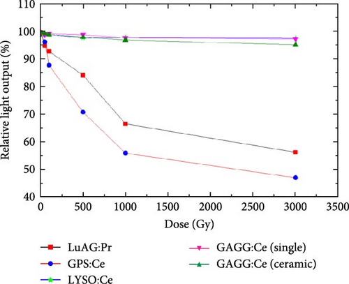
| Treatment | Relative light output (relative value, %)a |
Energy resolution before/after treatment (%)b |
|---|---|---|
| LuAG:Pr, 50 Gy | 95.1 ± 0.6 | 16.7 ± 0.3/17.4 ± 0.5 |
| LuAG:Pr, 100 Gy | 93.1 ± 0.3 | 16.3 ± 0.2/16.8 ± 0.3 |
| LuAG:Pr, 500 Gy | 85.6 ± 0.6 | 15.7 ± 0.3/17.4 ± 0.3 |
| LuAG:Pr, 1000 Gy | 72.3 ± 0.5 | 15.7 ± 0.3/18.6 ± 0.3 |
| LuAG:Pr, 3000 Gy | 66.2 ± 1.2 | 16.3 ± 0.2/18.6 ± 0.3 |
| GPS:Ce, 50 Gy | 95.6 ± 0.2 | 14.7 ± 0.2/12.8 ± 0.2 |
| GPS:Ce, 100 Gy | 87.7 ± 0.3 | 8.5 ± 0.1/8.5 ± 0.2 |
| GPS:Ce, 500 Gy | 73.2 ± 0.2 | 7.9 ± 0.1/8.1 ± 0.2 |
| GPS:Ce, 1000 Gy | 63.6 ± 0.4 | 8.7 ± 0.1/10.3 ± 0.3 |
| GPS:Ce, 3000 Gy | 56.9 ± 0.3 | 8.1 ± 0.1/10.7 ± 0.4 |
- aData (compared with the light output of the sample before radiation exposure) are the average and standard deviation of three measurements.
- bData are the average and standard deviation of three measurements.
The decrease in the light output can be attributed to light interference, light path impairment, and light absorption or scattering at specific locations. The decrease in the light output and energy resolution degradation under electron beam irradiation is attributed to defects generated within the scintillator; such defects change the surface color of the scintillator. In particular, the generation of color centers within the scintillator interferes with the planned generation and transportation of scintillator light through processes such as defect-induced light scattering or absorption [14, 15].
According to previous studies [16, 17], GAGG:Ce and LYSO:Ce scintillators exhibit significant signal losses due to a quenching effect at temperatures above 150°C. However, in contrast to the LuAG:Pr and GPS:Ce scintillators investigated in the present study, their performance degradation is minimal, even when exposed to 3 kGy electron beams. Therefore, the present study was focused on the identification of the mechanisms underlying the spectral performance degradation of LuAG:Pr and GPS:Ce scintillators, their mitigation strategies, and the factors to be considered in harsh environments.
3.2. Optical Performances
Transmittance measurements were conducted to demonstrate the changes in the optical performance corresponding to variations in the scintillator behavior. Specifically, we explored the influence of increasing the electron beam dose on the previously mentioned color changes in LuAG:Pr and GPS:Ce and their transmittances. Figure 3 shows the effect of electron beam irradiation on the transmittances of LuAG:Pr and GPS:Ce. Upon exposure to a beam dose of 3 kGy, the transmittance of LuAG:Pr decreases from 65.1% to 30.2% and that of GPS:Ce decreases from 68.8% to 46.9%. This decrease in the transmittance with the increasing electron beam dose is ascribed to the defects that affect the scintillation mechanism. LuAG scintillators have a wide bandgap of 5–6 eV, and scintillation occurs when electrons de-excite from an excited state to the ground state. However, this wide bandgap can reduce light output and energy resolution, which is why doping with praseodymium (Pr) is used. Pr ions act as activation centers, introducing intermediate energy levels within the bandgap, enhancing electron transitions, and improving scintillation performance through 5d–4f transitions. Pr doping also increases electron–hole recombination, improving light yield and energy resolution in LuAG scintillators.
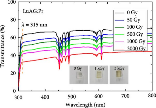
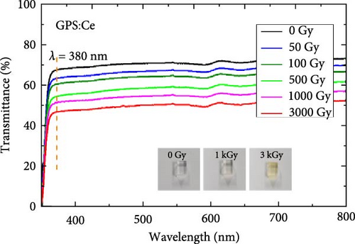
Electron beams cause elastic and inelastic scattering, leading to atom displacement through knock-on damage, where kinetic energy is transferred to atoms. The likelihood of defect formation depends on the crystal lattice structure [14, 18–20]. Pr ions can be displaced from the lattice if enough energy is transferred, creating vacancies and interstitial defects. These defects degrade scintillation performance by disrupting light generation and absorbing photons, resulting in reduced light yield and energy resolution. Therefore, maintaining crystal lattice integrity is crucial for optimal scintillation performance. Additionally, defect-induced nonradiative recombination processes broaden the emission spectrum, thereby degrading the energy resolution. Under electron beam irradiation, the results shown by GPS:Ce are similar to those of LuAG:Pr. Thus, the reduction in the transmittance of GPS:Ce is comparable to that observed in LuAG:Pr, highlighting the critical role of Ce ions in scintillation light generation. Furthermore, the diminished optical performance, as depicted in Figure 3, supports the deterioration of crystallinity that leads to defect formation. These defect centers affect the conversion of incident particle radiation into scintillation light through a series of radiative and nonradiative processes.
3.3. PL Spectral Analysis
The influence of performance degradation on PL due to electron beam irradiation was examined, and the corresponding spectra are shown in Figure 4. The intensities around the emission peaks (~310 and ~380 nm, respectively) of LuAG:Pr and GPS:Ce decrease as the electron beam dose is increased up to 3 kGy. Specifically, the relative intensities near the emission peaks of LuAG:Pr and GPS:Ce are reduced by approximately 79.1% and 56.4%, respectively.
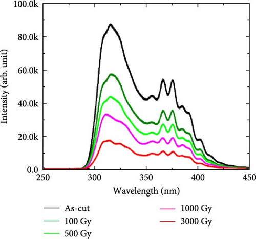
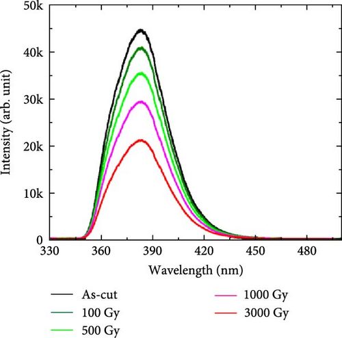
The defect-mediated deterioration of surface properties reduces the light collection efficiency as well as leads to optical performance degradation due to inhibition of transport of light to the photodetector. Defects can create optical barriers that scatter and absorb light as it travels from the surface through the bulk material, leading to its degradation. To ensure the transmission of a sufficient number of scintillation light photons to the photodetector, optimal conditions for surface properties, such as surface roughness, defect quantity, and refractive index matching, need to be established. However, exposure to high radiation doses inevitable generated various types of defects both within the bulk and the surface of the scintillator. These defects influence various factors that affect the conversion efficiency of absorbed light energy into luminescence during PL measurements. These factors include changes in the crystal structure and state due to the altered surface or internal conditions of the scintillator, which affect its PL intensity. The change in the PL intensity is ascribed to defect-induced variations in the bandgap energy, ultimately manifesting as specific peaks and changes in absorption peak intensities within the PL spectrum. Investigations based on carrier mobility and recombination processes provide insights into the origin and generation mechanism of the defects present on the scintillators’ surfaces as well as reveal their potential impact on the radiation detection performance of the scintillators. Figure 3 confirms that the defects generated by the irradiated electron beam cause various phenomena, such as nonradiative recombination, which reduce the PL intensity.
Electron beams can damage a scintillator by breaking the bond structure inside its crystal or by forming defects, which can affect the PL efficiency. Trapped electrons can also interact with the material and affect the PL characteristics by creating new energy levels that compete with those responsible for the PL, thereby reducing the PL intensity. Additionally, various defects can create interference factors, such as light absorption and scattering, that ultimately decrease the PL intensity.
The electrons irradiated on a scintillator material are trapped in the defects or impurity sites within the scintillator’s crystal lattice. Over time, these trapped electrons accumulate and hinder the movement of free electrons, resulting in the degradation of the crystal quality and luminescence efficiency of the scintillator. The effects of trapped electrons on the luminescence properties of a scintillator depend on several factors, such as the nature and location of the trap site. In some cases, the trapped electrons can act as energy donors or acceptors, contributing to luminescence. However, if the trapped electrons cannot be recovered over time, then they can affect the efficiency and performance of the scintillator. Therefore, understanding the underlying mechanisms of electron trapping and developing effective strategies to mitigate these effects are essential for optimizing the performance and efficiency of scintillator materials for various applications.
3.4. TL Spectral Analysis
Figure 5 shows the TL spectra corresponding to the electron beam-induced changes in the bulk sites. Both the LuAG:Pr and GPS:Ce scintillators exhibit significant performance degradation due to radiation, as evidenced by earlier reported investigations of surface changes and energy spectra. A similar trend is observed in the TL spectra. Upon irradiation with electron beam doses of up to 3 kGy, the TL intensities of both LuAG:Pr and GPS:Ce increase at approximately 300 and 150°C, respectively. Degradation of the scintillator’s optical performance due to electron beam irradiation occurs throughout the crystal, and the color centers that form in the bulk sites significantly deterioration the light transmission and collection efficiencies.
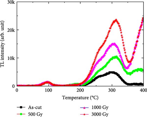
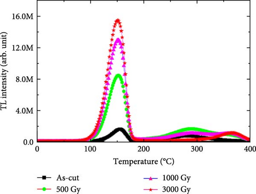
Color centers inhibit photon emission, thereby affecting the luminescence of scintillators owing to a quenching effect that traps excitons within the crystal. Ionizing radiation, such as X-rays or γ-rays, can create defects by trapping electrons, which block signal transmission [21]. However, the trapped electrons can recombine with holes and emit photons upon sample heating. Thus, this approach was employed in the present study to estimate the defect density by comparing the intensities of the emitted light signals. Compared with surface defects, mitigating the defects present in the bulk of materials, without applying external processes such as heat treatment, is more challenging. However, when exposed to photon or particle radiation, scintillators with a high radiation tolerance experience fewer processes in which energy is lost as heat rather than as light. This tolerance may be attributed to the solid structure of the crystal, its purity, or materials added to it.
3.5. Thermal Annealing
Thermal annealing, optical bleaching, and chemical treatment are commonly employed for removing color centers [22]. Among these methods, we adopted high-temperature annealing, which is a straightforward and efficient method. Oxide-based scintillators contain abundant oxygen atoms that contribute to scintillation crystal formation and thus increase the likelihood of defect generation during growth, postprocessing, and fabrication. LuAG:Pr and GPS:Ce, which are oxide materials, were subjected to heat treatment at 1000°C for 10 h in air to remove defects, especially oxygen-induced dangling bonds, generated within their crystals by the irradiated electron beam. This high-temperature annealing treatment restored their performances to levels observed before the irradiation (Figure 6).
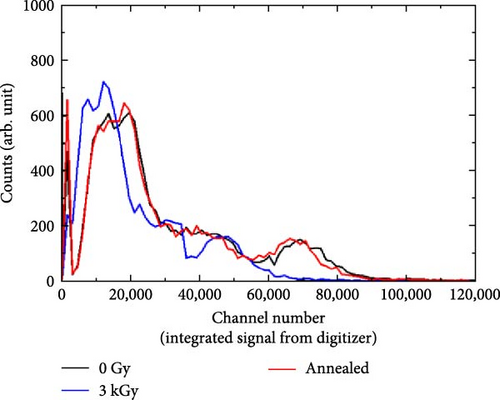
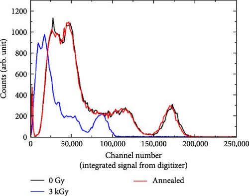
The reduced relative light outputs of both the LuAG:Pr and LuAG:Ce scintillators (66.2% and 56.9%, respectively), upon exposure to an electron beam dose of 3 kGy, are subsequently restored to their original values after the heat treatment. In addition, the optical transparency values of the scintillators return to their previous values, indicating the recovery of the scintillation performance due to the quenching-induced removal of sufficient defects from the crystals’ surfaces and bulk regions. Specifically, under electron beam irradiation: (1) the irradiated electrons collide with those present inside the scintillator; (2) the electrons themselves act as defect sources, causing irregularities in the crystal’s lattice structure; and (3) the density of the induced defects depends on the energy of the transmitted electron beam. These defects generate color centers within the crystal structure, and these resulting color centers hinder light emission from the scintillators or absorb the generated light, thereby inhibiting light transmission. Evidently, such defects can be successfully eliminated through the high-temperature heat treatment applied in this study.
4. Conclusions
Through this study, we identified promising inorganic scintillators suitable for medical and aerospace applications that involve exposure to high levels of particle radiation. This step highlights the importance of selecting radiation-tolerant scintillators to ensure quality assurance of medical devices. While LuAG:Pr and GPS:Ce showed poor radiation tolerance, LYSO:Ce and GAGG:Ce demonstrated robustness against electron beams. For LuAG:Pr and GPS:Ce scintillators, significant degradation of scintillation and optical properties occurred at MGy doses with photon beams [2], while similar degradation was observed at kGy doses with electron beams. The PL and TL comparative evaluation revealed a significant degradation in spectral performance due to defect formation at both the surface and bulk sites of the crystal. The observed changes in the PL and TL intensities of LuAG:Pr and GPS:Ce scintillators highlight the need to develop rigorous radiation tolerance criteria to maintain competitiveness in radiation detection efficiency. Thermal annealing was effective in mitigating the defects, particularly those at the nonradiative recombination sites. The findings of the study, along with those of previous photon beam investigations, are expected to guide the selection and advancement of inorganic scintillation crystals for diagnostics, therapeutic equipment, and aerospace research.
Conflicts of Interest
The authors declare no conflicts of interest
Author Contributions
Chansun Park and Sangsu Kim contributed equally to this work.
Funding
This work was supported by the National Research Foundation of Korea (RS-2023-00240040, RS-2023-00234651, RS-2022-00165164, NRF-2023R1A2C2007545, NRF-2020R1A2C2007376, and NRF-2022R1I1A1A01065484), the Korea Health Technology R&D Project grant, through the Korea Health Industry Development Institute (KHIDI), funded by the Ministry of Health & Welfare, Republic of Korea (RS-2024-00436472), and the Korea University Grant.
Open Research
Data Availability Statement
The data that support the findings of this study are available from the corresponding author upon reasonable request.


