EFFECTS OF STIFLE FLEXION ANGLE AND SCAN PLANE ON VISIBILITY OF THE NORMAL CANINE CRANIAL CRUCIATE LIGAMENT USING LOW-FIELD MAGNETIC RESONANCE IMAGING
Abstract
Low-field magnetic resonance imaging (MRI) is commonly used to evaluate dogs with suspected cranial cruciate ligament injury; however, effects of stifle positioning and scan plane on visualization of the ligament are incompletely understood. Six stifle joints (one pilot, five test) were collected from dogs that were scheduled for euthanasia due to reasons unrelated to the stifle joint. Each stifle joint was scanned in three angles of flexion (90°, 135°, 145°) and eight scan planes (three dorsal, three axial, two sagittal), using the same low-field MRI scanner and T2-weighted fast spin echo scan protocol. Two experienced observers who were unaware of scan technique independently scored visualization of the cranial cruciate ligament in each scan using a scale of 0–3. Visualization score rank sums were higher when the stifle was flexed at 90° compared to 145°, regardless of the scan plane. Visualization scores for the cranial cruciate ligament in the dorsal (H (2) = 19.620, P = 0.000), axial (H (2) = 14.633, P = 0.001), and sagittal (H (2) = 8.143, P = 0.017) planes were significantly affected by the angle of stifle flexion. Post hoc analysis showed that the ligament was best visualized at 90° compared to 145° in the dorsal (Z = −3.906, P = 0.000), axial (Z = −3.398, P = 0.001), and sagittal (Z = −2.530, P = 0.011) planes. Findings supported the use of a 90° flexed stifle position for maximizing visualization of the cranial cruciate ligament using low-field MRI in dogs.
Introduction
Cruciate ligament injury is one of the most common conditions in canine orthopedic practice.1 The presence of a positive cranial tibial drawer test in combination with radiological evidence of stifle soft tissue swelling are common diagnostic findings in patients with cranial cruciate ligament damage.1 However, in cases of partial cranial cruciate injury, the cranial drawer test may be negative. In these cases, cruciate injury could be suspected but further investigation may be required.2 An MRI technique that maximizes full visualization of the cranial cruciate ligament would be beneficial in a clinical setting, especially for cases where partial cranial cruciate ligament rupture is suspected. Additionally MRI of the cranial cruciate ligament may be beneficial in a research setting and provide useful insights into the mechanisms of cruciate disease. Research in human medicine has shown that images obtained with flexion of the knee more clearly differentiate normal from torn cranial cruciate ligaments vs. images obtained during extension.3 This improved visualization has been attributed to the increased separation of the cruciate ligament from the margin of the femoral intercondylar fossa in the flexed position.3-6 Previous canine studies have recommended varying stifle angles for MRI examinations.7-10 To the authors’ knowledge, the effect of stifle angle on MRI visualization of the cranial cruciate ligament has not been reported in dogs.
The aim of the current study was to compare the visualization of the cranial cruciate ligament at different stifle angles and for different scan planes. We hypothesized that, due to the similar anatomical relationship between the cranial cruciate ligament and the margin of the intercondylar fossa in people11 and dogs,12 stifle flexion would result in better visualization of the cranial cruciate ligament.
Material and Methods
The present study was performed in accordance with guidelines provided by the animal ethics committe. Six normal stifle joints were prospectively obtained from dogs scheduled to undergo euthanasia for reasons unrelated to the present study. Joints were determined to be normal based on physical examination before euthanasia, radiographic assessment after euthanasia, and gross pathologic inspection after MRI. On physical examination, included dogs had to have no evidence of lameness, articular swelling, or pain on manipulation of the stifle joint. On radiographic examination, included dogs had to have no evidence of joint effusion, periarticular osteophytes, enthesophytes or intra-articular mineralization in lateral and caudocranial projections. On gross pathological examination, included dogs had to have an intact cranial cruciate ligament, and no evidence of cruciate ligament fibrillation, joint effusion, capsule thickening, meniscal tears, or cartilage erosion.
Stifles selected for inclusion were disarticulated at the level of the coxofemoral joints. Either the right or the left stifles were randomly selected. Magnetic resonance (MR) scanning was performed on all stifle joints within 3 h of euthanasia, using the same low-field MR scanner (Esaote Vet Grande 0.25 Tesla system, Genoa Via A. Siffredi, 58 16153 Genova ITALY). Images were acquired using a linear volume (C1) coil and a fast spin echo (FSE) T2-weighted sequence. This sequence was chosen because it has been reported to provide excellent contrast resolution between the synovial fluid and the cranial cruciate ligament.13 Technical factors for the FSE sequence were: TR 4730 ms, TE = 100 ms, Nex = 4, ETL = 14, echo spacing = 20 ms, and included a T2 Refocusing pulse. Resolution parameters were: FOV 190 × 190, Matrix 190 × 190 pixels, 3-mm slices at 0 slice gap.
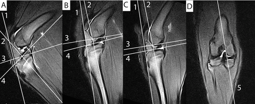
Disarticulated limbs were positioned with their lateral aspect against the table and the coil centered on the stifle joint, as would have been performed in a live patient. Stifles were imaged in three different angles: 90° (flexed position), 135°(physiological position), and 145°(extended position). These angles were defined using the angle measurement tool in the viewing software and sagittal scout images. Angles were measured between the long axis of the femur and the tibia. Pilot scans were acquired and repeated as needed until the correct stifle angulations were achieved. A custom positioning device was designed and used to acquire and maintain the desired stifle angle during the MR examination. This device consisted of one polyvinyl chloride (PVC) pipe, two adjustable rings, and two nylon belts (Fig. 1). The nylon belts were used to attach the limb to the rings. One belt was attached to the femoral neck through a drilled hole and then attached to one of the rings, and the other belt was attached to the calcaneus through a drilled hole and then attached to the second ring. The PVC pipe was then inserted through the hole of both rings. By running the rings along the PVC pipe, the stifle angle could be adjusted as desired. A series of FSE T2-weighted sequences were acquired in eight different planes for each of the stifle angles. Previous reports in veterinary medicine used either the patellar ligament or the tibial plateau as the anatomical landmark to plan the dorsal and axial planes.7, 14-16 Because we were interested in the effect of stifle angle on the visibility of the cranial cruciate ligament regardless of the landmark used for planning, we decided to include scans based on both anatomical landmarks. In addition, we included oblique scans aligned with the cranial cruciate in the dorsal, axial, and sagittal planes. Each stifle joint was therefore scanned in three dorsal, three axial, and two sagittal planes. One dorsal plane was aligned such that slices were parallel to the patellar ligament (Fig. 2A–C line1) on the sagittal scout while another dorsal plane was aligned such that slices were perpendicular to the tibial plateau (Fig. 2A–C line2) in the same scout. Both of these dorsal planes were aligned with the caudal aspect of the femoral condyles in the transverse scout. The third dorsal plane was aligned such that slices were parallel with the long axis of the cranial cruciate ligament on the sagittal and transverse scouts. This plane was named dorsal oblique. One axial plane was aligned such that slices were perpendicular to the patellar ligament on the sagittal scout (Fig. 2A–C line 3) while another axial plane was aligned such that slices were parallel to the tibial plateau (Fig. 2A–C line4) on the same scout. Both axial planes were aligned with the distal border of the femoral condyles in the dorsal plane. The third axial plane was aligned such that slices were perpendicular to the cranial cruciate ligament on the sagittal and dorsal scouts. This plane was named axial oblique. One sagittal plane was aligned such that slices were parallel to the medial condyle of the femur on the transverse and dorsal scouts (Fig. 2D line 5). The other sagittal plane was aligned such that the slices were parallel with the long axis of the cranial cruciate ligament on the dorsal and transverse scouts. This plane was named sagittal oblique.


Image Analysis
For each scan, two board certified veterinary radiologists with prior experience interpreting stifle MR studies (P.G. and T.S.) evaluated the cranial cruciate ligament based on a visual assessment score of 0–3 and subjective criteria. A score of 0 indicated that the cranial cruciate ligament was not visible. A score of 1 indicated the cranial cruciate ligament was visualized partially. A score of 2 indicated that the totality of the cranial cruciate ligament was identified however it was poorly demarcated. A score of 3 indicated a cranial cruciate ligament that was totally visualized and well demarcated. Visualization scores referred to the visibility of the cranial cruciate ligament using all available images in one scan sequence. Open access image analysis software (Osirix 4.1.2, Open Source) was used to project and process the images on a flat-screen liquid crystal display (LCD) monitor (30 inch Apple Cinema HD display, Cupertino, CA) during the evaluation. The studies were anonymized and all data indicating the stifle angles and planes were removed. The observers evaluated the sequences independently and were unaware of each other's scores. Observers were provided with scans of one of the stifle joints to familiarize them with the available scanning planes. These pilot study images were removed from analysis, resulting in a total of five stifle joints being analyzed. Observers had unlimited time to review the images and were allowed to adjust windowing levels and scale as desired.
Statistical Analysis
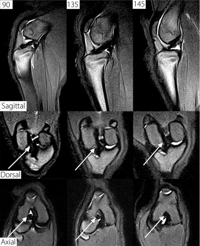
Statistical analyses were selected and performed by two of the authors (E.H. and M.M.). All calculations were performed using commercial software (IBM SPSS Statistics, version 20, International Business Machines Corp.). Visualization score rank sums of the cranial cruciate ligament were calculated for each plane in each joint angle and for each observer (Table 1). To compare visualization scores between different stifle angles, scan planes were pooled into three groups. One group included all dorsal planes (the dorsal plane parallel to the patellar ligament, the dorsal plane perpendicular to the patellar ligament, and the dorsal oblique plane). A second group included all axial planes (the axial plane parallel to the tibial plateau, the axial plane perpendicular to the patellar ligament, and axial oblique plane) and the last group included all sagittal planes (the sagittal plane parallel to the medial condyle of the femur and sagittal oblique plane; Fig. 3). The Friedman repeated measures analysis of variance on ranks was used to determine statistical differences in the visualization scores between the three different joint angles, for each group. Where significance (P < 0.05) was found between different joint angles, post hoc analysis with Wilcoxon signed-rank tests was conducted with a Bonferroni correction applied, resulting in a significance level set at P < 0.017, to determine which position differences accounted for the significance. Interobserver differences were tested using intraclass correlation coefficient analysis.
| 90° | 135° | 145° | |
|---|---|---|---|
| Planes\angles | Rank Sums | Rank sums | Rank sums |
| Sagittal parallel medial condyle | |||
| Observer 1 | 15 | 14 | 13 |
| Observer 2 | 15 | 13 | 12 |
| Combined | 30 | 27 | 25 |
| Transverse parallel tibial plateau | |||
| Observer 1 | 11 | 10 | 9 |
| Observer 2 | 11 | 8 | 8 |
| Combined | 22 | 18 | 17 |
| Transverse perpendicular patellar ligament | |||
| Observer 1 | 12 | 9 | 9 |
| Observer 2 | 13 | 12 | 7 |
| Combined | 25 | 21 | 16 |
| Dorsal parallel patellar ligament | |||
| Observer 1 | 15 | 14 | 11 |
| Observer 2 | 15 | 10 | 10 |
| Combined | 30 | 24 | 21 |
| Dorsal perpendicular tibial plateau | |||
| Observer 1 | 13 | 14 | 10 |
| Observer 2 | 15 | 13 | 10 |
| Combined | 28 | 27 | 20 |
| Sagittal oblique plane | |||
| Observer 1 | 14 | 13 | 14 |
| Observer 2 | 15 | 12 | 12 |
| Combined | 29 | 25 | 26 |
| Transverse oblique plane | |||
| Observer 1 | 12 | 12 | 9 |
| Observer 2 | 15 | 13 | 15 |
| Combined | 27 | 25 | 24 |
| Dorsal oblique plane | |||
| Observer 1 | 14 | 12 | 11 |
| Observer 2 | 12 | 14 | 9 |
| Combined | 26 | 26 | 20 |
- *Visual assessment score (0 = cranial cruciate ligament was not visible; 1 = cranial cruciate ligament visualized partially; 2 = cranial cruciate ligament visualized totally but poorly demarcated; 3 = cranial cruciate ligament totally visualized and well demarcated).

Results
Dog breeds included in the study were two Staffordshire Bull Terriers, two Australian Collies, and two cross breeds. Dog weights ranged from 18–30 kg. The median age ranged from 1–2 years. There were two entire males and four entire females. The visual assessment score rank sums for cranial cruciate visualization are summarized in Table 1. Visualization score frequencies for the combined dorsal, axial, and sagittal planes are shown in Fig. 3.
Visualization scores rank sums were higher when the stifle was flexed at 90° regardless of the plane. Visualization scores of the cranial cruciate ligament in the dorsal planes were significantly affected by the angle of flexion of the stifle, H (2) = 19.620, P = 0.000. Median visualization scores for the dorsal planes at 90°, 135°, and 145° stifle joint angles were 3, 3, and 2, respectively. Post hoc analysis with Wilcoxon signed-rank tests revealed statistically significant differences between cranial cruciate ligament visualization scores for the dorsal planes at 90° compared to 145° (Z = −3.906, P = 0.000), at 135° compared to 145° (Z = −2.826, P = 0.005) but not at 90° compared to 135° (Z = −1.807, P = 0.071) joint angles.
Visualization scores of the cranial cruciate ligament in the axial planes were significantly affected by the angle of flexion of the stifle, H (2) = 14.633, P = 0.001. Median visualization scores for the axial planes at 90°, 135°, and 145° stifle joint angles were 3, 2, and 2, respectively. Post hoc analysis with Wilcoxon signed-rank tests revealed statistically significant differences between cranial cruciate ligament visualization scores for the axial plane at 90° compared to 145° (Z = −3.398, P = 0.001), but not at 90° compared to 135° (Z = −2.357, P = 0.018) or at 135° compared to 145° (Z = −1.261, P = 0.207) joint angles.
Visualization scores of the cranial cruciate ligament in the sagittal planes were significantly affected by the angle of flexion of the stifle, H (2) = 8.143, P = 0.017. Median visualization scores for the sagittal planes at 90°, 135°, and 145° stifle joint angles were 3, 3, and 3, respectively. Post hoc analysis with Wilcoxon signed-rank tests revealed statistically significant differences between cranial cruciate ligament visualization scores for the sagittal plane at 90° compared to 145° (Z = −2.530, P = 0.011), but not at 90° compared to 135° (Z = −2.333, P = 0.020) or at 135° compared to 145° (Z = −0.333, P = 0.739) joint angles.
The intraclass correlation coefficient was 0.677 (95% CI: 0.54–0.78) for all measurements; therefore the interrater agreement was interpreted as good (Table 2).
| Observer 2 | ||||||
|---|---|---|---|---|---|---|
| Score 0 | Score 1 | Score 2 | Score 3 | Total | ||
| Observer 1 | Score 0 | |||||
| Number of counts | 0 | 0 | 0 | 0 | 0 | |
| Percentage of total | 0% | 0% | 0% | 0% | 0% | |
| Score 1 | ||||||
| Number of counts | 0 | 6 | 0 | 1 | 7 | |
| Percentage of total | 0% | 5% | 0% | 0.8% | 5.8% | |
| Score 2 | ||||||
| Number of counts | 0 | 9 | 26 | 20 | 55 | |
| Percentage of total | 0% | 7.5% | 21.7% | 16.7% | 45.8% | |
| Score 3 | ||||||
| Number of counts | 0 | 0 | 17 | 41 | 58 | |
| Percentage of total | 0% | 0% | 14.2% | 34.2% | 48.3% | |
| Total | Number of counts | 0 | 15 | 43 | 62 | 120 |
| Percentage of total | 0% | 12.5% | 35.8% | 51.7% | 100% | |
- *Visual assessment score (0 = cranial cruciate ligament was not visible; 1 = cranial cruciate ligament visualized partially; 2 = cranial cruciate ligament visualized totally but poorly demarcated; 3 = cranial cruciate ligament totally visualized and well demarcated
Discussion
Visualization scores of the cranial cruciate ligament were significantly affected by stifle angle in the dorsal, axial, and sagittal planes. The cranial cruciate ligament ligament was best visualized in all planes when stifles were positioned at a 90° angle. The better visualization of the cranial cruciate ligament at 90° may have been related to the anatomical relation between this ligament and the surrounding bony structures. Similar to studies performed in people,17 our study revealed that during extension the majority of the femoral portion of the cranial cruciate ligament is in contact with the proximal and the lateral margins of the intercondylar fossa (Fig. 4). Due to the similar signal intensity of ligament and bony structures, their contours cannot be clearly delineated when they are in contact. During flexion, the cranial cruciate ligament separates from these bony structures allowing synovial fluid to extend around the margins. Due to the high signal intensity of the synovial fluid on T2 FSE weighted images, the contours of the cranial cruciate ligament can therefore be better delineated.

Scanning the canine stifle in a flexed (90°) position required the use of larger receiver coils compared to scanning the same stifle in an extended position. Because using different coils for different angles of stifles flexion would have resulted in the addition of an undesirable variable, we decided to use only one coil for all examinations. This coil was large enough to scan the stifles both in flexion and in extension. Using this coil may have resulted in a reduction in signal while scanning the stifle in an extended position. For clinical studies in live dogs, we would recommend using the smallest coil that allows scanning of the stifle in flexion for best visualization of the canine cranial cruciate ligament.
Disarticulated limbs were positioned with their lateral aspect against the table and the coil centered on the stifle joint. This limb positioning is similar to the one that would be used in a live animal. A special device was used to position and maintain the stifles in the desired angle of flexion. In a clinical setting, positioning of the stifle in a flexed position could be achieved with restraining tools such as tape and foam wedges. The angle of stifle flexion was measured using the angle measurement tool from the viewing software on the scout images. This was a fast and easy procedure, which in most cases required only minor adjustements and repositioning. Based on our experience, we believe this technique could be replicated in a clinical setting.
There were some limitations in the current study. The small number of stifle joints did not allow statistical analyses comparing visualization scores of the cruciate ligament for each individual scan plane, and therefore data had to be pooled for each type of scan plane (axial, sagittal, dorsal). In addition, only the FSE T2 sequence was used for comparisons. The authors acknowledge the importance of using multiple sequences in a clinical context. The T2 FSE-weighted sequence was chosen because it has been reported to provide excellent contrast resolution between the synovial fluid and the cranial cruciate ligament, and to be less susceptible to magic angle artifacts.13 T2 FSE-weighted sequences have also been shown to be highly sensitive and specific for demonstrating cranial cruciate ligament injury in people.18 The presence and severity of magic angle artifacts were not tested in dogs of the current study. Changing the stifle angle may have also influenced the visualization of other clinically important structures such as the menisci, patellar ligament, collateral ligaments, long digital extensor tendon, or articular cartilage. The effect of stifle flexion on visualization of these structures was not tested in this study. Therefore, our conclusions should be limited to the effect of stifle angle of flexion on visualization of the cranial cruciate ligament only. Additionally, cruciate disease is particularly common in large- and giant-breed dogs such as the Labrador retriever, Rottweiler, and Saint Bernard. None of the dogs tested in this study were of those breeds. However, any breed of dog may be affected with cruciate ligament disease.19 Our sample population of dogs reflected the breeds that are commonly present with this disease at our institution. Authors acknowledge that caution should be exercised before applying our findings to other dog breeds.
In conclusion, findings from this canine cadaver study indicated that low-field MR images of the stifle at a 90° angle resulted in better visualization scores of the cranial cruciate ligament. Future studies are needed to evaluate the effect of stifle angle on the visibility of other structures such as the menisci, patellar ligament, collateral ligaments, long digital extensor tendon, or articular cartilage.


