Clinical and histopathological aspects of an alopecia syndrome in captive Andean bears (Tremarctos ornatus)
Abstract
enBackground
Captive Andean bears (Tremarctos ornatus) develop a distinct alopecic syndrome of unknown aetiology.
Hypothesis/Objectives
To describe the histological features of healthy Andean bear skin, to define the clinical and histopathological features of Andean bears with signs of alopecia, and to propose an aetiopathogenesis.
Animals
Eighteen healthy Andean bears housed in 12 European zoos and 13 Andean bears with mild to severe alopecia housed in nine European zoos.
Methods
Two surveys describing signalment and clinical features of affected bears; follicular density was measured in a single healthy bear using a dermatoscope; cytological samples were collected by tape stripping from two healthy and three alopecic bears; skin biopsies were collected for histological evaluation from healthy and alopecic bears; immunohistochemistry (CD3, AE1/AE3 cytokeratins) was performed when lymphocytic inflammation was observed.
Results
The syndrome is an acquired, slowly progressive alopecia. Bears are otherwise healthy. Histological features include a dermal inflammatory infiltrate composed of T lymphocytes and eosinophils; atrophy of hair follicles at the level of or below the isthmus, and lymphocytic infiltration of hair follicles and the epidermis. Multinucleated giant cells were present in the outer root sheaths of hair follicles in five bears.
Conclusion and clinical importance
Andean bear alopecia syndrome is an acquired, progressive alopecia with histological features consistent with a lymphocytic immune-mediated reaction directed against follicular sheaths and the epidermis. Trigger factors have not been identified. Further studies are indicated to define the features of this multifactorial syndrome.
Résumé
frContexte
Les ours à lunettes (Tremarctos ornatus) captifs développent un syndrome alopécique d’étiologie inconnue.
Hypothèses/Objectifs
Décrire les critères histopathologiques de la peau des ours à lunettes sains, définir les critères cliniques et histopathologiques des ours à lunettes avec alopécie et d'en proposer une étiopathogénie.
Sujets
Dix huit ours à lunettes sains de 12 zoos d'Europe et 13 ours à lunettes atteints d'alopécie modérée à sévère issus de neufs zoos européens.
Méthodes
Deux études décrivant le signalement et les données cliniques des ours atteints ; la densité folliculaire a été mesurée pour un seul ours à lunette sain à l'aide d'un dermoscope ; des échantillons cytologiques ont été collectés par cellophane adhésive de deux ours sains et trois ours alopéciques ; l'immunohistochimie (cytokératines CD3, AE1/AE3) a été réalisée quand une inflammation lymphocytaire a été observée.
Résultats
Le syndrome est une alopécie acquise lentement progressive. Les ours sont par ailleurs en bon état général. Les critères histologiques comprenaient un infiltrat inflammatoire dermique composé de lymphocytes T et d’éosinophiles ; une atrophie des follicules pileux au niveau ou au dessous de l'isthme, et un infiltrat lymphocytaire des follicules pileux et de l’épiderme. Les cellules géantes multinucléés étaient présentes dans la gaine épithéliale externe des follicules pileux de cinq ours.
Conclusion et importance clinique
Le syndrome alopécique des ours à lunette est une alopécie acquise, progressive avec des critères histopathologiques consistant en une réaction à médiation immune lymphocytaire dirigée contre les gaines folliculaires et l’épiderme. Les facteurs déclenchant n'ont pas été identifiés. Des études supplémentaires sont nécessaires pour définir les critères de ce syndrome multifactoriel.
Resumen
esIntroducción
Los osos andinos en cautividad (Tremarctos ornatus) desarrollan un síndrome alopécico distintivo de etiología desconocida.
Hipótesis/Objetivos
Describir las características histológicas de la piel sana del oso andino, definir las características clínicas e histopatológicas de los osos andinos con signos de alopecia y proponer una etiopatogenia.
Animales
Dieciocho osos andinos sanos alojados en 12 zoológicos europeos y 13 osos andinos con alopecia de leve a grave alojados en nueve zoológicos europeos.
Métodos
dos cuestionarios que describen la presentación y las características clínicas de los osos afectados; la densidad folicular se midió en un solo oso sano usando un dermatoscopio; se recogieron muestras citológicas mediante cinta adhesiva de dos osos sanos y tres alopécicos; se obtuvieron biopsias de piel para evaluación histológica de osos sanos y alopécicos; se realizó inmunohistoquímica (citoqueratinas AE1 / AE3 y CD3) cuando se observó inflamación linfocítica.
Resultados
El síndrome es una alopecia adquirida, lentamente progresiva. Los osos son saludables. Las características histológicas incluyen un infiltrado inflamatorio dérmico compuesto de linfocitos T y eosinófilos; atrofia de los folículos pilosos a nivel o debajo del istmo e infiltración linfocítica de los folículos pilosos y la epidermis. Se observaron células gigantes multinucleadas en las vainas radiculares externas de los folículos pilosos en cinco osos.
Conclusión e importancia clínica
el síndrome de alopecia del oso andino es una alopecia progresiva adquirida con características histológicas compatibles con una reacción linfocítica inmunomediada dirigida contra las envolturas foliculares y la epidermis. Los factores desencadenantes no han sido identificados. Se necesitan estudios adicionales para definir las características de este síndrome multifactorial.
Zusammenfassung
deHintergrund
Andenbären (Tremarctos ornatus)in Gefangenschaft entwickeln ein typisches Alopeziesyndrom unbekannter Ätiologie.
Hypothese/Ziele
Das Ziel dieser Studie war eine Beschreibung der histologischen Charakteristika gesunder Haut von Andenbären, um die klinischen und histopathologischen Merkmale der Haut der Andenbären mit Zeichen einer Alopezie zu definieren und um eine Ätiopathogenese vorzuschlagen.
Tiere
Achtzehn gesunde Andenbären, die in 12 europäischen Zoos und 13 Andenbären mit milder bis hochgradiger Alopezie, die in neun europäischen Zoos untergebracht waren.
Methoden
Zwei Studien, die Signalement und klinische Merkmale der betroffenen Bären beschreiben; die Dichte der Follikel wurde bei einem gesunden Bären mittels Dermatoskop gemessen; zytologische Proben wurden mittels Klebestreifen von zwei gesunden und von drei Bären mit Alopezie genommen; Hautbiopsien wurden zur histologischen Evaluierung von gesunden Bären sowie von Bären mit Alopezie entnommen; Immunhistochemie (CD3, AE1/AE3 Zytokeratin) wurde durchgeführt, wenn eine lymphozytäre Entzündung beobachtet wurde.
Ergebnisse
Es handelt sich bei dem Syndrom um eine erworbene, langsam progressive Alopezie. Die Bären sind ansonsten gesund. Die histologischen Charakteristika bestehen aus einem dermalen entzündlichen Infiltrat, welches sich aus T Lymphozyten und Eosinophilen zusammensetzt; eine Atrophie der Haarfollikel auf dem Level oder unter dem Isthmus, einem Lymphozyteninfiltrat der Haarfollikel und der Epidermis. Mehrkernige Riesenzellen kamen in den äußeren Schichten des Haarfollikelschafts bei fünf Bären vor.
Schlussfolgerung und klinische Bedeutung
Beim Alopeziesyndrom der Andenbären handelt es sich um eine erworbene, progressive Alopezie mit histologischen Charakteristika, die mit einer lymphozytären immun-mediierten Reaktion, die sich gegen die Follikelschäfte und die Epidermis richtet, einhergeht. Es wurden keine auslösenden Faktoren gefunden. Weitere Studien sind nötig, um die Charakteristika dieses multifaktoriellen Syndroms weiter zu definieren.
要約
ja背景
捕獲されたアンデスクマ(Tremarctos ornatus)は、原因不明かつ特有の脱毛症候群を発症する.
仮説/目的
健常なアンデスクマの皮膚の組織学的特徴を説明するとともに、脱毛症の兆候を示したアンデスクマにおける臨床的および病理組織学的特徴を定義し、その病因論について提唱する.
被験動物
欧州に存在する12施設の動物園に収容された健常なアンデスクマ18頭と、9施設に収容された軽度から重度の脱毛症を伴うアンデスクマ13頭.
方法
罹患したクマのシグナルメントおよび臨床的特徴に関する2種類の調査; 1頭の健常なクマにおける毛包密度のダーマスコープを用いた測定; 2頭の健康なクマおよび3頭の脱毛症のクマからテープストリッピングによる細胞診検体の収集;健常なクマおよび脱毛症のクマにおける皮膚生検の実施と組織学的評価;リンパ球性炎症が確認された場合は免疫組織化学染色(CD3、サイトケラチンAE1 / AE3).
結果
本症候群は、後天性で緩徐に進行する脱毛症である。本症候群に罹患したクマは、脱毛症を除き健康である。組織学的には真皮においてTリンパ球および好酸球からなる炎症細胞浸潤が認められ、毛包峡部または下部における毛包萎縮が認められ、また毛包および表皮にはリンパ球浸潤が認められる。5頭のクマの外毛根鞘では多核巨細胞が認められた.
結論と臨床的重要性
アンデスクマの脱毛症候群は後天性の進行性脱毛症であり、組織学的には毛嚢鞘および表皮に対するリンパ球成免疫介在成反応パターンと一致した特徴を有する。本症の誘因は特定されていない。今後はこの多因子症候群の特徴を明らかにするためのさらなる研究が必要である.
摘要
zh背景
圈养的安第斯熊(眼镜熊)发生不明原因的独特脱毛综合征.
假设/目的
描述健康安第斯熊皮肤的组织学特征,定义具有脱毛症状的安第斯熊的临床和组织病理学特征,并推测发病机理.
动物
十八头健康的安第斯熊,栖息在12所欧洲动物园内,十三头轻度至重度脱毛的安第斯脉熊,栖息在九所欧洲动物园中.
方法
两项调查阐述了发病熊的病征和临床特征。对单个健康熊使用皮肤镜测量毛囊密度。使用胶带对两个健康和三个脱毛熊进行细胞学采样。对健康和脱毛熊进行皮肤活检,用于组织学评估。当观察到淋巴细胞炎症时,进行免疫组化染色(CD3,AE1 / AE3 细胞角质)
结果
这是一种获得性、缓慢进行性的脱毛综合征。熊的其他方面是健康的。组织学特征包括由T淋巴细胞和嗜酸性粒细胞构成的皮肤炎性浸润;峡部或峡部以下毛囊萎缩,毛囊和表皮被淋巴细胞浸润。 5只熊的毛囊外根鞘中存在多核巨细胞.
结论和临床意义
安地斯熊脱毛综合征是一种获得性、进行性脱发,其组织学特征是针对毛囊鞘和表皮的淋巴细胞免疫介导反应。触发因素尚未明确。定义这种多因素综合征的特征,需要更多研究.
Resumo
ptContexto
Os ursos andinos (Tremarctos ornatus) criados em cativeiro podem desenvolver uma síndrome alopécica de etiologia desconhecida.
Hipótese/Objetivos
Descrever as características histopatológicas da pele hígida de ursos andinos, definir as características clínicas e histopatológicas da pele de ursos andinos com alopecia, e propor uma etiopatogênese.
Animais
Dezoito ursos andinos saudáveis de 12 zoológicos europeus e 13 ursos andinos com alopecia leve a grave de nove zoológicos europeus.
Métodos
Foram utilizados dois questionários descrevendo o histórico e os sinais clínicos dos ursos afetados; a densidade folicular foi mensurada em um único urso saudável usando um dermatoscópio; amostras para avaliação citológica foram coletadas de dois ursos saudáveis e dois ursos alopécicos por fita adesiva; biópsias cutâneas para avaliação histopatológica foram coletadas dos ursos saudáveis e dos alopécicos; nas amostras que apresentavam inflamação linfocítica, realizou-se imunohistoquímica (CD3 e citoqueratinas AE1/AE3).
Resultados
A doença constitui-se de uma síndrome alopécica adquirida de progressão lenta. Exceto pela alopecia, os ursos apresentavam-se saudáveis. As características histopatológicas incluem infiltrado inflamatório dérmico composto de eosinófilos e linfócitos T; atrofia dos folículos pilosos ao nível do istmo ou abaixo dele e infiltrado linfocítico nos folículos pilosos e na epiderme. Células gigantes multinucleadas estavam presentes na bainha externa da raiz do folículo piloso de cinco ursos.
Conclusões e importância clínica
A síndrome alopécica dos ursos andinos é uma doença alopécica progressiva adquirida, com características histológicas consistentes com reação imuno-mediada linfocítica direcionada à epiderme e bainhas foliculares. Não foram identificados os fatores desencadeadores. Mais estudos são necessários para caracterizar essa síndrome multifatorial.
Introduction
The Andean bear (Tremarctos ornatus) is the only extant member of Ursidae living in South America.1, 2 In the Andean bear, as in other Ursidae, cutaneous diseases are common and alopecic skin disease is caused predominantly by parasitic infestation.3, 4 Captive Andean bears, however, develop a specific alopecic syndrome leading to complete alopecia. The pathogenesis is unknown. This syndrome has been reported in bears located in Europe and North America, but has not been described previously in bears from South America.5-7 Female bears may be predisposed.8 Previous studies describing the clinical and histological features of this syndrome have not identified an aetiopathogenesis.3, 8-10 The objective of this study was to identify the normal histological features of the skin of Andean bears and to define the clinical and histopathological features of the alopecic syndrome, based on two surveys and evaluation of skin biopsies from healthy and affected bears.
Material and methods
Animal recruitment
The healthy group comprised 18 Andean bears (10 males and eight females) with no signs of dermatological disease housed in 12 different European zoos. The average age at the time of skin sampling was 13 years (standard deviation, SD 8): males were 7 to 29 years (mean 16 years), females were 1 to 20 years (mean 8 years). The alopecic group comprised 13 Andean bears (two males, 11 females) with mild to severe alopecia housed in nine different European zoos. The average age at the time of skin sampling was 20 years (SD 6). Females were 12 to 28 years, with an average age of 18 years, while males were 25 to 29 years, with an average age of 27 years.
Cytological evaluation
Cytological samples of the skin surface were collected by tape stripping (Scotch, 3M; Cergy-Pontoise, France)) from two healthy and three alopecic bears; stained with RAL 555® (RAL Diagnostics; Site Montesquieu-Martillac, France) and examined microscopically at ×1000, immersion.
Follicular density
Hair follicle density was measured in one healthy animal using a dermatoscope (Heine Delta® 20T Dermatoscope, Heine Optotechnik; Herrsching, Germany).
Skin biopsies
Histological features of normal skin were characterized by evaluating 55 skin biopsies collected from 11 different sites of the body, including perineal (n = 10), interdigital (n = 9) and supracaudal (n = 8) regions of healthy bears. From the alopecic group, 144 skin biopsies were collected from various body sites. For six of 144 skin samples, the body region was unknown, and 16 of 144 biopsies were collected from macroscopically healthy skin.
Skin biopsies were collected under general anaesthesia using a 6 mm diameter skin biopsy punch. No surgical preparation of the skin was undertaken. All samples were fixed in 10% neutral buffered formalin, routinely processed and stained with haematoxylin and eosin, and then evaluated.
Immunohistochemical investigation was performed on formalin-fixed, paraffin-embedded tissues from alopecic bears when histological examination revealed a lymphocytic inflammatory infiltrate. An anti-CD3 antigen rabbit monoclonal antibody (no dilution, CD3-12, AbCys; Paris, France) labelling T cells was used. AE1/AE3 anti-K1-6, K8, K10, K14/15, K16 and K17 cytokeratin antibodies (NeoMarkers; Fremont, CA, USA) were used on available formalin-fixed, paraffin embedded tissues from alopecic bears with histological evidence of multinucleated giant cells in the follicular outer root sheaths.
Surveys
Clinical and anamnestic data were collected using two different surveys. In 2004, a survey developed by the European Association of Zoos and Aquaria (EAZA) Bear Taxon Advisory Group was sent to all zoos holding Andean bears in Europe. This survey aimed at identifying alopecic bears and defining cutaneous lesions using a multiple-choice questionnaire. It also listed diagnostic investigations performed, treatments implemented and outcome. The dietary composition was also recorded.
A second clinical survey was conducted in 2012 and information based on a six-stage scoring system developed by the European Working Group was collected.8 Using this scoring system, Stage 0 defined a healthy bear with no alopecia. Stage A correlated with initial signs of alopecia affecting the caudal part of the body. Stage B defined a more extensive alopecia affecting the caudal aspect and Stage C was complete alopecia affecting the caudal aspect with a sparse coat affecting the anterior body. Stage D was defined by total alopecia except for the head and Stage E corresponded to complete alopecia. Additional information such as the date of skin biopsy sampling and treatments were included.
Statistical analysis
Means and standard deviations were used to describe age repartition in healthy and alopecic bears. Pearson's chi-square test was used to test the possible predisposition of females. The reference population for this test was the European population of captive Andean bears in May 2012, which was composed of 29 females and 31 males according to the European Studbook of the species. The level of significance was set at P < 0.05.
Results
Histological features of healthy skin
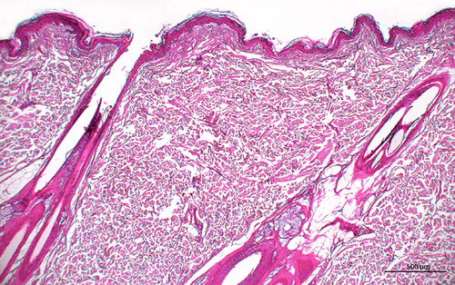
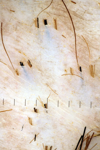
In normal Andean bear skin, the stratum corneum and the epidermis were thin (three to four cell layers thick), except in sites of friction such as the rump or the interdigital spaces, where orthokeratotic hyperkeratosis and mild acanthosis were observed. In the dermis, collagen bundles were well organized and perpendicular to each other (Figure 1), with very few inflammatory cells evident. Hair follicles were compound: the central primary follicle was surrounded by several lateral secondary follicles (Figure 1). The hair follicle density was 8–12 compound hair follicles per cm² on the lateral aspect of the chest and 18–22 compound hair follicles per cm² on the lateral aspect of the thigh and flank. Each compound hair follicle had three to seven hairs, giving a hair density of 40–85 hairs per cm² on the chest and 60–150 hairs per cm² on the thigh and the flank (Figure 2). Hair follicles were widely spaced (mean measured distance between hair follicles of 1,500 μm) (Figure 1). Sebaceous glands were well developed, whereas sweat glands were small. The dermal and follicular features were similar all over the body.
Alopecic syndrome
Signalment
The initial signs of alopecia developed in bears between two and 25 years of age. The average age of onset was 13 years (SD 6). All affected bears had enclosure mates and six of them lived with unaffected bears. A Pearson's chi-square test comparing the alopecic group to the European reference population (χ2 = 6.231, P < 0.05) demonstrated that female bears were predisposed.
Clinical features
The initial clinical signs (data available for 11 bears) included thinning of the hair coat on the flanks (n = 7), the caudal body (n = 2), the middle of the back (n = 1) or the caudal part of the abdomen (n = 1). Bilateral symmetric alopecia was an initial feature in eight bears and developed in four additional cases. Pruritus was reported in seven bears at the onset of alopecia and developed in an additional five cases.
Alopecia was generally extensive (n = 10), although seasonal fluctuations were described in seven bears and spontaneous recovery was noted for two others. For one of these bears, alopecia associated with pruritus recurred four years later in a more advanced stage. Alopecia did not recur for the second bear, but seasonal pruritus in the absence of alopecia occurred each summer for two years following the alopecic episode.
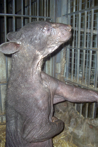
In advanced cases, diffuse hyperpigmentation with patches of depigmentation and lichenification were observed (Figure 3). Four bears progressed to complete alopecia (Stage E). General health was unaffected in the initial stages; in the long term in severely affected individuals there was a lack of appetite and decreased activity secondary to severe pruritus, and secondary microbial infections were observed that were associated with loss of body condition.
Preliminary diagnostic investigations
Physical and dermatological examinations were performed before the start of this study and were available for eight alopecic bears. A complete blood count (n = 4) revealed a mild eosinophilia for two bears. Serum biochemistry analysis (n = 3) revealed a moderate increase of aspartate aminotransferase, alanine aminotransferase and total bilirubin in one bear after several corticosteroid treatments. Total thyroxine (n = 1) was within the normal species reference range. Urinalysis (n = 1) did not reveal any abnormal findings. Urogenital ultrasound examination (n = 2) revealed cystic uterine structures attributed to age or long-term contraception in one female, and pyometra in another severely affected female.
Skin scrapings (n = 5), bacterial and dermatophyte cultures (n = 3) and trichograms (n = 3) were negative or unremarkable. Cytological evaluation by local veterinarians using skin impression smears (n = 3) were unremarkable; however, bacteria and Malassezia were observed in two of these bears several months later.
Therapeutic trials
There was no improvement with a variety of therapeutic interventions, summarized in Table 1.
| Treatment administered | Case numbers | Clinical response |
|---|---|---|
| Antibiotics | ||
| Cefalexin | 2 | None |
| Amoxicillin – clavulanic acid | 1, 3, 4, 9 | |
| Doxycycline (5 mg/kg p.o. once daily, 15 days) | 5 | |
| Tetracycline | 5 | |
| Ampicillin (15 mg/kg p.o. twice daily, 15 days) | 5 | |
| Clindamycin (7.5 mg/kg p/o/ twice daily, three weeks) | 5 | |
| Antiparasitic drugs | ||
| Moxidectin | 5 | None |
| Ivermectin | 1, 2, 3, 4, 6 | |
| Antifungal drugs | ||
| Ketoconazole (3 weeks) | 5 | None |
| Griseofulvin (2 weeks) | 5 | |
| Enilconazole | 3, 4, 5 | |
| Terbinafine (p.o. once daily, 8 weeks) | 7 | |
| Glucocorticoids | ||
| Prednisolone (p.o. once daily, 2–4 weeks) | 1, 2, 3, 4, 6, 9 |
Slight decrease of pruritus (Case 3) No effect on alopecia |
| Prednisolone (11 weeks) + oxatomide (8 weeks) | 3, 4 |
Resolution of pruritus No effect on alopecia |
| Flumethasone (once daily, 15 days) | 5 | None |
| Triamcinolone (every 3 months, 12 years) | 5 | |
| Anti-histamine drugs | ||
| Chlorpheniramine | 1, 7, 8 | None |
| Clemastine (3 weeks) | 5 | |
| Oxatomide | 1 | |
| Oxatomide (8 weeks) + prednisolone (11 weeks) | 3, 4 |
Resolution of pruritus No effect on alopecia |
| Hormones | ||
| Aglepristone | 9 | None |
| Deslorelin (4.7 mg) | 10 | |
| Megestrol acetate (3 weeks) | 3, 4 | |
| Metenolone acetate (once) | 5 | |
| Food additives | 2, 3, 4, 5, 6, 7, 8, 11 | None |
- p.o. per os.
Cytological features
In our study, direct cytological examination by the authors of tape strips revealed rare micro-organisms in healthy bears. In the alopecic bears, numerous bacteria – mainly cocci – and Malassezia yeasts were observed.
Histopathological features of the alopecic bears
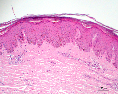
Mild to severe orthokeratotic hyperkeratosis was described in 123 of 144 biopsies. Mild to severe acanthosis, with epidermal ridge formation, was observed in 127 of 144 samples (Figure 4). The changes were extremely severe in bears that were entirely alopecic. Epidermal spongiosis was present in 92 of 144 samples, and exocytosis of lymphocytes associated with vacuolar degeneration of individual keratinocytes of basal and suprabasal layers of the epidermis was observed in 107 of 144 biopsies. Pustules, colonies of cocci in crusts and Malassezia yeasts were present on the skin surface and were particularly abundant in the six most severely affected bears.
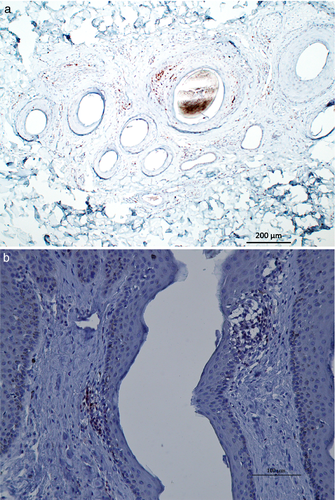
The main lesion observed in the dermis was a perivascular to interstitial inflammatory infiltrate, present in 130 of 144 samples. The intensity was highly variable. This infiltrate was composed primarily of lymphocytes, with some plasma cells and histiocytes. Eosinophil populations were highly variable and were more abundant and predominant in the more severely affected bears.
There was a mild to moderate perifollicular inflammatory infiltrate, composed primarily of lymphocytes, observed in 115 of 144 samples. Exocytosis of lymphocytes with vacuolar degeneration of individual keratinocytes of the outer root sheath at the level of the isthmus or below was seen in 97 of 144 biopsies. CD3 staining indicated that T lymphocytes were present (Figure 5).
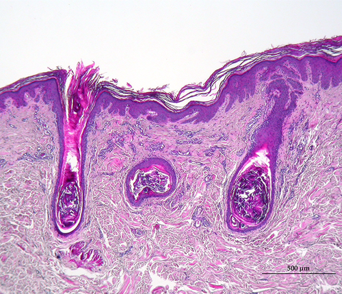
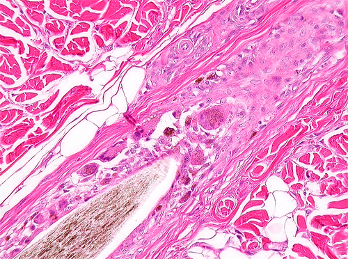
Follicular lesions included moderate to severe follicular atrophy at the level or below the isthmus in 97 of 144 samples. Thickening of the fibrous sheaths was observed in 87 of 144 biopsies. Dystrophic hair fragments or follicular plugging dilating the infundibulum in atrophic hair follicles was observed in 86 of 144 samples (Figure 6). In two severely affected bears, follicular comedones were evident. Multinucleated giant cells were seen in the outer root sheath in five animals (Figure 7) at various clinical stages: four were Stage E and one was Stage B. Immunohistochemical investigation, using AE1/AE3 cytokeratin antibodies, demonstrated that the multinucleated giant cells were not of epithelial origin. Orphan sebaceous glands, devoid of any region of the hair follicle, were present in 46 of 144 samples, and orphan sweat glands were seen in 20 of 144 biopsies and attributed to follicular atrophy.
Some of the histopathological features described above were found in five of 16 skin samples from macroscopically healthy areas. Mild orthokeratotic hyperkeratosis and acanthosis were observed in four biopsies. Mild epidermal spongiosis was described in one sample and epidermal exocytosis of lymphocytes was observed in two samples. A mild perivascular mononuclear cell infiltrate was observed in five samples. In hair follicles there was local exocytosis of lymphocytes and thickening of fibrous sheaths observed in two biopsies. None of the 16 skin samples from healthy areas showed signs of follicular atrophy.
Discussion
Previous studies of alopecic Andean bears have evaluated clinical and epidemiological aspects8; those that have performed histological evaluation have done so only in end-stage affected females.9 Our study has combined both clinical and histological data from affected bears. In our study, female Andean bears were significantly more affected than males, consistent with previous reports.8 Six of 13 alopecic bears lived with non-alopecic congeners, suggesting that the condition is not contagious.
In the healthy Andean bear the stratum corneum and epidermis are thin, similar to domestic carnivores.11 Hair follicles are less abundant and compound follicles contain less hair, leading to a lower hair density when compared to dogs. In the dog, follicular density is 100–600 hair follicles per cm², each compound follicle has two to 15 hairs, and hair density is 1,500–4,000 hairs per cm².12 Consequently, skin biopsy samples collected with small biopsy punches from bears could contain insufficient hair follicles and be nondiagnostic. Thus, the authors recommend the use of a large skin biopsy punch when collecting skin biopsy samples.
The clinical features and histopathological evaluation of skin biopsies from affected Andean bears enables several causes of alopecia to be ruled out. A widespread, diffuse alopecia is not consistent with the clinical presentation for localized alopecic skin diseases such as dermatophytosis, demodicosis or bacterial pyoderma. Demodicosis has not been described previously in Andean bears and multiple skin scrapings were negative for Demodex mites. Other parasitic infestations or dermatophytosis would seem unlikely due to the lack of contagion between housed bears.
Traumatic alopecia as a result of allergic pruritus seems unlikely. Despite the presence of eosinophils in the infiltrate, the histological features of the affected Andean bears were not considered consistent with an allergic aetiology; a lymphocytic mural folliculitis and follicular atrophy are not features of allergic skin disease, at least in dogs and cats, and the alopecia did not improve with the administration of antihistamines and corticosteroids.12
A hormonal cause of alopecia also seems improbable. Affected bears were systemically well and spontaneous resolution, as well as fluctuation of alopecia, was described. These features are not consistent with hormonal imbalance such as hypothyroidism, Cushing's disease or sex hormone imbalance. Furthermore, the histological features were not consistent with a hormonal aetiology.13 A neoplastic or paraneoplastic aetiology can also be ruled out due to a lack of systemic disease and inconsistent histological features.
The clinical and histopathological features of the alopecic bears may be most consistent with an immune-mediated pathogenesis. It is proposed that the primary mechanism of disease is a lymphocytic attack directed against keratinocytes of the basal and suprabasal layers of the outer root sheath and/or the epidermis, leading to vacuolar degeneration and follicular atrophy at the level or below the isthmus. Multinucleated giant cells are part of the inflammatory response. Secondary bacterial and/or yeast infections may increase lymphocyte recruitment and eosinophil infiltration, leading to pruritus.14 Furthermore, alopecic bears may be vulnerable to insect bites which could further explain the eosinophil infiltrates.
In domestic carnivores, cutaneous lupus erythematosus, alopecia areata or pseudopelade may result in clinical signs and histological features similar to those described for the alopecic bears.12 Immune-mediated skin diseases have not previously been described in Ursidae. If the alopecic syndrome is immune-mediated then the trigger factors are unknown. It is possible that nutritional and social stress factors may be involved, but this remains unproven. In our study, all alopecic bears had shared or were sharing their enclosure with at least one other bear. Andean bears are normally, in the wild, solitary animals and it is plausible that chronic social stress could be a potential factor contributing to the alopecia; further studies are indicated to clarify this hypothesis. The reproductive status and genetic background of affected bears need to be explored as potential contributing factors to any putative immune-mediated syndrome.15
Acknowledgments
The authors would like to thank Lydia Költer, Alberto Barbon, Nadine Bechstein, Gabby Drake, Almuth Einspanier, Klaus Eulenberger, François Huyghe, Sandra Langguth, William Magnone, Eva Martinez Nevado and Martina Schachtner, members of the European working group on the Andean bear alopecia syndrome, for their long-term personal involvement in trying to understand this disease and their help in gathering data for the present study. The authors are very grateful to their colleagues Guillaume Douay, Eva Gruenz, Maëlle Guezenec, Jean-Michel Hatt, Kathrin Jaeger, Tobias Knauf, Dorothée Ordonneau, Harald Schwammer, Marie Simon, Francis Vercammen and Chris Wenker for their contribution to this study by sending skin biopsies from their healthy or alopecic bears and by returning clinical surveys. Monika Welle is acknowledged for her insight on these cases at the beginning of the study. The authors thank Guillaume Douay and the staff of Lyon Zoo for their active participation in the determination of follicular density in healthy bears. Finally, the authors would like to thank the AFVPZ (Association Francophone des Vétérinaires de Parcs Zoologiques) for supporting this work through a DVM thesis grant.


