What is your diagnosis? Glossal imprint from a yellow-footed tortoise (Chelonoidis denticulata)
1 CASE PRESENTATION
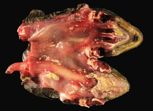
A 5-year-old, male, yellow-footed tortoise (Chelonoidis denticulata) was presented to the Aquatic, Amphibian, and Reptile Pathology Service at the University of Florida for postmortem examination after being found deceased following a 2-day history of lethargy and decreased appetite. Postmortem examination identified numerous, frequently coalescing, approx. 1-6 mm diameter areas of mucosal ulceration overlain by thick, yellow, caseous debris within the oral cavity, choana, and proximal esophagus (Figure 1). Touch impressions of the surface of the tongue were collected and stained with a Wright-Giemsa stain for routine cytologic examination (Figure 2). No significant gross lesions were detected in the other examined organs.
Cytologic interpretation:
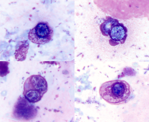
Mild heterophilic, histiocytic, and lymphoplasmacytic glossitis and epithelial cell hyperplasia with the presence of occasional intranuclear inclusions suggestive of an infectious origin (presumptive coccidial infection)
Tissue imprint preparations of the glossal lesion were moderately cellular, adequately preserved, with erythrocytes on a medium basophilic background containing abundant extracellular bacilli and few diplococci, consistent with oral bacterial microflora. Inflammatory cells appeared mildly increased with degranulated heterophils, histiocytes, small well-differentiated lymphocytes, and rare plasma cells dispersed throughout the preparation. Admixed within inflammatory cells were low numbers of hyperplastic epithelial cells. Occasionally, within the free nuclei of lysed epithelial cells, were distinct, approx. 5-10 μm diameter, round to oval, granular, variably refractile, pale basophilic intranuclear inclusions suggestive of coccidial gametes.
2 ADDITIONAL RESULTS
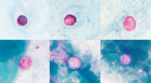
On cytology, the intranuclear inclusions exhibited discrete, consistently positive acid-fast staining with Ziehl-Neelsen staining (Figure 3). The majority of the intranuclear inclusions showed variable, pale to variably granular pink, staining with periodic acid-Schiff (PAS) (Figure 3).
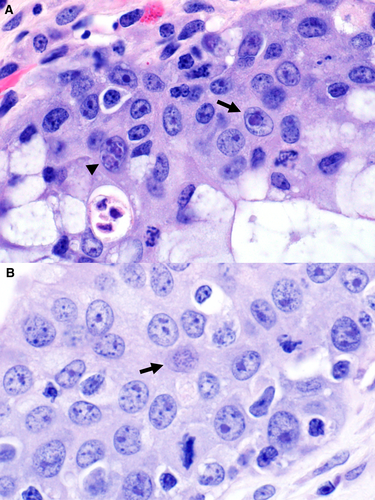
Within representative histologic sections of the tongue, there was severe lymphohistiocytic and heterophilic, necrotizing glossitis with mucosal hyperplasia and hyperkeratosis. Occasionally, within the nucleus of epithelial cells, there were one to two spherical, 5-10 µm diameter gametes with lightly basophilic cytoplasm and a small, slightly eccentric nucleus (Figure 4A). With less frequency within epithelial cell nuclei, there were low numbers of spherical, approx. 6-10 µm diameter, meronts containing several, sometimes >10, basophilic merozoites (Figure 4B). Ziehl-Neelsen and PAS staining of glossal histological lesions did not highlight any acid-fast or PAS-positive intranuclear organisms, respectively. This disparity in positive staining by cytology and lack of staining in tissue sections is unknown, but is consistent with previous reports and should be considered when attempting special stains for this organism.1, 2 Additionally, microscopic examination identified severe stomatitis, esophagitis, and severe pneumonia associated with the presence of intranuclear coccidia. There was also extensive meningitis and mild rhinitis and tracheitis. Although the organisms were not apparent in these tissues on histopathology, given the similar patterns of inflammation and consistency with previously reported findings in other chelonian species, these lesions were still presumed to be associated with coccidial infections.1-4
An oral/choanal swab was positive by 18S rRNA qPCR for intranuclear coccidia of Testudines (TINC), while ranavirus and herpesvirus PCR results were negative.4
3 DISCUSSION
Intranuclear coccidiosis of TINC is an important emerging infectious agent of tortoises and turtles that causes systemic disease with high mortality in several threatened and endangered species.1, 3 Over 30 different species of coccidia have been identified in chelonians; however, only a few, including TINC, have been reported to cause significant lesions and potentially result in mortality.4 Currently, TINC has not been assigned to a genus, and complete phylogenetic characterization has not been reported in the literature to date. It is known that the parasite infects epithelial cells, which can cause multisystemic disease associated with lymphocytic or mixed inflammation with variable degrees of tissue necrosis and epithelial cell hyperplasia, most commonly throughout the digestive and respiratory tracts.1, 3 Given the unique intranuclear localization, distribution across various types of epithelia, and distinct morphology as round to oval organisms with granular, pale basophilic cytoplasm and a variable refractile appearance, no other coccidian organisms would be considered as differential diagnoses. In contrast to gametes of TINC, classically recognized coccidian species of chelonians, that is, Eimeria sp., are often morphologically larger (>10 μm diameter), infect specific target organs of the host, and produce life stages that are located either within the cytoplasm of host cells as distinctly recognizable intracytoplasmic sexual stages (microgametes and macrogametes) or extracellularly as oocysts.5 Oral transmission of TINC via sporulated oocysts has been recently confirmed in five Old World tortoise species with subsequent development of endogenous sexual stages (e.g., gametes) following experimental exposure.6 In this tortoise, the distribution of lesions primarily in the oral cavity and respiratory tract was interesting, as there was apparent sparing of tissues classically involved with intranuclear coccidial infections, such as the intestine and pancreas; however, similar respiratory-centric lesions have been reported in other New World tortoise species, specifically red-footed tortoises (Chelonoidis carbonaria).1, 3 TINC has predominantly been documented in Old World species, and only a few reports of New World species with complete histologic examinations exist at this time.1, 3 Despite this, TINC should be a differential diagnosis for respiratory disease and oral lesions in chelonians, especially as the understanding of the known host range continues to evolve. Other differential diagnoses based on clinical presentation and gross lesions, in this case, included herpesvirus, ranavirus, mycoplasma, and nutritional imbalances.
This report characterizes the cytologic and special staining features of TINC, an infectious organism of emerging concern, with a concurrent histologic correlation. There has been very limited information on anticipated cytologic findings of TINC, and tissue imprints have proven to be of great diagnostic benefit as an adjunct diagnostic tool in both antemortem diagnosis and at the time of postmortem examination.2 Cytologically, various TINC stages could be misinterpreted for prominent nucleoli, with this case providing an opportunity to demonstrate the distinct morphology of cytologically recognizable gametes. The recognition of distinct life stages by cytology might be more challenging than by histopathology, mainly due to variable exfoliation of cytologic specimens. In the authors' experience, cytology is most useful in the identification of gametes allowing for the cytodiagnosis of TINC; however, their absence in both cytologic preparations and tissue sections does not exclude the possibility of infection. Furthermore, consistently acid-fast positive and variable PAS staining of TINC gametes in cytologic preparations might aid in the diagnosis of TINC in clinical cases and in decision-making regarding the pursuit of molecular diagnostics.
DISCLOSURE
The authors have indicated that they have no affiliations or financial involvement with any organization or entity with a financial interest in, or in financial competition with, the subject matter or materials discussed in this article.


