What is your diagnosis? Mass in the abdomen of a cat
1 CASE PRESENTATION
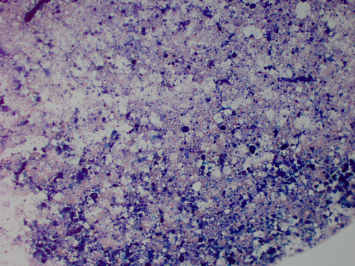
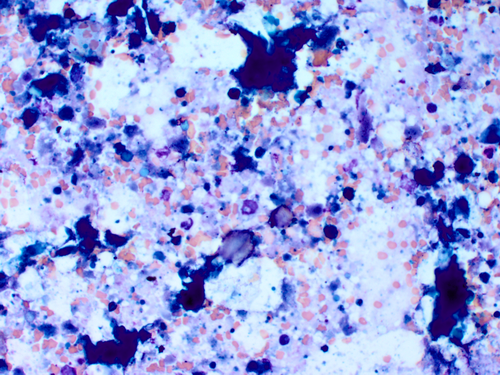
A 6-year-old spayed female domestic shorthair cat was presented to the North Carolina State Veterinary Hospital Small Animal Primary Care service for a routine annual examination. History revealed the owner had changed the patient to an over-the-counter reduced-calorie diet 9 months prior. Physical examination revealed weight loss from 6.87 kg (body condition score [BCS] 8/9) to 5.73 kg (BCS 6/9) since the previous visit 9 months before. Two round, movable, firm abdominal masses were also found. A CBC and a chemistry panel were performed. The CBC was within normal limits. The chemistry panel revealed elevations in ALT (125 IU/L, RI: 22-105 IU/L) and AST (140 IU/L, RI: 12-44 IU/L). Abdominal radiography and ultrasound were performed and confirmed the presence of two masses. A fine-needle aspirate from one of the two masses was collected and prepared for cytologic evaluation. (Figures 1 and 2).
Cytologic interpretation:
Pyogranulomatous inflammation with necrosis and probable mineral deposition
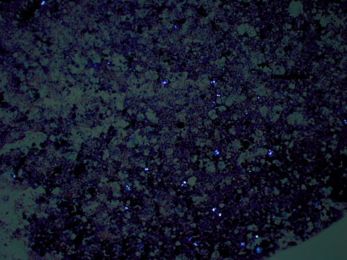
The cellularity was adequate on a basophilic background with blue cellular debris, degenerate cells, and amorphous material (likely necrotic cells). Few RBCs can be seen. The majority of intact cells were scattered nondegenerate to mildly degenerate neutrophils and macrophages often containing phagocytized cellular debris. Scattered refractile clear to slightly blue to green material that could be polarized was also present (Figure 3). No evidence of overt neoplasia or etiologic agents were identified.
2 ADDITIONAL DIAGNOSTICS
Radiographic interpretation
Round mixed mineral and fat opaque mass-like structures within the right mid-abdomen, most consistent with Bates bodies. (Figure 4).
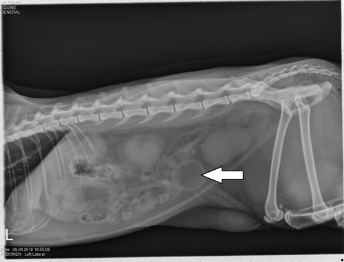
Final diagnosis: Nodular fat necrosis or Bates Bodies.
3 DISCUSSION
Nodular fat necrosis, also known as Bates bodies or Bates floaters, present as focal, benign masses located in the abdominal fat of feline and canine patients. Typically, these lesions present as singular or multiple, firm, nodular masses. Each mass varies in size from approximately 5 to 40 mm.1 These lesions are usually incidental findings; however, one report documented a case of acute abdomen in a feline patient due to pedunculation and detachment of the nodular fat necrosis.2 In addition, it is important to differentiate these masses from neoplasia.
Nodular fat necrosis is typically diagnosed by radiology, ultrasound, and/or histologic evaluation. On histologic examination, these lesions usually represent areas of necrotic fat tissue surrounded by a fibrous or mineralized capsule.1 In some cases, saponification, dystrophic calcification, and cholesterol crystallization can be present.1, 2 Lesions can also have varying levels of inflammatory cells, as well as hemorrhage.1 Necrosis of adipose tissue has a variety of causes; however, the exact pathogenesis of nodular fat necrosis is poorly understood.3 These lesions are most common in older, obese feline patients. It has been theorized that pressure ischemia due to excessive body fat is a potential factor.1, 3 Hydrolysis of lipids due to metabolism is another possible cause.1 In this case, the patient had a history of obesity and rapid weight loss over 9 months, which provides evidence for both theories.
To the best of the author's knowledge, this article is the first publication to discuss cytologic findings of nodular fat necrosis. As discussed above, surgical biopsy is a commonly used diagnostic approach. However, most cases of nodular fat necrosis occur in older, obese patients. Surgical removal might not be ideal in these cases. Therefore, ultrasound-guided aspirates of the mass or masses in question could be a safer alternative to surgical biopsy when nodular fat necrosis is suspected.
DISCLOSURE
The authors have indicated that they have no affiliations or financial involvement with any organization or entity with a financial interest in, or in financial competition with, the subject matter or materials discussed in this article.
ACKNOWLEDGMENTS
We thank the NC State Veterinary Hospital Small Animal Primary Care service and the NC State Veterinary Clinical Pathology Laboratory for collecting and preparing the samples, and the NC State Veterinary Diagnostic Imaging service for evaluating radiographic and ultrasonographic images.


