RETRACTED: Evaluating the results of eyebrow lift by combining methods of subcutaneous flap and thread support in patients with droopy eyebrows
Abstract
Background
Brow lift also known as eyebrow lift was first described in 1919, and since then, many changes have been made in the methods of doing it, although there is still no agreed method of absolute superiority for eyebrow lift. Most previous studies have reported the results generally qualitatively and based on patient or surgeon satisfaction. In this study, by combining two less complicated methods of eyebrow lift, we have evaluated the quantitative results.
Method
Before the surgery, a standard photograph of the face was taken. The vertical distance between the tail of the eyebrow and interpupillary line was determined.
Results
This study was performed on 15 females with a mean age of 38.27 ± 6.82 years. The mean distance between the eyebrow and interpupillary line by photographic measurement before surgery, 3 weeks, and 6 months after surgery was, respectively, 10.45 ± 1.74, 15.72 ± 1.77, and 13.53 ± 1.69 mm using the tail of the eyebrow and 18.47 ± 1.67, 23.33 ± 1.57, and 21.55 ± 1.66 mm using the crown of the eyebrow. In the clinical measurement, the eyebrow tail was 11.98 ± 1.75, 19.22 ± 1.73, 17.35 ± 1.68 and 15.13 ± 1.76 mm away from the pupil line, and the crown of eyebrow was 20.45 ± 1.90, 27.12 ± 1.58, 25.00 ± 1.80, and 23.35±1.78 mm. There is a significant difference between the distance of the tail of the eyebrow and the crown of the eyebrow in both measurement methods (photographic and clinical) at different times (p-value <0.001).
Conclusion
Performing eyebrow lift with the Pretrichial method has many comparative advantages to other methods. Additionally, eyebrow lift with the thread support is a less invasive method.
1 INTRODUCTION
Eyebrow lifts were first described by Passot in 1919 and since then, many changes have been made to the methods of eyebrow lifts and newer methods are associated with fewer incisions and complications. While these methods are more effective, there is still no method with absolute superiority for eyebrow lift agreed by experts.1 There are various techniques for raising the height (lift) of the forehead and eyebrows. Current methods include direct browlift, internal browpexy, Midforehead lift, Pretrichial browlift, endoscopic browlift, coronal browlift, and Temporal Browlift.2, 3
More recently, less invasive suture techniques have been used that can be lifted precisely with sutures on any area of the eyebrow with small incisions above the growth line. Although this method can be done with high mastery and can have significant initial results, the durability of the results is not clear. This method is more indicated in patients who want small surgery.4
Another method of lift is the peritrachial method, which is mostly performed in patients with drooping eyebrows and wrinkles on the forehead at a low to moderate level and can be performed simultaneously with blepharoplasty. Advantages of this method include being less aggressive, providing a proper dissection plan, lowering the risk of supraorbital nerves and arteries damage, alopecia, and occult scars.5
In each of the different eyebrow lift methods, we face advantages and disadvantages. In general, the main complications of different methods are: alopecia, forehead anesthesia, hematoma, skin necrosis, receding hairline, visible scars, and low durability of eyebrow lift results.6-9
None of the lift methods alone can have lasting and uncomplicated results and can be considered as a definitive method. In most previous studies, the results are generally qualitative and based on patient or surgeon satisfaction. Performing this lift operation with outpatient surgery has been used in fewer studies and less has been reported.10 In patients with low to moderate eyebrow drooping, results have been acceptable. Therefore, we intend to combine the two less complicated methods of eyebrow lift with subcutaneous flap at the border of the hairline (Pretrichial) and the thread lift method to support the amount of displacement. Evaluation of quantitative results was done in three-period times including before, 3 weeks, and 6 months after eyebrow lift (even in patients with severe drooping eyebrows) and its persistence with statistical analysis. Introduction of this method as a recombinant method with high capability in lateral eyebrow lift in patients with different intensities of drooping eyebrows and with lasting results can help maintain the benefits of the main method and improve the quantitative and qualitative results.
2 MATERIAL AND METHOD
In this project, diagnosis was performed in collaboration with the specialized clinics of Shariati hospitals and Tehran School of Dentistry. After providing the necessary explanations about the surgical and follow-up procedures and obtaining written consent, 15 patients with drooping eyebrows whom were candidates for this study were selected. According to a 2007 article by Mavrikakis et al, a standard deviation of 1.25 mm was considered. For a test of a difference of 2.7 mm with changes in the overhead over time and assuming a 50% return, lift (SM = 1.35) using PASS 11 software and ANOVA menu with a minimum sample size of 10 samples for the first type error was calculated at 0.50 and statistical power above 80%. Assuming a fall of 30% of the samples, the final volume of 15 people was considered. To compare the amount of eyebrow lift changes in three time periods; immediately before and after surgery, 3 weeks, and 6 months after surgery were considered. Male or female patients with a drooping corner of the eyebrow are operated on at the discretion of the maxillofacial surgeon and patients in the age range of 20–70 years were included in the study. Patients with a history of eyebrow surgery or other previous cosmetic procedures on the forehead or eyebrows, patients who did not want to participate in the relevant follow-up, patients who had contraindications to surgery due to systemic medical problems, and patients who were simultaneous candidates for blepharoplasty and eyebrow lift were all excluded from the study.
2.1 Interventions and surgical process
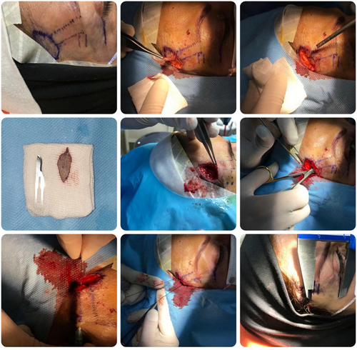
Patients underwent outpatient surgery under local anesthesia after consultations and necessary tests before surgery. An oval incision line was designed by a surgical marker in the area of the hair growth line (Figure 1A). Horizontal lines were drawn the lateral canthus of the eye and tail of eyebrow (end of the eyebrow) and the crown of the eyebrow. The measurement of the vertical distance of the horizontal line to the perimeter of the canthus to the tail of the eyebrow and the crown of the eyebrow was performed with a high-precision digital gauge on each eyebrow and recorded.

Prophylactic use of a 5 mg diazepam tablet for sedation (in anxious patients) and a 2 g cephalexin capsule was performed 1 h before surgery. First, the eyebrows and forehead were Prepped and draped to the skin incision area, the whole forehead area, and some of the upper eyelids. Local anesthesia with 2% lidocaine containing epinephrine 1.100000 was used in the corners of the eyebrows and above the eyebrows up to the skin incision line (at the site of hair growth). If necessary, some of the hair in the area of the skin incision line was trimmed and shortened with a razor blade No. 15 for better visibility to design the flap. The skin and subcutaneous tissue in the area of the incision line were removed in an oval shape with a razor blade No. 15, proper homeostasis was performed with a surgical catheter, and a pressure pack was performed with sterile gauze (Figure 1B–D). Subcutaneous flap dissection was performed through oval incisions around the hairline (depending on the case) and perpendicular to the projected vector to move the corner of the eyebrow with scissors. Dissection was performed in the subcutaneous plane and on the frontalis muscle up to the line of eyebrow hair growth while maintaining the appropriate thickness of the skin and subcutaneous tissue to prevent excessive thinning of the skin and possible complications such as skin necrosis (Figure 1E,F). If necessary, after controlling the bleeding with vascular cauterization, the appropriate amount and location of the eyebrow corner are determined and, using an angiocatheter or needle under the flap and its exit, attached to the eyebrow border at the highest point to establish the new position of the eyebrow corner. Then, 3/0 sutures were passed and in a return path, both ends of the thread were sutured by taking a deep tissue in the upper part of the skin incision to remove the skin tension under the flap. If necessary, depending on the size of the raised eyebrow and its displacement, this suspension process was repeated. two nylon threads of three zeros in two vectors predicted for lifting were used. Furthermore, the skin tissue, in addition to imitating the initial incision, was removed if necessary, and the initial incision was closed with four nylon suture threads (Figure 1G,H). After surgery, the forehead and eyebrows were cleaned, and vertical measurements of the distance between the lines to the lateral canthus and eyebrow crown were performed and recorded by a digital gauge, which indicates the amount of lift. Finally, the skin incisions were dressed with 3% tetracycline. After ensuring the stability of the patient's vital signs, the patient was discharged with oral antibiotics, analgesics, necessary recommendations regarding the use of pressure dressings and cold compresses, and the usual instructions after surgery. The skin sutures were removed 7 days later.
Evaluation of results is done by two methods of analysis; standard patient photographs and clinical evaluation. In standard photography, a graph of the patient's face was taken in a vertical sitting position facing up. The vertical distance from the corner of the eyebrow or the same point of the patient's eyebrow tends to be raised, in two parts of the eyebrow crown, the eyebrow ends in each eyebrow were determined by the digital gauge in millimeters from the line passing through the center of the pupils in millimeters. The amount of eyebrow lift was measured 3 weeks and 6 months after surgery by taking standard photographs in natural head position. In clinical evaluation, horizontal lines were drawn on the periphery and along the lateral canthus of the eye, tail, eyebrow, and crown of the eyebrow. The vertical distance of these lines was measured with precision gauge before and after surgery, 3 weeks, and 6 months after surgery.
(Figure 1I). Changes in the vertical distance of the lift area in the mentioned periods were used with both photographic and clinical evaluation to evaluate the adequacy and efficiency of the combined eyebrow lift method in order to change the results over time over time to extract descriptive results. After collecting the data, it was entered into SPSS 25. Frequency and percentage of frequency were used to describe qualitative variables and mean and standard deviation were used to describe quantitative variables in case of normal distribution, otherwise the mean and distance between quartiles were used. Generalized estimating equations (GEE) test was used to analyze the data and compare preoperative measurements with the values measured in the follow-up. In these tests, the differences in measurements at different times were compared (p-value ˂0.05).
Limitations of the project were: limitations due to COVID-19 disease, patients' lack of cooperation to evaluate the results at the exact time of the Follow-up, and age and gender variations in the eyebrow area and forehead length of the participants.
3 RESULTS
This was study was performed on 15 females with a mean age of 38.27 ± 6.82 years, minimum age of 31, and a maximum age of 54 years.
3.1 Photographic examination of the eyebrow tail
The distance from the tail of the eyebrow to the interpapillary line measured by photographic method for the left eyebrow before, 3 weeks after, and 6 months after surgery are 10.23 ± 1.79, 15.47 ± 1.82, and 13.33 ± 1.87 mm, respectively. The right eyebrow is 10.67 ± 1.95, 15.97 ± 1.88, and 13.73 ± 1.80 mm. The results of the GEE statistical test show that the side (left or right) of the eyebrow has a statistically significant relationship with the distance between the tail of the eyebrow and the interpapillary line in the photographic method (p-value = 0.006) (Table 1). Also, there was a statistically significant relationship between the time (before, 3 weeks, or 6 months after surgery) and the distance between the eyebrow tail and the interpapillary line in the photographic method (p-value<0.001). Patient age was not significantly associated with this distance (p-value = 0.281). By removing the effect of age as an accompanying variable, the mean distance between the eyebrow tail and the interpapillary line is shown in the photographic method for three times (before, 3 weeks, and 6 months after surgery). Due to the significant time-size relationship and extent of this distance, a post hoc test was used to compare pairs of different times, and this time was significantly different from other times (p-value <0.001) (Tables 2 and 3).
| 95% Wald confidence interval | |||||
|---|---|---|---|---|---|
| p-Value | Upper | Lower | Std. error | Mean | Eyebrow side |
| 0.006 | 13.8222 | 12.2000 | 0.41381 | 13.0111 | Left |
| 14.2861 | 12.6250 | 0.42377 | 13.4556 | Right | |
| 95% Wald confidence interval | |||||
|---|---|---|---|---|---|
| p-Value | Upper | Lower | Std. error | Mean | Measurement time |
| <0.001 | 11.32 | 9.58 | 0.44321 | 10.45 | Before surgery |
| 16.60 | 14.83 | 0.45128 | 15.72 | 3 weeks after surgery | |
| 14.38 | 12.69 | 0.43250 | 13.53 | 6 months after surgery | |
| 95% Wald confidence interval for difference | ||||||
|---|---|---|---|---|---|---|
| Upper | Lower | p-Value | Std. error | Mean difference1, 2 | Measurement time 2 | Measurement time 1 |
| −4.60 | −5.93 | <0.001 | 0.34032 | −5.27 | 3 weeks after surgery | Before surgery |
| −2.60 | −3.56 | <0.001 | 0.24457 | −3.08 | 6 months after surgery | |
| 2.69 | 1.68 | <0.001 | 0.25762 | 2.18 | 6 months after surgery | 3 weeks after surgery |
3.2 Photographic examination of the eyebrow crown
This distance is 18.37 ± 1.85, 23.03 ± 1.83, and 21.43 ± 1.66 mm for the left eyebrow at the three times measured and 18.57 ± 1.56, 23.63 ± 1.76, and 21.67 ± 1.85 mm for the right eyebrow, respectively (Figure 2). The results of the GEE statistical test show that the measurement time has a statistically significant relationship with the distance between the crown of the eyebrow and the interpapillary line in the photographic method (p-value <0.001) (Table 4) while the side (left or right) of the eyebrows (p-value = 0.065) and the patient's age (p-value = 0.624) were not significantly associated with this distance. By removing the effect of age as an accompanying variable, the average distance between the crown of the eyebrow and the interpapillary line in the photographic method photographic method for the right and left eyebrow is 20.94 mm and 21.29 mm, respectively. Due to the significant relationship between measurement time and the amount of this distance, a post hoc test was used to compare pairs of different times, which at this time was significantly different from other times (p-value <0.001) (Tables S1 and S2).

| 95% Wald confidence interval | |||||
|---|---|---|---|---|---|
| p-Value | Upper | Lower | Std. error | Mean | Eyebrow side |
| 0.065 | 21.6403 | 20.2486 | 0.35505 | 20.9444 | left |
| 21.9842 | 20.5936 | 0.35477 | 21.2889 | right | |
3.3 Clinical examination of eyebrow tail
Clinically measured distance from eyebrow tail to interpapillary line for left eyebrow at previous, immediately after, 3 weeks, and 6 months after surgery visits were: 11.77 ± 1.76, 19.20 ± 1.71, 17.10 ± 1.84, and 14.97 ± 1.82 mm and for the right eyebrow: 12.20 ± 2.01, 19.23 ± 1.96, 17.60 ± 1.72, and 15.30. 1.97 mm. There was a statistically significant relationship between eyebrow tail and interpapillary line in the clinical method, while the patient's age was not significantly associated with this distance (p-value = 0.253) (Table S3).
Eliminating the effect of age as an accompanying variable, the average distance from the tail of the eyebrow to the interpapillary line in the clinical method is 15.76 mm for the left eyebrow and 16.08 mm for the right eyebrow. Due to the significant relationship between measurement time and the amount of this distance, a post hoc test was used to compare pairs of different times, which at this time was significantly different from other times (p-value <0.001) (Table S4 and S5).
3.4 Clinical examination of eyebrow crown
This distance, for the left eyebrow, measured before, immediately after, 3 weeks, and 6 months after surgery was: 20.10 ± 2.03, 27.00 ± 1.78, 24.73 ± 2.02, and 23.20 ± 1.77 mm, respectively, and for the right eyebrow: 20.80 ± 1.81, 27.23 ± 1.67, 25.27 ± 2.03, and 23.50 ± 1.94 mm. The results of the GEE statistical test showed the side (left or right) of the eyebrow (p-value <0.001) and the time measurement (before, immediately after, 3 weeks, and 6 months after surgery) (p-value <0.001) with the amount the distance between the crown of the eyebrow and the interpapillary line in the clinical method was statistically significant, while the age of the patient was not significantly related to this distance (p-value = 0.530) (Table S6).
Eliminating the effect of age as an accompanying variable, the average distance between the crown of the eyebrow and the interpapillary line in the clinical method is 23.76 mm for the left eyebrow and 24.20 mm for the right eyebrow. By removing the effect of age as a variable due to the significant relationship between measurement time and the amount of this distance, a post hoc test was used to compare pairs of different times that this time was significantly different from other times (p-value <0.001) (Table S7 and S8).
4 DISCUSSION
The present study was designed and performed to periodically evaluate the results of eyebrow lift intervention by combining two subcutaneous flap methods at the perimeter of the hairline (pretracheal) and thread support in patients with drooping eyebrow angles. The outcome of this study was the distance between the tail and the crown of the eyebrows to the line connecting the pupils and lateral canthus, which was measured three times (before, 3 weeks, and 6 months after surgery). Also, this study was conducted in two ways comprised of clinical and photographical measurements. All surgical eyebrow lift techniques have relatively common complications including alopecia, anesthesia of the operated area, infection, hematoma, skin necrosis, receding hairline, visible scar, low durability, vertical eyebrow asymmetry, and sensory and motor nerve damage.
In our study, two patients had 1–3-mm vertical asymmetry of the eyebrows in a 3-week follow-up, which were re-operated on due to the patients' insistence in which were corrected and removed. There were no cases of infection, hematoma, or skin necrosis in our study. A small number of patients complained of numbness or slight lateral pain in the forehead in the first 2–3 weeks after surgery, which resolved over time. This study did not presume motor nerve damage, and no damage to the eyebrow-raising motor nerves was observed. Regarding the surgical incision scar, since the skin incision scar was placed was placed inside and slightly behind the hairline, it was relatively, it was relatively visible in the first weeks, but gradually disappeared completely during the 6-month follow-up, and none of the patients complained. Patients did not have any surgical scars, which is one of the strengths of the surgical method of this study. Another strength of this technique is that it can be performed on an outpatient basis without the need for hospitalization and general anesthesia. It was noteworthy that a small number of patients with slight asymmetries of 1–2 mm did not have asymmetry in the 6-month follow-up study. Most patients also complained of dimples at the top of the eyebrows where the floss passes through the upper eyebrows during the first few weeks after surgery, which faded over time. In general, no significant or specific complications were observed in this surgical technique in the long run and most of the complications that did occur, were low in prevalence Were low in prevalence and within the first 3 weeks after surgery, which resolved over time, which resolved over time.
According to the results obtained, this new surgical technique can be considered as a method with both clinically desirable results and patient satisfaction due to its outpatient possibility with no need for anesthesia and very few and negligible complications.
The ideal eyebrow position has always been debated among cosmetic and plastic surgeons. In 1974, Westmore simply described the proper position of the eyebrows as follows: The inner part of the eyebrow should start from the vertical plane corresponding to the outer part of the nasal ala and the inner canthus, and the outer part of the eyebrow along the sloping line between the outermost part of the nasal ala and the outer canthus.18 The inner and outer part of the eyebrow should be almost on a horizontal plane with the highest part of the eyebrow arch in the upper part of the outer limbus. In general, the eyebrow should be above the supraorbital rim in women and along it in men.
McKinney et al. States that the minimum distance between the mid pupil and the upper part of the eyebrow should be 2.5 cm and if it is less than this, it indicates eyebrow ptosis, which means complete or partial drooping of the eyebrow.11 For about a century, eyebrow lifting surgery has been popular as a result of aging.12 There are several methods for eyebrow lift in these cases including: browpexy, direct brow lift, preitrachial brow lift, mid-forehead brow lift, coronal brow lift, thread support, etc.8 The patients evaluated in this study were all female with a mean age of 38.27 years. Although in most previous studies, almost all subjects were female, the mean age of the subjects was usually higher than in our study.
The results of this study show that both the distance between the tail of the eyebrow and the crown of the eyebrow are significantly different in both methods of examination (photographic and clinical) at different times. Although in the photographic method, the distance between the tail and the crown in the follow-up of the sixth month is significantly less than in the third week, it is still significantly higher than before the surgery. The same trend was observed in clinical measurements; at each time after surgery, we saw a significant reduction in the distance from the tail and crown to the line of pupil connection compared to the previous fall (third week compared to immediately after surgery, the sixth month compared to immediately after surgery, and 3 weeks later from surgery). In general, the amount of this gap in the follow-up of the sixth month compared to before surgery is significant, which indicates the high durability and long-term effectiveness of this recombinant method. In a study to evaluate the amount of eyebrow lift following upper eyelid blepharoplasty and pretrachial lift, the amount of lift after 3 months in lateral canthus, lateral limbus, mid-pupil, and medial limbus were, respectively, 1.85, 1.54, 1.31, and 1.07 mm.10 The results of our study show that the amount of tail and eyebrow lift after 6 months with photographic measurements were 3.08 and 3.08 mm, respectively, and in clinical measurements are 3.15 and 2.9 mm. This indicates the high durability of the combined method of the subcutaneous flap and thread support so that even in longer follow-up, the effect of this recombinant method is higher than performing peritrachial lift alone.
Vikšraitis et al.: The amount of eyebrow lift after 6 months in the endoscopic method: 2.9 mm in the outer corner of the eye, 2.5 mm in the outer part of the iris, 2.3 mm in the inner part of the iris, and 2.2 mm in the corner internal ores of the eye which reported that the rate of eyebrow lift was still higher in the method performed in our study at the same time.13
In another study, the amount of eyebrow lift by the endoscopic method after 1 year was reported to be 2.1 mm in the external canthus, 1.9 mm in the external limbus, and 2.4 mm in the internal canthus.14 In another study, the amount of eyebrow lift after 6 months by the endoscopic method was 2.81 mm in the inner canthus, 1.13 mm in the outer canthus, and 2.05 mm in the middle part of the papilla.15 Given the high popularity of endoscopic eyebrow lifts in recent years, the recombinant method used in our study is competitive in terms of effectiveness and even more effective than endoscopic lifts, while requiring less equipment.
In another study that examines the lift of the eyebrows in the internal and external browpexy, the amount of eyebrow center after four to 5 months of surgery was 2.29 and 1.47 mm in the internal browpexy and 2.97 mm and 1.90 mm in the external browpexy.16
Another study evaluated the effect of Botox injection on eyebrow lift with the average eyebrow height before injection at 15.9 mm and 2 weeks after Botox injection at 16.9 mm. Based on the results of this study, the height of the eyebrows before the intervention in the outer cantus part was 13.8 and then 18.6 mm after injection. The eyebrow lift lasted in all patients for 3–4 months. The mechanism of the effect of this method is probably due to the paralysis of eyebrow depressors followed by botulinum injection. The results of our study show a 40% return in the sixth month of the eyebrow lift rate compared to the third week and immediately after surgery.
Although studies have been conducted to investigate the effectiveness of thread help, the success of this method and the satisfaction of patients in short-term follows have been reported, but the last-term effect of lift with yarn has not been satisfactory.17, 18 However, the combination of lift with yarn and other methods of lift have more successful results than this method alone.19 The contact of the eyebrow (left or right) with the distance of the eyebrows to the line between pupils in the photographic method and its relation with the rate of the tail and an eyebrow crown to line between pupils clinically was statistically significant. But the left and right differences in all of these comparisons are less than 0.5 mm (in all measurements, the right side was slightly higher). As a result, this difference was statistically significant but clinically insignificant.
5 CONCLUSION
Performing eyebrow lift with the pretrichial method has many comparative advantages to other methods. Also, eyebrow lift with the thread support is a less invasive method. The combination of these two techniques as a recombinant method was the aim of this study in the hopes that good results will be obtained. The results of photographic measurements in this study showed that there was an increase in vertical height in the eyebrow tail area after surgery. The clinical measurement showed an increase in vertical height in the eyebrow tail area and the crown of the eyebrow. In the crown area of the eyebrow, both in clinical and photographic measurements, the return of eyebrow height within the 3 weeks after surgery was more than the eyebrow tail.
CONFLICT OF INTEREST
The authors declare that there is no conflict of interest.
Open Research
DATA AVAILABILITY STATEMENT
The data will be available on request.


