Bone Healing and Clinical Outcome Following Medial Opening-wedge High Tibial Osteotomy Using Wedge-Shaped Cancellous Allograft
Jinlun Chen, Jiahao Li and Haitao Zhang contributed equally to this work.
Grant sources: The current study received funding from High-Level Hospital Construction Project of the First Affiliated Hospital of Guangzhou University of Chinese Medicine(Grant Number 211010010722), National Natural Science Foundation of China(Grant Number 82104882) and Science Fund of Guangdong Traditional Chinese Medicine Bureau (Grant Number 20201099).
Disclosure: The authors declare that they have no conflict of interest.
Abstract
Objective
Medial opening-wedge high tibial osteotomy (MOWHTO) is considered to be an effective treatment for symptomatic knee osteoarthritis (KOA) of isolated the medial compartment with varus alignment of the lower extremity. However, the choice of material to fill the void remains controversial. This study aims to evaluate the bone union of the osteotomy gap using a novel wedge-shaped cancellous allograft after MOWHTO and its effect on clinical outcomes.
Methods
All patients who underwent MOWHTO using a novel wedge-shaped cancellous allograft combined with TomoFix locking compression plate (LCP) fixation between January 2016 and July 2020 were enrolled. The radiographic parameters including hip-knee-ankle angle (HKAA), medial proximal tibial angle (MPTA), femorotibial angle (FTA) and posterior tibial slope angle (PTSA) were measured between pre-operative and post-operative radiographs. Knee Society score (KSS) and range of motion (ROM) were assessed preoperatively and at last follow-up. Patients included in this study were divided into two groups according to the correction angle: small correction group (< 10°; SC group) and large correction group (≥ 10°; LC group). The modified Radiographic Union score for tibial fractures (mRUST) was used to assess the difference in bone healing between the two groups at 1, 3, 6, and 12 months postoperatively and at the final follow-up. A paired student's t test was conducted for comparison of differences of the relevant data pre-operatively and post-operatively.
Results
A total of 82 patients (88 knees) were included in this study. The HKAA, MPTA, FTA and PTSA increased from −6.4° ± 3.0°, 85.1° ± 2.6°, 180.1° ± 3.2° and 7.7° ± 4.4° preoperatively to 1.2° ± 4.3° (p < 0.001), 94.4° ± 3.3° (p < 0.001), 171.0° ± 2.8° and 11.8° ± 5.8° (p < 0.001) immediately postoperatively, respectively. However, no significant statistic difference was found in above-mentioned parameters at last follow-up compared to immediate postoperative data (p > 0.05). All patients in this study achieved good bone healing at the final follow-up and no significant differences in mRUST scores were seen between the SC group and LC group. The KSS-Knee score and KSS-Function score improved significantly from 55.4 ± 3.7 and 63.3 ± 4.6 preoperatively to 86.4 ± 2.8 (p < 0.001) and 89.6 ± 2.9 (p < 0.001) at last follow-up, respectively. Nevertheless, there was no significant difference in ROM between pre-operation and last follow-up (p > 0.05).
Conclusion
For MOWHTO, the wedge-shaped cancellous allograft was a reliable choice for providing good bone healing and clinical outcomes.
Introduction
Knee osteoarthritis (KOA) is one of the most common degenerative diseases of osteoarticular system1-3 and a leading cause of disability in middle-aged and elderly patients worldwide4, 5 resulting in heavy social and economic burdens.6 Unicompartmental KOA is the relatively early stage of knee joint degeneration which occurs in the medial compartment mainly.7
Medial opening-wedge high tibial osteotomy (MOWHTO) is considered to be an effective treatment for symptomatic KOA of isolated medial compartment with varus alignment of the lower extremity, especially the extra-articular deformity.8, 9 This therapy option gained increased popularity with the development of surgical techniques and the improvement of fixation materials.10 The main surgical principle of MOWHTO is opening a gap in the medial proximal tibia by osteotomy to shift the weight-bearing line (WBL) of the knee joint laterally in order to decompress the degenerated compartment and delay osteoarthritis.11-13 Bone healing and prevention of correction loss are two problems that attract attention from surgeons both related to osteotomy gap filling.14-18
However, the necessity of filling the void remains debatable continuously. Although some studies have showed acceptable gap union without bone grafting after MOWHTO with a small correction (opening height <14mm).19-22 Implantation material is another controversial point that arouses a multitude of researches.18 Varieties of gap fillers have been used for grafting such as autologous bone, allogeneic bone and synthetic bone materials.23-25 Autologous iliac crest is considered to be the standard material for implantation with the property of osteogenesis and the advantage of biocompatibility and becomes a remedial action in nonunion cases.26-29 Nevertheless, additional morbidities at the doner site and prolonged operation time have limited its application.30, 31 Synthetic bone materials, which mainly include demineralized bone matrix, composite materials and ceramics, may be the bone substitutes for MOWHTO which deserve intensive research despite some defects like infection and poor remodeling.18, 21, 24, 27, 32
Recently, bone allograft has become the most widely used material and good results in terms of bone healing as well as fast rehabilitation were shown by some studies.33-35 Several types of allogeneic bone graft material can be used, such as tricortical iliac crest allograft, allogenous bone chip or a structural allograft processed from allogenic femoral head intraoperatively.33, 35-37 At the same time, a wedge allograft has also been successfully used in high tibial osteotomy (HTO), and its wedge-shaped structure fitting into the osteotomy gap is considered to have adequate stabilization and osteoconductive potential. There are a few reports in the literature to date regarding its use in this procedure. The postoperative radiologic bone healing of the wedge allograft and its clinical outcomes remain to be demonstrated.
The aim of the current study was to: (i) investigate the bone healing of the osteotomy gap in MOWHTO using a novel designed wedge-shaped allograft made up of pure allogeneic cancellous bone; (ii) examine its effect in maintaining the correction angle as well as: (iii) investigating the clinical results. We hypothesized that the wedge cancellous allograft could be a satisfactory implantation option with adequate healing and correction maintenance in opening-wedge high tibial osteotomy (OWHTO) with TomoFix plate fixation and lead to good clinical outcomes at early to mid-term follow-up.
Materials and Methods
This study was approved by the Ethical Committee of the First Affiliated Hospital of Guangzhou University of Chinese Medicine (Approval Number: K-2020-080) and was conducted in accordance with the declaration of Helsinki.
Patient Enrollment
This was a retrospective study of patients diagnosed with KOA who underwent biplanar MOWHTO using a wedge-shaped cancellous allograft (Shanghai Yapeng Biological Technology, Shanghai, China) combined with TomoFix LCP fixation (Depuy Synthes, Raynham, MA, USA) by a single senior orthopedist at our institution according to the same indications from January 2016 to July 2020.
We included all patients that fulfilled the following criteria: (i) treatment for isolated medial compartmental symptomatic KOA between January 2016 and July 2020 with OWHTO; (ii) Kellgren–Lawrence (K-L) grade 2 or 3; (iii) knee ROM ≥100° with flexion contracture ≤10°; and (iv) no or only mild chondral damages in the lateral compartment and patellofemoral joint, as confirmed by MRI. Exclusion criteria consisted of: (i) symptomatic lateral or/and patellofemoral compartment osteoarthritis; (ii) significant ligament laxity; (iii) active inflammatory arthritis; (iv) previous knee surgery and injury; (v) concurrent cartilage/arthroscopic/bone tumor procedures; (vi) follow-up time <12 months; and (vii) incomplete patient records or inconsistent radiographic follow up. In all patients eligible for bilateral MOWHTO surgery, staged bilateral operation was performed. For further analysis, all enrolled patients were divided into two groups according to the correction angle: (i) small correction group (<10°; SC group); and (ii) large correction group (≥10°; LC group). Demographic data were extracted through patient's medical records.
Preoperative Planning and Surgical Technique
Correction planning was performed on a bilateral AP long-leg weight-bearing radiograph preoperatively. The target correction angle was measured based on the Miniaci technique38, 39 to shift the weight-bearing line laterally to the Fujisawa point.12 Nonetheless, we reduced the degree of correction relatively if the joint space of overloaded medial compartment was mainly intact. The wedge allografts were available in five sizes (3 cm *2 cm *1 cm, 3 cm *2 cm *1.2 cm,4 cm *2 cm *1.5 cm,4 cm *2cm *2 cm, 4 cm *2 cm *2.5 cm), and prepared accordingly to the preoperatively planned correction angle. Preoperative intravenous tranexamic acid and antibiotic prophylaxis were administered. One senior surgeon (YRZ) performed all OWHTO procedures following the standardized surgical technique introduced by Staubli et al.40 with a sterile tourniquet.
Operation Procedure
At the start, the patients were placed in the supine position and a balloon tourniquet was applied to the upper part of the affected limb after the combined spinal-epidural anesthesia is satisfactory. The surgeon placed the knee in 90 degrees of flexion and made an anteromedial longitudinal incision. After partial release of the gracilis and semitendinosus tendons and superficial medial collateral ligament (sMCL), the medial tibial plateau was exposed via layer-by-layer cutting.
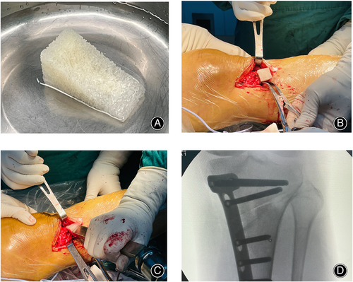
Two Kirschner wires were inserted parallel to the tibial slope toward the tip of the fibular head for indication of the direction of osteotomy. The ascending osteotomy plane was at 110° to the coronal osteotomy plane and lay posterior to the patellar tendon, and the tibial tuberosity bone block was at least 15 mm.41 Adjustment of the mechanical shaft was accomplished under fluoroscopy according to the preoperative plan, and the osteotomy gap was maintained with a bone spreader. A wedge-shaped allogeneic cancellous bone block of different height was then selected based on the measurement of the final opening gap for impaction grafting using a rectangular metal impactor (Figure 1). At last, internal fixation was achieved with TomoFix LCP system. A drainage tube was indwelled before incision closure and a cocktail solution consisted of 75 mg ropivacaine hydrochloride and 1000 mg tranexamic acid was injected through the drainage tube after finishing the suture procedure. A long-leg cotton pad and elastic bandage was used for compression bandage to reduce postoperative swelling.
Postoperative Management and Rehabilitation
Prophylactic antibiotics were administered within 24 h after surgery. For prevention of lower extremity deep vein thrombosis (DVT), all patients were recommended to wear graduated compression stockings 3 months postoperatively and patients without contraindications were given rivaroxaban 10 mg per day from the first postoperative day until 2 weeks after. We suggested early functional rehabilitation including ankle pump exercise, isometric contraction of the quadriceps and free motion of the knee. Full weight bearing with walking frame was allowed in patients with intact lateral hinge and Takeuchi type I fracture under the guidance of the physiotherapists and the activity level was dependent on pain.42
Radiographic Analysis and Clinical Outcome Evaluation
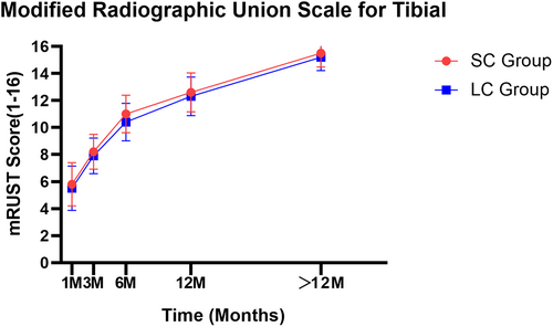
All enrolled patients underwent a whole-leg standing anteroposterior (AP) view of bilateral lower extremities with the knee in full extension and the patella facing forward and a standing lateral radiograph of the knee preoperatively, postoperatively and at last follow-up. Radiological assessment was performed by measuring the hip-knee-ankle angle (HKAA), medial proximal tibial angle (MPTA) as well as femorotibial angle (FTA) in the anterior–posterior (AP) radiographs and posterior tibial slope angle (PTSA) in the lateral radiographs. A loss of correction was defined as a change in the mechanical axis of ≥ 3° during follow up.43 To assess bone healing of the osteotomy gap, the modified RUST was adopted by two orthopedic surgeons at 1, 3, 6, 12 months postoperatively and every 3 months after 1 year follow-up until adequate union (Figure 2). If defined as mRUST 16 points, this means complete union at the medial, lateral and posterior cortex of the tibia.16, 44
Clinical outcome was evaluated based on the range of motion (ROM) and Knee Society score (KSS) which is consisted of KSS-Knee score and KSS-Function score preoperatively and at last follow-up.
Statistics
We accomplished the statistical analysis using IBM SPSS software version 22.0 (SPSS, Chicago, IL, USA). The normality of the distribution was checked for all parameters by the Kolmogorov–Smirnov test. When the normal distribution criteria were met, a paired student's t test was conducted for comparison of differences of the relevant data in this study pre-operatively and post-operatively. Mean ± standard deviation (SD) were calculated for the different measurements and presented for continuous variables. Two-sided p values of <0.05 is considered statistically significant. The intra-observer and inter-observer agreement of each measurement was assessed using the intra-class correlation coefficient (ICC).
Results
General Results
Eighty-two patients met the inclusion criteria (26 males and 62 females; 76 unilateral tibias and six bilateral tibias) contained 88 knees were enrolled in this study after elimination. Among the excluded patients, two patients did not have complete radiological data, and one patient was lost to follow-up, one patient was diagnosed with a comorbid giant bone cyst of the proximal tibia. Demographic characteristics of the patients are shown in Table 1.
| Parameter | Data |
|---|---|
| Age (years) | 60.75 ± 6.98 |
| Body mass index (kg/m2) | 25.63 ± 3.19 |
| Right/left | 43/45 |
| Male/female | 26/62 |
| Follow time (months) | 37.02 ± 12.85 |
| Correction angle (°) | 11.3 ± 3.2 |
| ASA | |
| I | 9 |
| II | 75 |
| III | 4 |
| Kellgren-Lawrence classification | |
| Grade2 | 62 |
| Grade3 | 26 |
- Abbreviation: ASA, American Society of Anesthesiology.
Radiologic Outcomes
The angle of the osteotomy wedge was 11.3° ± 3.2°(range 6°–22°). In the radiological evaluation of the coronal planar alignment, the HKAA, MPTA and FTA increased significantly from −6.4° ± 3.0°, 85.1° ± 2.6° and 180.1° ± 3.2° preoperatively to 1.2° ± 4.3° (p < 0.001), 94.4° ± 3.3°(p < 0.001) and 171.0° ± 2.8° immediate postoperatively, respectively. However, there was no significant statistic difference between the above parameters at immediate-postoperative and at last follow-up (p > 0.05). Radiological findings on lateral radiographs showed that the immediate-postoperative PTSA had a significant increase compared to the preoperative data (11.8° ± 5.8° vs. 7.7° ± 4.4°, p < 0.001). Similarly, no significant statistic difference was found in the PTSA at last follow-up compared to immediate postoperative data (p > 0.05); that is, no loss of correction was found after MOWHTO with a wedge-shaped cancellous allograft (Table 2).
| Preoperatively | Immediate postoperative | Latest follow-up | p value† | p value* | |
|---|---|---|---|---|---|
| HKAA (°) | −6.4 ± 3.0 | 1.2 ± 4.3 | 1.2 ± 4.5 | <0.001 | 0.903 |
| MPTA (°) | 85.1 ± 2.6 | 94.4 ± 3.3 | 94.3 ± 3.6 | <0.001 | 0.432 |
| FTA (°) | 180.1 ± 3.2 | 171.0 ± 2.8 | 171.3 ± 3.3 | <0.001 | 0.163 |
| PTSA (°) | 7.7 ± 4.4 | 11.8 ± 5.8 | 12.1 ± 4.3 | <0.001 | 0.554 |
- Abbreviations: FTA, femorotibial angle; HKAA, hip–knee–ankle angle; MPTA, medial proximal tibial angle; PTSA, posterior tibial slope angle.
- * Comparison of immediate postoperative and latest follow-up.
- † Comparison of preoperative and immediate postoperative.
Bone Healing and Clinical Outcomes
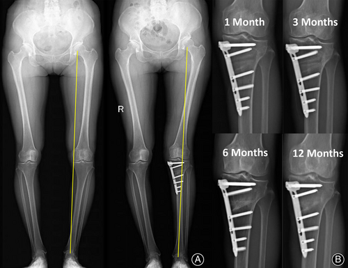
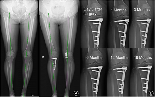
All patients in this study achieved good bone healing at the final follow-up and no significant differences in mRUST scores were seen between the SC and LC groups (Figure 3). Seven cases of lateral hinge fractures occurred postoperatively, six of which belonged to Takeuchi type I and one to type II. After a conservative rehabilitation program with delayed weight-bearing time on the ground, no adverse outcomes such as delayed healing or change in correction angle occurred at the final follow-up. It is notable that the only case of Takeuchi type II fracture was seen at 1 month postoperatively at the time of X-ray review and was considered as delayed lateral hinge fracture. And the osteotomy gap had completely healed 12 months after the operation (Figure 4).
The KSS-Knee score and KSS-Function score improved significantly from 55.4 ± 3.7 and 63.3 ± 4.6 preoperatively to 86.4 ± 2.8 (p < 0.001) and 89.6 ± 2.9 (p < 0.001) at last follow-up, respectively (Table 3).
| Pre-operative | Final follow-up | p value† | Mean change (91%CI) | |
|---|---|---|---|---|
| Knee society score | ||||
| Knee | 55.4 ± 3.7 | 86.4 ± 2.8 | <0.001 | 31.0 (30.2,31.8) |
| Function | 63.3 ± 4.6 | 89.6 ± 2.9 | <0.001 | 26.3 (24.9,27.7) |
| ROM° | 116.4 ± 14.7 | 117.9 ± 6.3 | 0.285 | 1.4 (−1.221,4.1) |
- Abbreviation: ROM, range of motion.
- † Comparison of preoperative and final follow-up.
Discussion
The principal finding of our study was that medial opening-wedge high tibial osteotomy using a novel wedge-shaped cancellous allograft impaction brings about good clinical outcomes and gap healing. Furthermore, such a type of bone grafting was also seen to be effective in maintaining the osteotomy gap in early to mid-term follow-up. The present study is the first to investigate clinical outcomes and gap healing after MOWHTO using a novel wedge-shaped cancellous allograft.
Potential Bone Healing Advantages of Wedge-Shaped Cancellous Allograft
Bone grafting is considered to be a good option to fill the osteotomy gap after OWHTO. Among these, autografts were regarded as the “gold standard” for filling bone voids in OWHTO due to their osteoinduction and osteocompatibility.45 However, the use of autografts was accompanied by complications such as prolonged operative time and severe donor site pain. Therefore, allografts have become a common choice for surgeons and patients.18
Previous studies have shown that the augmentation of a wedge-shaped bone block taken from hemispherical allogeneic femoral head into the osteotomy gap seems to facilitate bone healing to a satisfactory degree in larger correction group.46, 47 In a clinical study, Yacobucci et al.48 found that the use of corticocancellous proximal tibial wedge allograft in HTO resulted in satisfactory bone healing with a mean healing time of 12.1 weeks and only two patients suffered from nonunion. The wedge-shaped bone blocks used in our study were slightly different from those used by Yacobucci et al. They used proximal tibial corticocancellous wedge allograft, whereas our bone blocks consisted entirely of cancellous bone. In this situation, the cancellous portion of this graft served as an ideal osteoconductive substitute, and creeping substitution, a well-established bone healing process, can be observed at the osteotomy site.49 Other substitutes such as hydroxyapatite, β-tricalciumphosphate, and acrylic bone cement that have been reported in the literature and used in the clinic lack this characteristic.50, 51 In our series, the modified RUST in two groups above all came up to 15–16 points at last follow, and no bone nonunion was found in the patients. Therefore, wedge-shaped cancellous allografts have shown satisfactory results in bone healing.
Reliability in Terms of Maintaining Corrective Angles and Mechanical Stability
On the other hand, the role of different implant materials and bone grafts in maintaining the stability of the osteotomy gap was also a matter of particular concern. A mechanical study by Belsey et al.52 reported that OWHTO with a wedge allograft sourced from the proximal tibia of a donor can achieve a more stable construct and a lower incidence of valgus malrotation. In a vitro study, Yacobucci et al. have shown that the use of wedge-shaped synthetic bone graft and TomoFix had the potential to improve the initial axial and rotational stability of the osteotomy site and to reduce the stress concentration on the plate or at the lateral cortical hinge.53 In addition, previous studies have found that OWHTO using autogenous tricortical iliac bone graft was effective in preventing changes in the posterior tibial slope.
We chose wedge-shaped cancellous allograft as the implant material mainly based on the following reasons. First, the implantation of the wedge-shaped cancellous allograft provided additional intrinsic stability to the osteotomy gap, which allowed patient to begin weight bearing as early as the second to third day after surgery. The advantages of the wedge allograft in terms of mechanical strength and stiffness had been proved in an in vitro mechanical test by Belsey et al..54 Second, axial loading forces can be redistributed by this press-in graft and therefore it can be used as a load-sharing implant with the same structural properties as the recipient bone.
Meanwhile, our results were similar to the above study. No statistically significant differences were found for HKAA, FTA, MPTA and PTSA at the last follow-up compared to the immediate postoperative data (p > 0.05), which demonstrates the reliability of the wedge-shaped cancellous allograft in maintaining corrected angle and mechanical stability.
Strengths and Limitations
This is a mid-term retrospective study of 82 patients (88 knees) of open wedge HTO using wedge-shaped cancellous allograft. It not only demonstrated the clinical and radiologic outcomes of HTO using a wedge-shaped bone block, but also confirmed their advantages in bone healing.
There are a few limitations to this current study. Our study only analyzed patients who were implanted with an allogeneic wedge bone alone and did not compare the results with patients who were not implanted or who used a different implant material. Second, an objective and quantitative assessment of gap filling status on a simple radiograph is a difficult task with potential bias. Nevertheless, both intra- and interobserver agreement (ICC) for bone union in our study was higher than 0.8. In addition, there is still a lack of studies comparing the clinical outcomes of wedge-shaped allogeneic cancellous grafts with other implant materials. The advantages of wedge-shaped cancellous allograft need to be confirmed in subsequent studies with larger sample sizes and compared with other types of implant materials.
Conclusion
MOWHTO with a novel wedge-shaped cancellous allograft brought about good clinical outcomes and bone union. Such kind of allograft bone transplantation was also seen to be effective in maintaining the osteotomy gap in early to mid-term follow-up.
Author Contributions
Conceptualization: JLC, JHL and YRZ. Literature search: HTZ, WJF, and YWH. Data extraction and quality assessment: PCY, PD, and YJL. Measurement: HTZ and JHL. Validation: JL, XYQ and JCZ. Writing: JLC and JHL. All authors read and approved the final manuscript.
Ethics Statement
Ethics and Academy Committee of The First Affiliated Hospital of Guangzhou University of Chinese Medicine reviewed and approved the study protocol (K-2020-080).
Open Research
Data Availability Statement
The datasets used and/or analyzed during the current study are not publicly available due to feasibility but are available from the corresponding author on reasonable request.


