Collagen-Based Hydrogels for Cartilage Regeneration
Abstract
Cartilage regeneration remains difficult due to a lack of blood vessels. Degradation of the extracellular matrix (ECM) causes cartilage defects, and the ECM provides the natural environment and nutrition for cartilage regeneration. Until now, collagen hydrogels are considered to be excellent material for cartilage regeneration due to the similar structure to ECM and good biocompatibility. However, collagen hydrogels also have several drawbacks, such as low mechanical strength, limited ability to induce stem cell differentiation, and rapid degradation. Thus, there is a demanding need to optimize collagen hydrogels for cartilage regeneration. In this review, we will first briefly introduce the structure of articular cartilage and cartilage defect classification and collagen, then provide an overview of the progress made in research on collagen hydrogels with chondrocytes or stem cells, comprehensively expound the research progress and clinical applications of collagen-based hydrogels that integrate inorganic or organic materials, and finally present challenges for further clinical translation.
Introduction
Cartilage defects are very common and can cause joint pain, effusion, and limited function, resulting in a significant medical burden. Cartilage tissues are classified as elastic cartilage, fibrocartilage, and hyaline cartilage. Unlike elastic and fibrous cartilage, articular cartilage acts as hyaline cartilage with an ECM composed of type II collagen (COL II) fibrils (15%–25%), proteoglycans (5%–10%), and water (5%–10%).1 However, different from other hyaline cartilage, articular cartilage is composed of four parts: the superficial zone (SZ), the middle zone (MZ), the deep zone (DZ), the calcified zone (CZ) (Figure 1A).2 The number and orientation of the cells in each layer of the structure are different in articular cartilage; also mature articular cartilage has no blood supply and the subchondral bone is porous and sparse (Figure 1B).3 Being an avascular tissue, mature articular cartilage hardly recovers itself if it is not treated properly, cartilage defects may penetrate deep into the subchondral bone and ultimately require joint replacement.4 There are numerous classifications of cartilage defects, for example, Noyes and Stabler Methods,5 histological classification,6 International Osteoarthritis Cartilage Histopathology Assessment System (OOCHAS),6 and International Cartilage Repair Society grading system (ICRS).7 The most widely used is Outerbridge classification system (Figure 1C,D).8 Traditionally, cartilage defects have been managed clinically in a number of ways, that include non-steroidal anti-inflammatory drugs (NSAIDs) and physical fixation, and surgical strategies like osteotomy, arthroscopic polished arthroplasty, autologous osteochondral grafting, microfracture, autologous chondrocyte implantation (ACI), matrix-assisted autologous chondrocyte transplantation (MACT). In the short term, these treatments provide pain relief, but in the long term, the results are not satisfactory because ACI, MACT, and microfractures often lead to fibrocartilage proliferation rather than hyaline cartilage regeneration. Therefore, the most optimal treatment option available for cartilage defects remains inconclusive.

As we all know, collagen is one of the most studied biopolymers in medicine. The disadvantages of collagen are its weak mechanical properties and low thermal stability. To solve this problem for its clinical application, collagen can be combined with other biomaterials to make collagen-based hydrogels. Collagen-based hydrogels are used for cardiovascular disease, dental restoration, neurological disorders, bone defects, cartilage regeneration, wound healing, and tendon injuries. In recent years, cartilage tissue engineering (CTE) has been recognized as the most promising option for the therapy of cartilage defects due to imitating a three-dimensional (3D) ECM microenvironment resulting in the regeneration of cartilage. The desirable material should have not only the physical properties for cell proliferation, but also the chemical properties to regulate cell differentiation. Hydrogels have highly hydrated properties similar in composition and structure to ECM, and have gained wide applications as CTE materials. Hydrogels are also injectable and shield host cells from mechanical damage during the injection process. In additional, some hydrogels have fast gelation properties, which significantly reduces operation time. Materials used for CTE mainly include natural polymers (collagen, chitosan, silk proteins, hyaluronic acid, alginates), as well as synthetic polymers (polycaprolactone (PCL), polyglycolic acid (PLGA), polyethylene glycol (PEG)). In general, natural polymer hydrogels have superior cytocompatibility, but inferior mechanical properties, while synthetic polymer hydrogels have better mechanical strength but weak cytocompatibility. Therefore, it is challenging to invent hydrogel materials that meet all the needs of cartilage regeneration. Collagen is one of the natural biomaterials that exist in a variety of animal tissues, and they are cytocompatibility, bioadaptive, toxicity-free, and degradable.9-11 Notably, collagen also possesses good hemostatic properties by promoting platelet aggregation and plasma coagulation. In addition, specific acid sequence in the collagen structure provides many adhesion sites for cells, which enables the delivery of chondrocytes or stem cells for chondrocyte derivatization and ECM composition. Furthermore, increases in collagen content can boost the swelling rate of hydrogels.12 Many studies had shown that collagen hydrogels are the most prospective material for cartilage regeneration.10, 13 Although collagen is a major component of natural connective tissue, in the absence of covalent cross-linking, collagen hydrogels have weak mechanical properties. It is not conducive to regenerate stiffer tissues. To overcome these defects, cross-linking of collagen hydrogels increases the stiffness of the matrix and stimulates differentiation of mesenchymal stem cells (MSC) into the osteogenic spectrum best at Young's modulus above 25 kPa.14 In addition to the strength of the crosslink or the presence of polyfunctional groups, the crosslink density is also a major factor affecting the physical properties of collagen hydrogels. As well, blending with other materials (inorganic nanomaterials and organic polymers) is also required to enhance the properties of collagen hydrogels to fit the different needs of CTE.15 The reader can consult the comprehensive reviews by Thoniyot et al. and Tozzi et al.16, 17 and Fathi-Achachelouei et al.,18 which provide an overview of examples of collagen hydrogels combined with other materials to enhance their stiffness.
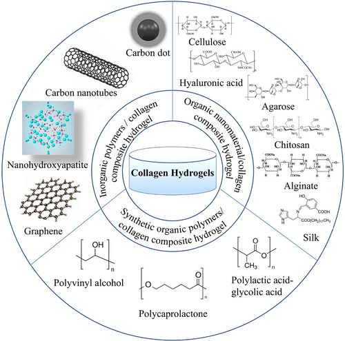
The aim of this review is to systematically present recent advances and application of collagen-based hydrogels for CTE (Figure 2). We first briefly introduce the structure of articular cartilage, cartilage defects classification and the collagen, and summarize the developments in the study of pure collagen hydrogels as scaffolds loaded with chondrocytes and stem cells in detail. The applications of hybrid collagen-based hydrogels for cartilage repair are then highlighted. Finally, the status and challenges of these collagen hydrogel-based cartilage repairs are summarized, and the perspectives of its development are discussed.
Collagen
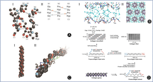
As we all know, collagen is the most abundant protein in animals, accounting for 25%–35% of the total protein content.19 The most common triplet in collagen is Gly-Pro-Hyp, which accounts for about 10.5%. The secondary structure of collagen consists of (Gly-X-Y)n, the C-terminus is associated with the triple helix structure and the N-terminus regulates the diameter of collagen fibers (Figure 3A).19 The non-helical ends are connected by covalent cross-linking and the CO groups and hydroxyl groups on the main chain of Hyp residues provide multiple anchoring points for water molecules (Figure 3B).20 In the collagen structure, the right triple helix structure (tertiary structure) is composed of two A1 chains and one A2 chain (Figure 3C).19 The tertiary structure of collagen can autonomously assemble into well-ordered collagen fibers (quaternary structure) (Figure 3D).19 Type I collagen (COL I) hydrogel is very effective for cartilage regeneration.21-24 Notably, COL II hydrogels containing chondrocytes produce more cartilage-specific matrix proteins than COL I hydrogels containing chondrocytes.10 However, COL II tends to promote fibrocartilage rather than hyaline cartilage regeneration, this significantly limits its clinical application. In fact, if the induction of fibrocartilage regeneration by COL II can be addressed, then it is better to use COL II rather than COL I.
Collagen Hydrogel
Collagen Hydrogel
In 2009, Erggelet et al. first published the cell-free matrix cartilage regeneration technique, which is derived from MACT and belongs to matrix-induced autologous cartilage regeneration (MACR).25 Collagen is often used as a matrix for MACR because their gap structure can provide guidance for cell migration and their chemotactic properties can selectively recruit cells.
Cell-free collagen hydrogel has been shown to be effective in the treatment of cartilage defects smaller than 1 cm2. Later, the researchers treated patients with cartilage defects of 3.71 ± 1.93 cm2 with small arthrotomy implants of cell-free COL I hydrogel and found promising short-term results. OtaŠeviČ et al. used a combination of basal microfracture and cell-free COL I hydrogel to repair knee cartilage with a mean defect area of 8 cm2, the MOCART score at 18-month follow-up was 74.67 ± 14.08.26 The migration of chondrocytes from healthy cartilage through the matrix into the collagen hydrogel is the key to the success of this approach. Although studies have confirmed the presence of migrating chondrocytes in cell-free collagen hydrogels, the process of chondrocyte migration and the factors that influence cell settlement are not fully understood. Nachtsheim et al. developed an in vitro model in which cell-free COL I hydrogels were placed on a collagen matrix containing human chondrocytes, and after a period of cultivation simulating the in vivo environment, it was found that chondrocytes existed in all cell-free COL I hydrogels, but the chondrocytes mainly spread in the bottom layer of the collagen hydrogels, and the difference in the distribution of the chondrocyte population might be influenced by the mechanical properties of collagen hydrogels, such as the sensitivity strain rate.27
Although encouraging results have been obtained for the treatment of cartilage defects in animal models and clinical patients with cell-free collagen hydrogels, recently Horbert et al. found that cell-containing collagen hydrogels were more effective than cell-free collagen hydrogels for cartilage defects.28 However, the presence of a mild immune response is a problem that cannot be ignored.
Pure Collagen Hydrogels Loaded with Cells
Combining hydrogels with cells offer a promising approach for repairing cartilage defects. On the other hand, collagen has been used as a cell-loaded CTE material due to the ability to modulate cellular immune responses and has a high density of cellular adhesion sites.
Pure Collagen Hydrogels Loaded with Chondrocytes
Pure collagen hydrogels containing chondrocytes for cartilage defects belong to the MACT technique. Earlier, Ochi et al. successfully repaired cartilage defects in the human knee using COL I hydrogel loaded with autologous chondrocytes.29 To enable more efficient cell settlement in hydrogels, researchers have developed 3D printing hydrogel. A previous report indicated that the survival rate of chondrocytes remained high after printing a collagen hydrogel with a chondrocyte density of 10 million cells mL−1.30 However, the cell density in CTE is typically between 100 and 100 million cells mL−1. Recently, Diamantides et al. found collagen hydrogels had the best storage modulus and viscosity at a cell density of 100 million cells mL−1, 14-day culture results showed no harmful effects of the printing process on cell viability or function, indicating that the printability of collagen hydrogels not only related to rheology, but also increases with the increase of cell density.31
One issue that must be considered is the influence that the physical properties of collagen hydrogels can have on chondrocyte metabolism. For example, hardness (Young's modulus >20 kPa) hydrogels are more appropriate for chondrocyte regeneration. Ogura et al. exploited chondrocyte-loaded collagen hydrogels and demonstrated that chondrocyte metabolic function is modulated by hydrostatic pressure (HP), deviatoric stress (DS), and stress loading time, with HP contributing to the stimulation of ECM synthesis by chondrocytes and DS possibly effecting the catabolism of ECM.32 Dong et al. synthesized three different DS of photo-crosslinked COL I (CM) by modifying the amino group of COL I with methacrylic anhydride (MA), and then used CM to synthesize a hydrogel whose degradation rate could be adjusted by modifying the DS of CM, and the contractility of the CM hydrogel could also be adjusted to promote the synthesis of cartilage-specific matrices.33
Another thought-provoking issue is the dedifferentiation of chondrocytes in chondrocyte-containing collagen hydrogels. Chondrocyte dedifferentiation occurs during the 2D expansion phase of chondrocyte culture in vitro. Typically, autologous human serum (HS) is added to the culture matrix to induce chondrocyte redifferentiation, whereas in most cases, supplementation with HS causes chondrocytes to differentiate into fibrocartilage rather than hyaline cartilage. Jeyakumar et al. added hyperacute serum (HAS) and platelet-rich plasma (PRP) to chondrocyte-containing collagen hydrogels and found that the addition of PRP exceeding 14 days decreased IL-6 secretion and increased platelet derived growth factor (PDGF) production by chondrocytes (p < 0.05) and enhanced gene expression of COL II A1 and SOX9 (p < 0.05), whereas addition of HAS enhanced the generation of COL I A1.34 Similarly, Mohd et al. demonstrated that the incorporation of Pseudomonas aeruginosa aqueous extract (SCAE) into chondrocyte collagen-containing hydrogels stimulated chondrocyte proliferation, decreased chondrocyte dedifferentiation, and contributed to the synthesis of cartilage matrix.35
To overcome the limited source of autologous chondrocytes, Lim et al. added human nasal septum-derived chondrocytes (hNCs) to a COL I hydrogel and treated cartilage defects in rats.36 Dasargyri et al. used COL I hydrogel enriched with a high density of fetal chondrocytes to culture cartilage tissue in vitro, but the disadvantage of this novel material is the characteristic of auto-contraction.37
Pure Collagen Hydrogel Loaded with Stem Cells
In CTE, stem cells have induced differentiation and have become a good substitute for autologous chondrocytes in recent years. Collagen hydrogels interact with bone mesenchymal stem cells (BMSCs) to boost cellular signal transduction as well as production of ECM. In addition to promoting the synthesis of ECM, these natural materials can maintain the phenotype of chondrocytes. It is not certain which source of stem cells is better suited for cartilage regeneration. Earlier, Wakitani et al. synthesized injectable collagen-based hydrogels by embedding BMSCs into calf skin COL I for the treatment of cartilage defects in rabbits, and showed that COL I promotes the differentiation of BMSCs into chondrocytes.38 Sakura et al. added induced pluripotent stem cells (iPSs) to collagen hydrogel and introduced differentiation of iPSs, and after 8 weeks green fluorescent protein (GFP) immunohistochemistry demonstrated cartilage repair by iPSs.39 Similarly, Elizabeth et al. reported that iPSs-loaded collagen hydrogels were a promising CTE hydrogel.40 Fabre et al. recently employed a microscopic mass model to research the chondrogenic differentiation potential of bone, adipose, dental pulp (DP), and Wharton jelly (WJ)-derived MSCs. and found that only BMSCs showed chondrogenic induction, concluding that BMSCs are most beneficial for cartilage regeneration.41 In CTE, besides pluripotent stem cells (iPSs) and bone mesenchymal stem cells (BMSCs), other stem cells also exist, such as synovial mesenchymal stem cells (SMSCs) and human umbilical cord blood mesenchymal stem cells (hUCB-MSCs). The origin of SMSCs was unclear and was generally believed to be derived from synovium, type B synovial lining, or infrapatellar pad. It has been shown that the amount of SMSCs in the knee joint cavity was increased approximately 100-fold in patients with OA.42 Therefore, the concentration of SMSCs may be positively correlated with the degree of joint damage. Lee et al. found that SMSCs had a greater chondrocyte differentiation capacity compared to BMSCs in vitro.43 A large number of studies have shown that the use of SMSCs to treat cartilage injuries may be feasible in the future.44, 45 However, no clinical studies have been reported on the use of SMSCs for cartilage regeneration. The lack of clinical evidence makes it difficult to draw conclusions about the efficacy and safety of SMSCs for cartilage regeneration. hUCB-MSCs have a higher chondrogenic potential compared to BMSCs. Ibrahim et al. demonstrated that differentiation of hUCB-MSCs into chondrocytes was possible in vitro.46 Park et al. repaired critical sized full thickness cartilage defects in mice joints with hydrogel-loaded hUCB-MSCs and showed that the combination of hUCB-MSCs and 4% hyaluronic acid hydrogel was most effective in repairing cartilage defects in vivo.47 Park et al. found that undifferentiated hUCB-MSCs (undiff-MSCs) was more favorable for cartilage repair compared to chondrogenic pre-differentiated hUCB-MSCs (chondro-MSCs) in rat model, which indicated that chondrogenic pre-differentiation of hUCB-MSCs did not enhance cartilage repair.48 Gómez-Leduc et al. considered hypoxia as a key parameter for chondrogenic differentiation of hUCB-MSCs in type I/III collagen sponges.49 Therefore, hUCB-MSCs can be considered as a promising MSC source for cartilage regeneration.
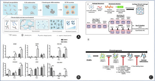
The technical difficulty of treating cartilage defects with stem cell-containing collagen hydrogels lies in the efficient differentiation of stem cells into hyaline chondrocytes. The chondrogenic differentiation of BMSCs depends on the type and properties of the hydrogel, the presence of soluble bioactive factors and external factors.10 The properties of hydrogels affect the cell-matrix interaction and thus the differentiation of BMSCs, and collagen-based hydrogels can provide a natural environment for the differentiation of BMSCs into chondrocytes. Wang et al. designed and synthesized three groups of collagen hydrogels based on articular cartilage ECM from rabbits of all ages. The research results indicated that all three groups of hydrogels could stimulate chondrogenesis in BMSCs, but the neonatal group synthesized the most cartilage matrix in BMSCs resulted in severe matrix calcification, while the adult group synthesized the least cartilage matrix in BMSCs had the least calcification, and overall the juvenile group collagen hydrogels were the most effective.50 Wu et al. demonstrated that heterologous collagen hydrogels were more capable of cartilage regeneration than decellularized porcine articular cartilage (DPC) microparticles in an experimental model of knee osteoarthropathy in rabbits.51 Stem cell multiplication and differentiation are influenced by the physical properties of collagen hydrogels, including stiffness, 3D pore size and pore topology form. Chen et al. designed a collagen-based hydrogel containing stromal cell-derived factor 1 (SDF-1) possessing channels in both horizontal and vertical directions and evaluated the effect of the hydrogel on in vivo osteochondral regeneration and in vitro MSC migration, found that the mechanical properties of the randomly oriented hydrogel were inferior to those of the radially oriented hydrogel, but both favored cell proliferation.52 Yang and Li et al. prepared two collagen-based hydrogels with different structures but similar physical and chemical properties and found that in the absence of growth factors, the collagen hydrogel with a fibrous network structure better promoted the differentiation of BMSCs and reduced the calcification of ECM; however, chondrocyte hypertrophy induced by BMSCs was present in both structural hydrogels.24 Yang and Tang et al. prepared three kinds of different hydrogels (hyaluronic acid methacrylate hydrogel, self-assembled collagen hydrogel, self-assembled collagen hydrogel cross-linked with kynurenine), and showed that the early collagen hydrogel were strong in cartilage induction, but the later self-assembled collagen hydrogel, self-assembled hydrogel cross-linked with kynurenine collagen hydrogels were severely contracted resulting in insufficient space for cell proliferation within the hydrogel. In comparison, the continued production of cartilage-associated matrix is facilitated by the stable structure of hyaluronic acid methacrylate hydrogels (Figure 4A).13
Soluble bioactive factors are favorable for the synthesis of ECM and the derivatization of BMSCs. The most recent studied soluble factor is the transforming growth factor TGF-β, which is a recognized bioactive factor for inducing chondrocyte formation, but their immune rejection and unexpected side effects (such as carcinogenesis, cartilage hypertrophy and production of an X-type collagen-rich matrix and expression of osteogenic marker genes) limit their clinical application. Gene editing enables BMSCs to produce cartilage growth factor autonomously and durably. Recently, the transcription factor SOX9 has been used to induce chondrogenesis in BMSCs by gene delivery via adenovirus and significantly reduced the level of chondro-hypertrophy in vitro culture.53 Weissenberger et al. induced cartilage regeneration with different degrees of hypertrophy in human mesenchymal stem cells (hMSCs) in collagen hydrogels by adenovirus-encoded genes for the SOX9, TGF-β1 and BMP2. TGF-β1 resulted in a significant rise in COL II A1 expression compared to SOX9 and BMP2, and BMP2 induced a remarkable increase in the hypertrophy marker gene alkaline phosphatase (ALP) more than TGF-β1.54
The selective addition of other bioactive factors such as n-cadherin, autologous PRP, COL II, and microRNAs (miRNAs) are also able to stimulate cartilage regeneration in collagen hydrogels loaded with BMSCs. Wang et al. showed that the early cartilage differentiation of MSCs was significantly slower in the presence of n-calcine mucin antibodies, and the later collagen microenvironment counteracted the inhibitory effect of n-calcine mucin antibodies on n-calcine mucin, suggesting that collagen hydrogels further promoted the late differentiation of BMSCs (Figure 4B).55 Fang et al. showed that PRP stimulated cell proliferation and hyaline cartilage formation through the TGF-β1/SMAD signaling pathway, while inhibiting the synthesis of the fibrocartilage marker gene COL I.56 Rajagopal et al. reported cartilage differentiation in BMSCs with mir-140-activated collagen hydrogels, which consistently provided mir-140, reduced COL I A1 synthesis, and promoted hyaline cartilage production.57
External factors closely affect the cartilage differentiation of BMSCs in stem cell-loaded collagen hydrogels, such as mechanical stimulation, negative electrical microenvironment, etc. Mechanical stimulation, including dynamic compressive and shear stresses, can mimic the natural joint loading environment and facilitate cartilage formation for BMSCs (Figure 4C).40 Yang et al. prepared three collagen hydrogels with different electrical properties and observed that the secretion of COL II was high in collagen hydrogels containing a higher negative charge.58 Notably, it was recently shown that hypoxia facilitates hMSCs differentiation towards cartilage and tendon,59 however, there were no such studies in cartilage regeneration with stem cell-loaded collagen hydrogels.
In summary, the main stem cells currently used for cartilage repair are mesenchymal stem cells of various origins, among which bone mesenchymal stem cells are the most common. The collagen hydrogels loaded with cells have the advantage of being non-cytotoxic and can repair cartilage defects to regenerative cartilage. However, low mechanical strength of collagen hydrogels may lead to cell death during the squeezing process.
Inorganic Nanomaterial/Collagen Composite Hydrogel
Nano Hydroxyapatite/Collagen Composite Hydrogel
Cartilage are complex biomineralized layered structures containing nanohydroxyapatite (nHA) and collagen. As a natural inorganic component with good biocompatibility and bio-integration, hydroxyapatite is often used as a tissue engineering material for cartilage and bone regeneration, with more applications in bone regeneration. Hydroxyapatite itself is hard, therefore, its addition to collagen hydrogels can improve the hardness of collagen hydrogels. Hydroxyapatite releases calcium and phosphate ions, which have an effect on certain mineralization signaling pathways. Previous studies have revealed that adding of hydroxyapatite nanoparticles in moderate amounts to hydrogels increased the Young's modulus of hydrogels and decelerated the degradation rate.60 Martincic et al. found lower infection rates in hydroxyapatite collagen hydrogels than alginate agarose hydrogels in the treatment of knee cartilage injuries.61 The biomimetic mineralization approach is the assembly of nHA onto COL I, a strategy that mimics natural bone at the molecular level. Mg2+ facilitates the binding of nHA and collagen.62 However, the drawback of most current hydroxyapatite/collagen hydrogels remains the weak mechanical properties, mainly caused by too rapid degradation of collagen and insufficient mechanical strength. Previous studies have shown that ribose can stabilize the collagen matrix by non-enzymatic glycosylation of collagen.63 Mg2+/HA/collagen hydrogels cross-linked with ribose have higher enzyme resistance and histocompatibility,64 and non-cytotoxicity.65 Although these studies have made significant progress, the relevant mechanisms are not precisely understood. Dellaquila et al. synthesized ribose cross-linked Mg2+/HA/collagen hydrogels and systematically investigated their properties by a full factorial design of experiment and found that glycosylation of collagen increased the swelling and porosity of hydrogels, while the degradation rate of composite hydrogels was influenced by the hydroxyapatite content and ribose content.66
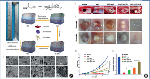
With the discovery of the role of subchondral bone in articular surface injuries, the focus of research has shifted to mimicking the structure of subchondral bone during the repair process. These scaffolds are assembled as double or triple-layered scaffolds to accommodate the simultaneous regeneration of cartilage and bone, depending on the structural characteristics of the osteochondral bone. Yudoh et al.67 manufactured an autologous cartilage complex with hydroxyapatite, proteoglycan, and COL II, which showed that hydroxyapatite-containing material was superior to the same material without hydroxyapatite, and that hydroxyapatite improved the viscoelasticity of the autologous cartilage material. The hydroxyapatite/collagen composite hydrogel had a gradient structure that induced BMSCs differentiate into cartilage tissue. Fan et al. synthesized a porous hydrogel that hydroxyapatite nanoparticles were filled in the pores of the hydrogel, and the different content of hydroxyapatite nanoparticles in different layers of the hydrogel could lead to different differentiation of BMSCs in the different layers of the hydrogel (Figure 5A).68 Yan et al. used hydroxyapatite/COL I and PLGA-PEG-PLGA hydrogel to make porous and gradient mineralized hydrogels to address the cartilage defects in rabbit femoral condyles, and stimulated BMSCs encapsulated in the hydrogel with electromagnetic fields (EMF), showing that EMF enhanced the cartilage repair properties of the hydrogels by coordinating PI3K/AKT/mTOR and Wnt1/LRP6/β-associated protein signaling pathways (Figure 5B).69 Thus, carbon nanoparticles are able to provide more mechanical strength to collagen hydrogels and even achieve cartilage and bone repair at the same time.
Carbon Nanomaterial/Collagen Composite Hydrogel
Currently, the common carbon nanomaterials in CTE are graphene, carbon nanotubes (CNTs), and carbon dot nanoparticles (CD NPs). Among them, graphene family is the oldest 2D carbon nanomaterial, which was first isolated in 2004.70 The oxygen-containing functional groups enhance the hydrophilicity and adsorption of graphene, so graphene oxide has good biocompatibility. An X-ray microimaging-based analysis of porous hydrogels showed that amino-functionalized graphene as a nano-crosslinker promoted the formation of uniform pore size and increased the interconnected microporous structure in collagen hydrogels. For hydrogels containing MSCs, the role of growth factors that induce chondrogenic differentiation is significant. To maintain the release of growth factors in the hydrogel, a second carrier needs to be added to the hydrogel, and graphene is a good choice because it not only improves the mechanical properties of the hydrogel, but also promotes the proliferation and differentiation of hMSCs by adsorbing and releasing bioactive factors. Zhou et al. found that graphene oxide tablets released transforming growth factor β3 (TGF-β3) more effectively than the addition of exogenous TGF-β3 without the need for repeated TGF-β3 supplementation.71
CNTs have a diameter of about 10–100 nm, and they have superior thermodynamic, electrical, and adsorption properties, so they are expected to be used for CTE. CNTs are commonly used as materials to increase the hardness of hydrogels, and it can change the rheological properties of the polymer matrix. CNTs can be classified into single-walled carbon nanotubes (SWCNT) and multi-walled carbon nanotubes (MWCNT). Mao et al. studied the cellular uptake of SWCNT using a 3D cell culture system and showed better proliferation of bovine articular chondrocytes in SWCNT/collagen composite hydrogels compared to collagen hydrogels (Figure 6A).72 Surface modification of SWCNT with -COOH but not -PEG has been shown to increase the expression of COL II A1 in SWCNT collagen composite hydrogels, suggesting that the charging of SWCNT by -COOH may regulate the metabolic activity of chondrocytes.73 Similarly, Wang et al. used fish collagen and SWCNT-COOH to prepare COL/SWCNT-COOH hydrogels with the addition of TGF-1, which were superior to pure collagen hydrogels for cartilage regeneration.74 Among multilayer carbon nanotubes MWCNT, vertically aligned carbon nanotubes (VACNT) have been shown to be very promising in cartilage regeneration (Figure 6B).75 Earlier, nanostructured scaffolds of MWCNT combined with poly-L-lactic acid (PLLA), which is highly stiff and conducive to the chondrogenic differentiation of MSCs, were fabricated using electrostatic spinning technology.76 Recently, Henao et al. used sodium riboflavin phosphate to modify multilayer carbon nanotubes and cross-link them with collagen to synthesize photosensitive collagen nanocomposites, and the results showed that MWCNT-riboflavin (MWFMN) nanoparticles improved the structural and thermal properties of the collagen matrix.77
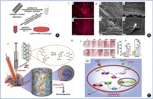
CD NPs have low cytotoxicity and good biocompatibility and can be applied as nanomaterials to enhance the mechanical stability of collagen hydrogels. Many researchers have shown that CD NPs can be applied as photosensitizers for photodynamic therapy (PDT), generating reactive oxygen species (ROS).78, 79 Lu et al. used genipin to combine collagen with CD NPs to develop a novel injectable hydrogel, and the CD NPs enhanced the stiffness of the collagen hydrogel, reduced degradation rate of collagen, and promoted cartilage regeneration in BMSCs by generating ROS under light (Figure 6C).80 The same strategy to increase the stiffness of collagen hydrogels as well as to increase the production of ROS is the incorporation of cadmium selenide (CdSe) quantum dots into collagen hydrogels using ginepin.81 In summary, both CD NPs, graphene, CNTs, and hydroxyapatite are able to enhance the physical properties of collagen hydrogels and favorably promote cartilage regeneration.
Organic Polymers/Collagen Composite Hydrogel
Natural Organic Polymers/Collagen Composite Hydrogel
Cellulose/Collagen Composite Hydrogel
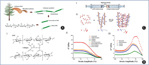
Cellulose, a natural hydrophilic polymer, has gained great attention in the biomedical field. Because of excellent characteristics, such as high tensile strength, modulus of elasticity (130–150 GPa), biocompatibility, and biodegradability (Figure 7A).82 Cellulose is mainly divided into cellulose nano-objects, cellulose microcrystals, and cellulose microprimary fibers. Cellulose nano-objects include cellulose nanocrystals (CNCs) as well as cellulose nanofibers (CNFs). Cellulose nano-objects have larger surface area and more uniform particle size distribution, which are more suitable for synthesizing hydrogels. Torabizadeh et al. compared the effects of CNCs and CNFs on physical properties and cellular properties of collagen hydrogels. As far as physical properties of collagen hydrogels, both CNFs and CNCs optimized the physical properties of collagen hydrogels (reduced gelation time, increased compressive strength, enhanced injectability, increased swelling rate, and reduced degradability), but the effect of CNCs was greater in terms of cellular properties. CNFs were harmless to fibroblasts, whereas CNCs improved cell viability.83 Liu et al. studied the characteristics of CNCs/collagen hydrogels with different pore orientations (horizontal, random, vertical, multi-structured), hydrogels arranged horizontally with the largest stiffness and energy dissipation capacity, and multi-structure hydrogels exhibited a larger storage modulus compared to conventional hydrogels with random pore (Figure 7B).84 Acidic collagen is able to bind to chemical cross-linking agents. Cellulose is easily surface modified, so the modified cellulose can be combined with collagen hydrogel and the properties of collagen hydrogel can be changed. Yang et al. mixed collagen solution with dialdehyde carboxymethylcellulose (DCMC) in a biphasic manner and showed that the DCMC/collagen hydrogel facilitated cell adhesion and proliferation.85 Unfortunately, above experiments were performed in fibroblast cell lines and not in chondrogenic cells. Later, Zhang et al. incorporated aldehyde-functionalized CNCs (a-CNCs) into collagen hydrogels (Figure 7C). Compared with CNCs/collagen hydrogels without Schiff base bonds, the shear stress of a-CNCs/collagen hydrogels were lower and more favorable for encapsulating MSCs, and a-CNCs/collagen hydrogels can be performed by 18-gauge needle subcutaneous injection, which is expected to deliver MSCs to the site of cartilage defects by injection.86
Hyaluronic Acid/Collagen Composite Hydrogel
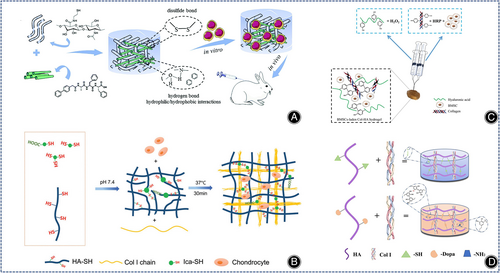
Hyaluronic acid (HA) is one of the main components of the ECM of cartilage and as glycosaminoglycans (GAGs) it has good water retention capacity and binds to various cell surface receptors. However, the hydrophilic and polyanionic properties of hyaluronic acid inhibit cell adhesion. The lack of adhesion sites is detrimental to the connection between nascent cells. Synergistic mechanical effect between collagen and HA enhance the hardness of the complex. In addition, collagen has self-assembly characteristics, and sulfur-HA (HA-SH) can be used to synthesize injectable hydrogels. The injectable composite hydrogels provide an excellent environment for in vivo cell growth and have been extensively studied and developed for CTE (Figure 8A).87 Previous studies have reported that injectable HA/collagen composite hydrogel relieved collagen contraction and promoted chondrocyte adhesion and proliferation.88-90 The physicochemical properties of HA/collagen composite hydrogel facilitate cartilage differentiation. Yang et al. showed that the chemical and physical microenvironments (microstructure, mechanical properties, electrical properties, protein adsorption) all affect the cartilage regeneration of BMSCs.13
Different cross-linking methods can also affect the performance of collagen hydrogels. Kong et al. designed and prepared three COL I/HA hydrogels and found that chondrocytes in HA-sNHS/COL I (HSC) and HA-CHO/COL I (HCC) produced more ECM, concluding that the chemical cross-linking method mainly affected the ECM secretion of chondrocytes and had no associated with chondrocyte differentiation.91 Gao et al. prepared different types of collagen-based hydrogels by varying the ratios of COL I and modified hyaluronic acid (HACHO) and chondroitin sulfate (CS-ADH), and found that C8CH hydrogels (4:1:1 volume ratio of COL I, HA-CHO and CS-ADH) had the best biological properties.92
Icariin (Ica) promotes cartilage regeneration in MSCs. Yang et al. showed that Ica-HA/collagen hydrogels is more effective in promoting cartilage regeneration in mice than HA/collagen hydrogels.88 To increase the content of Ica, Ica is often chemically incorporated into hydrogels by chemical crosslinking of methacrylate-modified Ica (Ica-MA). However, because Ica-MA is cytotoxic, new methods need to be developed. Liu et al. prepared thioglycoside-functionalized HA/collagen hydrogels with high Ica content by self-assembly of collagen molecules and disulfide bonding between thiol groups, which promoted chondrocyte proliferation, cartilage ECM secretion, and, most importantly, reduced the cytotoxicity of Ica compared with HA/collagen hydrogels (Figure 8B).93
Enzymatic cross-linking reactions enable rapid hydrogel formation under physiological conditions. Ren et al. invented a maleimide-modified bionic collagen-based hydrogel using matrix metalloproteinase (MMP) as a cross-linking agent, which contained both Arg-Gly-Asp (RGD) sequences and functional peptide sequences and was able to induce the differentiation of BMSCs into cartilage and inhibit their hypertrophic phenotypic differentiation.94 Zhang et al. used hydrogen peroxide (H2O2) and horseradish peroxidase (HRP) to synthesize a COL I tyramine (COL I-TA) complex hyaluronan tyramine (HA-TA) injectable hydrogel loaded with BMSCs and TGF-β1, providing an advantageous microenvironment for chondrogenic differentiation of BMSCs (Figure 8C).95 Also using COL I and HA-TA, Schwab et al. synthesized novel enzymatic cross-linked 3D printed hydrogels that had a more uniform distribution of collagen protofibrils with the ability to directionally regulate cell alignment and migration, allowing 3D printed collagen hydrogels to resemble natural cartilage tissue more.96 Kim et al. synthesized a composite collagen hydrogel using recombinant human transglutaminase 4 (rhTG-4), rhTG-4 was able to increase the stiffness of the composite hydrogel and induce the production of biomolecules that facilitate the differentiation of synovial mesenchymal stem cells (SDSCs) in rabbits in vivo.97
Yao et al. synthesized an injectable hydrogel using HA-SH, hyaluronic acid, and COL I, which promoted cartilage matrix production and hyaline cartilage-related gene expression by encapsulating exogenous chondrocytes.98 These hydrogels containing exogenous tissue cells increased the chances of an immune response.99 Injectable engineered bionic cartilage matrix hydrogels that recruit stem cells can solve this challenge. Li et al. synthesized dopamine-containing collagen-based hydrogels (HD-C) using COL I and hyaluronic acid derivatives (HA-DN). HD-C hydrogels with tighter pore structure, higher mechanical properties and water retention significantly promoted the proliferation of rabbit bone marrow stem cells (rBMSCs) and the synthesis of cartilage matrix through a potentially functional protein adsorption mechanism (Figure 8D).100
Chemically cross-linked initiators or catalysts may be harmful to the cells encapsulated in the hydrogels. Therefore, to meet the demand for cell encapsulation in CTE, new methods need to be explored to prepare hydrogels with better physical properties and biocompatibility. Yao et al. prepared injectable biomimetic double self-crosslinked HA-SH-Mal-HA (HSMSSA)/COL I hydrogels that enhanced the expression of genes related to hyaline cartilage formation and multi-proteoglycan secretion.98 Gao et al. prepared self-crosslinked injectable hydrogels using COL I and N-hydroxysulfosuccinate-activated hyaluronic acid (HA-sNHS), and showed that chondrocytes in the 32% DS hydrogel secreted more specific cartilage matrix.101 Wang et al. created an injectable bionic hydrogel that overcame the immunogenicity of animal-derived collagen, and the bionic composite hydrogel improved chondrocyte proliferation compared to HA-SH hydrogel.87
In conclusion, the current research direction for HA/collagen hydrogels is focusing on both overcoming the shortcomings of cell adhesion ability and avoiding the toxicity of chemical cross-linking agents to cells.
Agarose/Collagen Composite Hydrogels
Agarose is a linear polysaccharide extracted from red algae. Agarose is biocompatible, has strong mechanical properties and maintains round cell morphology. Research on agarose has centered on enhancing biological properties and reducing cellular immunotoxicity. On the one hand, agarose is biologically inert. The biological properties of agarose can be improved by covalent modification, but covalent modification is very time-consuming and cytotoxic. Another approach is the addition of collagen, where the two components interact to form both the biological function of natural ECM and the tunable mechanical properties. Ulrich et al. showed that the addition of agarose to COL I hydrogels greatly improved their elastic modulus and reduced cell migration.102 Later studies showed that the typical concentration of agarose to achieve mechanical stability in dynamic compression was equal to or higher than 2% w/v.103 Cambria et al. mixed two concentrations of COL I with 2% wt/vol agarose showing that agarose-collagen hybrid hydrogels were superior to pure agarose hydrogels. This result is identical to previous studies of collagen hydrogels mixed with lower concentrations of agarose.104
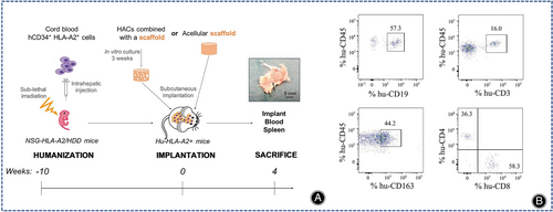
Another study on agarose showed variable results on host cell immune rejection by agarose. Rahfoth et al.105 demonstrated that embedding of rabbit allogeneic chondrocytes in agarose hydrogel did not produce an immune rejection reaction. However, Kawabe and Yoshinao showed the contrary result, they transplanted cartilage into rabbits and after 2~3 weeks the transplanted cartilage started to degenerate, partly due to humoral immune response and partly due to cytotoxicity.106 Recently, Perrier et al. comparatively studied the responses of mice to collagen hydrogels and agarose hydrogels, the result showed that collagen hydrogels triggered a weak inflammatory response, whereas agarose hydrogels had no inflammatory response (Figure 9).107
Chitosan/Collagen Composite Hydrogel
Chitosan is a polyatomic polysaccharide that is found in insects, crustaceans. It is synthesized from chitin by deacetylation reaction. Chitosan is widely used in tissue engineering owing to it being non-toxic, antifungal, antibacterial, as well as having good biocompatibility. The biological properties are similar to those of GAG in articular cartilage, chitosan has been commonly utilized to create materials that mimic cartilage. However, using only chitosan, the mechanical strength is not enough. Modification of natural hydrogels by blending or cross-linking has been reported to improve their properties.108 The addition of chitosan to collagen not only improved the mechanical properties and reduced the degradation rate of the composite hydrogel, but also enhanced the cell attachment and cell proliferation.
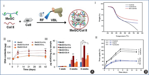
The development of multilayer hydrogels is an effective strategy to model the multilayered structure of osteochondral tissue. Chitosan, collagen, and nano-hydroxyapatite were combined in a bionic way to form a multilayer hydrogel, and these nanocomposite hydrogels were promising strategies for regenerating articular cartilage tissue.109, 110 ATDC5 chondrocytes can survive well in this multilayer hydrogel without growth factors.109, 110 Chitosan can be easily modified.9 Modification of chitosan by natural chemical cross-linking agents optimizes the conjugation of chitosan to collagen. For example, tannins and genipin were used as crosslinkers of chitosan and collagen for cartilage regeneration.111 Choi et al. used photopolymerizable chitosan (methacrylated glycol chitosan, MeGC), COL II and riboflavin (RF) to synthesize injectable modified chitosan collagen hydrogels under visible blue light (VBL), avoiding the disadvantages associated with ultraviolet (UV) irradiation and toxic initiators, and this hydrogel system enhanced cell-matrix interactions through the interaction of integrin α10 with COL II and was able to support the proliferation of pericytes and mesenchymal stem cells and the production of cartilage matrix (Figure 10A).112 Recently, Shah et al. prepared collagen-chitosan hydrogels for the first time using dual cross-linkers (tannic acid and genipin) (Figure 10B), but this hydrogel has only been used for corneal repair and skin regeneration, and has not been used for CTE.113
Alginate/Collagen Composite Hydrogel
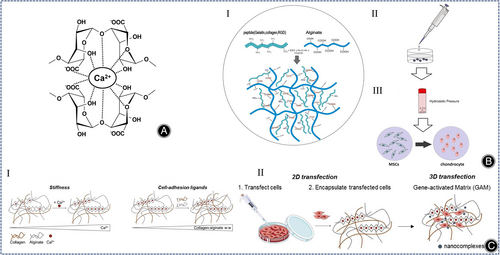
Alginate belongs to water-soluble linear polysaccharides, copolymers of β-D-mannate (M) and α-L-glutamate (G) were randomly arranged to form poly-GG, poly-MG, and poly-MM fragments linked by 1,4-glycosidic bonds. Alginates are easily cross-linked with divalent cations (Ca2+) (Figure 11A).9 Therefore, changing the calcium concentration can adjust the hardness of alginate-based complexes. Since there are no cell attachment sites or specific receptors in alginate, and collagen has more integrin structural domains, alginate can be used in combination with collagen to synthesize cell culture scaffolds in CTE. More than a decade ago, in a study of cartilage reconstruction in rats, Zheng et al. demonstrated that alginate collagen hydrogel (CAH) had superior cytocompatibility to genipin cross-linked collagen hydrogel (CGH).114 Later, Jahanbakhsh et al. used alginate collagen hydrogels for in vitro cartilage regeneration of MSCs and concluded that the use of hydrostatic pressure (HP) (5 MPa) on COL I -encapsulated MSCs had the potential to replace the role of TGF-β (Figure 11B).115 The important effect of HP on cartilage regeneration in MSCs was also demonstrated by Ledo et al. In their design of a COL I and alginate interpenetrating polymer network (IPN), the hardness of IPN was varied by changing the number of calcium carbonate nanoparticles, while the number of ligands was varied by changing the weight ratio of sodium alginate and collagen, and the hydrogel was stiffer and more similar to a real ECM than a biologically inert polymer hydrogel in the presence of cell adhesion ligands (Figure 11C).116 Another method for the improvement of cell adhesion of alginate collagen hydrogels is the enzymatic cross-linking method. Saghati et al. synthesized a phenolic alginate collagen hydrogel and investigated the effect of this collagen-based hydrogel on the chondrogenic differentiation of human amniotic mesenchymal stem cells and found that collagen increased the elasticity of the composite hydrogel by 10% and also improved the survival of human amniotic mesenchymal stem cells in the composite hydrogel.117
Silk/Collagen Composite Hydrogel
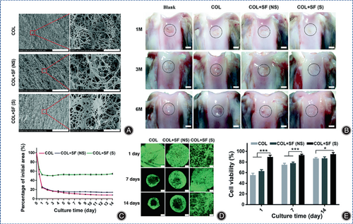
Silk fibroin (SF) is a natural fibronectin composed of 18 amino acids, of which Gly, Ala, and Se account for more than 80%, and these amino acids are linked by peptide bonds. Remarkably, the amino acid composition gives SF satisfactory biological properties and the hierarchical structure of SF contributes to its excellent mechanical properties and degradability. SF can be made into hydrogels by various methods. However, the poor mechanical properties and low cell affinity of SF limit the further application of SF in cell culture. Collagen/SF hybrid hydrogels are a promising material for CTE.118 Due to the poor interaction between collagen and SF, the composite hydrogels exhibit poor mechanical strength and rapid degradation. Ultrasound sonication is an eco-friendly technology that can regulate SF degradation, and has been used to induce gelation of SF to achieve uniform cell encapsulation prior to gelation.119 Long et al. invented a silk collagen (COL) composite hydrogel (COL+SF(S)) and evaluated its ability to regenerate cartilage in rabbit knee defects, producing intact articular hyaline cartilage after 6 months in the absence of any exogenous growth factors (Figure 12).120
A material with similar properties to SF is silk glue (SG), but most CTE uses SF instead of SG because of the immunogenic character of SG. However, recent studies have shown no significant difference in the immunogenicity of SF and SG.121 In addition, several studies have shown that SG is a good performing biomaterial because it is beneficial for mesenchymal cells.122 Hector et al. synthesized 3D printable composite hydrogels with stiffness close to that of natural cartilage using cocoons and rat COL I. SG was preserved by using physical solubilization, a process that ensures the integrity of the structure of COL I, more than two-fold increase in stiffness of collagen/SG hydrogels compared to pure collagen hydrogels, and the method is more friendly to BMSC cell culture and bioprinting as it does not require toxic chemicals, additional cross-linking agents, or complex physical methods.12
Synthetic Organic Polymers/Collagen Composite Hydrogels
Polyvinyl Alcohol/Collagen Composite Hydrogels
Polyvinyl alcohol (PVA) belongs to the synthetic biomolecular material. PVA hydrogels are rapidly developing in cartilage regeneration because of their high swelling, porosity, and viscoelasticity. However, its poor biocompatibility and microstructure impede its further application. Pure PVA hydrogels lack cell adhesion sites and, like many synthetic polymer hydrogels, are biologically inert. PVA hydrogels can be physically cross-linked. As well, Huang et al. produced a novel magnetic nanohydrogel using Fe3O4, PVA, and COL II in an optimal ratio of 5:95:5. Thus, PVA combined with collagen can be used in different ratios to synthesize new composite hydrogels with different functions for cartilage regeneration.123 Xie et al. utilized 3D printing to synthesize a novel hybrid PVA collagen hydrogel that mimics the properties of natural cartilage and provides mechanical support in which the collagen scaffold allows the hydrogel to integrate with the surrounding natural cartilage.124
Polycaprolactone/Collagen Composite Hydrogels
Polycaprolactone (PCL) is a synthetic polymer, which has certain rigidity, good compatibility with polymer materials, can also be used as a modifier for other polymers, and is widely used in biomedical field. And yet, disadvantages such as low hardness, few cell binding sites, poor bioactivity, and poor hydrophilicity limit its further promotion. PCL/collagen composite hydrogels exhibit good biochemical properties and cytocompatibility in bone tissue engineering,125 corneal tissue engineering,126 artificial blood vessels,127 wound healing,128 and nerve repair,129 but not in CTE.
Polylactic Acid-Glycolic Acid/Collagen Composite Hydrogels
Polylactic acid-glycolic acid (PLGA), which is polymerized from lactic acid and hydroxyacetic acid, is a degradable polymeric organic compound with good biocompatibility, non-toxicity, and good film-forming properties. Lamparelli et al. synthesized injectable PLGA microcarriers (PLGA-MCs)- collagen hydrogels containing hTFG-β1and performed in vitro culture of hBMSCs and found that the immunomodulatory activity of the cells was mainly related to the commitment rather than the synthetic environment, providing a fresh direction for the application of injectable collagen hydrogels.130
Conclusion and Prospects
This review describes recent advances in the characterization, synthesis, and application of collagen-based hydrogels for CTE. Overall, collagen-based hydrogels, as analogous polymers of natural ECM, can be used as cell-loaded materials for cartilage repair and has shown good results in vivo and in vitro tests. Due to the similarity to natural ECM, collagen-based hydrogels have the advantages of low immunogenicity, good biocompatibility, and biodegradability, while the disadvantages are their weak mechanical strength and their inability to induce cell differentiation well. In addition, the effectiveness of cartilage damage repair is influenced by the synthesis of ECM, and how to synthesize hydrogels with similar properties to natural cartilage is the key to hydrogel design. Despite the advantages of collagen hydrogels, their weak mechanical strength, and high shrinkage limit their use in CTE and also affect their clinical usability. To alleviate these problems, several strategies have been developed to improve the mechanical properties of collagen hydrogels by combining them with other materials or by physical or chemical modifications that are more favorable for their application in CTE. In CTE, several therapeutic strategies exist. Chondrocytes can be encapsulated in a collagen hydrogel to fill the cartilage defect site. BMSCs can also be encapsulated in a collagen-based hydrogel to induce differentiation of BMSCs into chondrocytes. In the clinic, for early cartilage defects in the knee joint, collagen-based hydrogel can be injected into the cartilage defect site through arthroscopy originally. Therefore, collagen-based hydrogel has a good application prospect in the clinic.
In the future, there are several issues to be considered to synthesize desirable collagen hydrogels as well as to accelerate clinical translation. (1) How to obtain collagen in a more efficient and less contaminated way to overcome the potential immunogenicity as well as the risk of virus transmission. (2) How to organically combine collagen and other natural or synthetic polymers to form a CTE material capable of inducing long-term growth of hyaline chondrocytes. (3) How to truly achieve organic integration of tissue-engineered cartilage with subchondral bone and even with surrounding tissues. (4) How to protect the loaded cells during the injection or printing of collagen-based hydrogels. (5) How to facilitate the batch preparation of collagen-based hydrogels, which is one of the key topics for cartilage regeneration materials to be widely used in clinical applications. (6) How to develop 3D printing of collagen-based hydrogels for cartilage defects to achieve precise treatment. (7) How to establish a precise and reliable clinical diagnosis standard, efficacy assessment principle, and risk control mechanism to achieve precision medicine.
Author Contributions
Conceptualization, supervision—Jianguo Liu, Chunsheng Xiao, Dongsong Li, Bo Cai; Fund Acquisition—Dongsong Li ; Original draft preparation—Lihui Sun, Yan Xu, Yu Han; Review and editing—Jing Cui, Zheng Jing, Dongbo Li; All authors have read and agreed to the published version of the manuscript.
Funding Information
Jilin Scientific and Technological Development Program (20230204077YY).
Conflict of Interest
The authors declare no conflict of interest.
Ethics Statement
Not applicable.


