Association of vascular endothelial growth factor and renal thrombotic microangiopathy-like lesions in patients with Castleman's disease
Ping-Ping Sun
Renal Division, Peking University First Hospital; Peking University Institute of Nephrology; Key Laboratory of Renal Disease, Ministry of Health of China; Key Laboratory of Chronic Kidney Disease Prevention and Treatment (Peking University), Ministry of Education, Beijing, China
Renal Pathology Center, Peking University Institute of Nephrology, Beijing, China
Search for more papers by this authorXiao-Juan Yu
Renal Division, Peking University First Hospital; Peking University Institute of Nephrology; Key Laboratory of Renal Disease, Ministry of Health of China; Key Laboratory of Chronic Kidney Disease Prevention and Treatment (Peking University), Ministry of Education, Beijing, China
Renal Pathology Center, Peking University Institute of Nephrology, Beijing, China
Search for more papers by this authorCorresponding Author
Su-Xia Wang
Renal Division, Peking University First Hospital; Peking University Institute of Nephrology; Key Laboratory of Renal Disease, Ministry of Health of China; Key Laboratory of Chronic Kidney Disease Prevention and Treatment (Peking University), Ministry of Education, Beijing, China
Renal Pathology Center, Peking University Institute of Nephrology, Beijing, China
Laboratory of Electron Microscopy, Pathological Center, Peking University First Hospital, Beijing, China
Correspondence
Dr Li Yang, Renal Division, Peking University First Hospital, Peking University Institute of Nephrology, Key Laboratory of Renal Disease, Ministry of Health of China, Key Laboratory of Chronic Kidney Disease Prevention and Treatment (Peking University), Ministry of Education, Beijing, People's Republic of China, No. 8, Xishiku Street, Xicheng District, Beijing 100 034, China. Email: [email protected]
Dr Su-Xia, Laboratory of Electron Microscopy, Pathological Center, Peking University First Hospital, Beijing, China, No. 8, Xishiku Street, Xicheng District, Beijing 100 034, China. Email: [email protected]
Search for more papers by this authorXu-Jie Zhou
Renal Division, Peking University First Hospital; Peking University Institute of Nephrology; Key Laboratory of Renal Disease, Ministry of Health of China; Key Laboratory of Chronic Kidney Disease Prevention and Treatment (Peking University), Ministry of Education, Beijing, China
Renal Pathology Center, Peking University Institute of Nephrology, Beijing, China
Search for more papers by this authorLei Qu
Renal Division, Peking University First Hospital; Peking University Institute of Nephrology; Key Laboratory of Renal Disease, Ministry of Health of China; Key Laboratory of Chronic Kidney Disease Prevention and Treatment (Peking University), Ministry of Education, Beijing, China
Renal Pathology Center, Peking University Institute of Nephrology, Beijing, China
Search for more papers by this authorFan Zhang
Renal Division, Peking University First Hospital; Peking University Institute of Nephrology; Key Laboratory of Renal Disease, Ministry of Health of China; Key Laboratory of Chronic Kidney Disease Prevention and Treatment (Peking University), Ministry of Education, Beijing, China
Renal Pathology Center, Peking University Institute of Nephrology, Beijing, China
Search for more papers by this authorYi-Yi Ma
Renal Division, Peking University First Hospital; Peking University Institute of Nephrology; Key Laboratory of Renal Disease, Ministry of Health of China; Key Laboratory of Chronic Kidney Disease Prevention and Treatment (Peking University), Ministry of Education, Beijing, China
Renal Pathology Center, Peking University Institute of Nephrology, Beijing, China
Search for more papers by this authorGang Liu
Renal Division, Peking University First Hospital; Peking University Institute of Nephrology; Key Laboratory of Renal Disease, Ministry of Health of China; Key Laboratory of Chronic Kidney Disease Prevention and Treatment (Peking University), Ministry of Education, Beijing, China
Renal Pathology Center, Peking University Institute of Nephrology, Beijing, China
Search for more papers by this authorCorresponding Author
Li Yang
Renal Division, Peking University First Hospital; Peking University Institute of Nephrology; Key Laboratory of Renal Disease, Ministry of Health of China; Key Laboratory of Chronic Kidney Disease Prevention and Treatment (Peking University), Ministry of Education, Beijing, China
Renal Pathology Center, Peking University Institute of Nephrology, Beijing, China
Correspondence
Dr Li Yang, Renal Division, Peking University First Hospital, Peking University Institute of Nephrology, Key Laboratory of Renal Disease, Ministry of Health of China, Key Laboratory of Chronic Kidney Disease Prevention and Treatment (Peking University), Ministry of Education, Beijing, People's Republic of China, No. 8, Xishiku Street, Xicheng District, Beijing 100 034, China. Email: [email protected]
Dr Su-Xia, Laboratory of Electron Microscopy, Pathological Center, Peking University First Hospital, Beijing, China, No. 8, Xishiku Street, Xicheng District, Beijing 100 034, China. Email: [email protected]
Search for more papers by this authorPing-Ping Sun
Renal Division, Peking University First Hospital; Peking University Institute of Nephrology; Key Laboratory of Renal Disease, Ministry of Health of China; Key Laboratory of Chronic Kidney Disease Prevention and Treatment (Peking University), Ministry of Education, Beijing, China
Renal Pathology Center, Peking University Institute of Nephrology, Beijing, China
Search for more papers by this authorXiao-Juan Yu
Renal Division, Peking University First Hospital; Peking University Institute of Nephrology; Key Laboratory of Renal Disease, Ministry of Health of China; Key Laboratory of Chronic Kidney Disease Prevention and Treatment (Peking University), Ministry of Education, Beijing, China
Renal Pathology Center, Peking University Institute of Nephrology, Beijing, China
Search for more papers by this authorCorresponding Author
Su-Xia Wang
Renal Division, Peking University First Hospital; Peking University Institute of Nephrology; Key Laboratory of Renal Disease, Ministry of Health of China; Key Laboratory of Chronic Kidney Disease Prevention and Treatment (Peking University), Ministry of Education, Beijing, China
Renal Pathology Center, Peking University Institute of Nephrology, Beijing, China
Laboratory of Electron Microscopy, Pathological Center, Peking University First Hospital, Beijing, China
Correspondence
Dr Li Yang, Renal Division, Peking University First Hospital, Peking University Institute of Nephrology, Key Laboratory of Renal Disease, Ministry of Health of China, Key Laboratory of Chronic Kidney Disease Prevention and Treatment (Peking University), Ministry of Education, Beijing, People's Republic of China, No. 8, Xishiku Street, Xicheng District, Beijing 100 034, China. Email: [email protected]
Dr Su-Xia, Laboratory of Electron Microscopy, Pathological Center, Peking University First Hospital, Beijing, China, No. 8, Xishiku Street, Xicheng District, Beijing 100 034, China. Email: [email protected]
Search for more papers by this authorXu-Jie Zhou
Renal Division, Peking University First Hospital; Peking University Institute of Nephrology; Key Laboratory of Renal Disease, Ministry of Health of China; Key Laboratory of Chronic Kidney Disease Prevention and Treatment (Peking University), Ministry of Education, Beijing, China
Renal Pathology Center, Peking University Institute of Nephrology, Beijing, China
Search for more papers by this authorLei Qu
Renal Division, Peking University First Hospital; Peking University Institute of Nephrology; Key Laboratory of Renal Disease, Ministry of Health of China; Key Laboratory of Chronic Kidney Disease Prevention and Treatment (Peking University), Ministry of Education, Beijing, China
Renal Pathology Center, Peking University Institute of Nephrology, Beijing, China
Search for more papers by this authorFan Zhang
Renal Division, Peking University First Hospital; Peking University Institute of Nephrology; Key Laboratory of Renal Disease, Ministry of Health of China; Key Laboratory of Chronic Kidney Disease Prevention and Treatment (Peking University), Ministry of Education, Beijing, China
Renal Pathology Center, Peking University Institute of Nephrology, Beijing, China
Search for more papers by this authorYi-Yi Ma
Renal Division, Peking University First Hospital; Peking University Institute of Nephrology; Key Laboratory of Renal Disease, Ministry of Health of China; Key Laboratory of Chronic Kidney Disease Prevention and Treatment (Peking University), Ministry of Education, Beijing, China
Renal Pathology Center, Peking University Institute of Nephrology, Beijing, China
Search for more papers by this authorGang Liu
Renal Division, Peking University First Hospital; Peking University Institute of Nephrology; Key Laboratory of Renal Disease, Ministry of Health of China; Key Laboratory of Chronic Kidney Disease Prevention and Treatment (Peking University), Ministry of Education, Beijing, China
Renal Pathology Center, Peking University Institute of Nephrology, Beijing, China
Search for more papers by this authorCorresponding Author
Li Yang
Renal Division, Peking University First Hospital; Peking University Institute of Nephrology; Key Laboratory of Renal Disease, Ministry of Health of China; Key Laboratory of Chronic Kidney Disease Prevention and Treatment (Peking University), Ministry of Education, Beijing, China
Renal Pathology Center, Peking University Institute of Nephrology, Beijing, China
Correspondence
Dr Li Yang, Renal Division, Peking University First Hospital, Peking University Institute of Nephrology, Key Laboratory of Renal Disease, Ministry of Health of China, Key Laboratory of Chronic Kidney Disease Prevention and Treatment (Peking University), Ministry of Education, Beijing, People's Republic of China, No. 8, Xishiku Street, Xicheng District, Beijing 100 034, China. Email: [email protected]
Dr Su-Xia, Laboratory of Electron Microscopy, Pathological Center, Peking University First Hospital, Beijing, China, No. 8, Xishiku Street, Xicheng District, Beijing 100 034, China. Email: [email protected]
Search for more papers by this authorABSTRACT
Aim
Renal thrombotic microangiopathy (TMA) is a common pathological manifestation of Castleman's disease (CD)-associated renal lesions. Increased level of plasma vascular endothelial growth factor (VEGF) has been shown in single-case reports. We aimed to investigate the dysregulation of VEGF in the pathogenesis of CD-associated TMA-like lesions (CD-TMA) in a larger cohort.
Methods
Nineteen patients with clinico-pathologically diagnosed CD with renal involvement were enrolled. Ten patients with pregnancy TMA or TMA of unknown reasons were enrolled as TMA control group. The plasma levels of VEGF, soluble Flt-1 and interleukin-6 (IL-6) were detected using enzyme-linked immunosorbent assay kits. The expression of VEGF in the kidney biopsied tissue sections and the lymph node specimens were detected by immunostaining.
Results
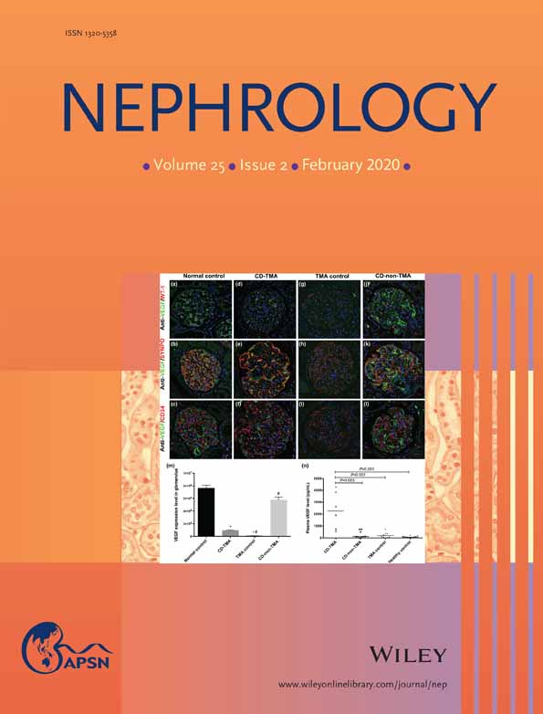
The plasma levels of VEGF and IL-6 levels were the highest in CD-TMA group compared to TMA control group and healthy controls. The levels of plasma VEGF was positively correlated with that of IL-6, and increased expression of VEGF and IL-6 was also observed in the lymph nodes from CD-TMA patients. However, the expression of VEGF in the glomerular podocytes was significantly decreased in CD-TMA group as well as in the TMA control.
Conclusion
Our findings suggest that renal VEGF expression might be important in the pathogenetic mechanism of CD-associated TMA-like lesions.
Supporting Information
| Filename | Description |
|---|---|
| NEP_13630-sup-0001-Supinfo.docxWord 2007 document , 1.4 MB |
Fig. S1 Flowchart of patients enrolment in the current study Fig. S2 Fluorescence in situ hybridization of interleukin-6 (IL-6) in biopsied lymph node. (a–c) Fluorescence in situ hybridization of IL-6 in a biopsied lymph node sample from patients with CD-non-TMA, CD-TMA and normal lymph node were all positive. (D) Comparison of IL-6 positive rate among the three groups. CD, Castleman's disease; TMA, thrombotic microangiopathy-like lesion. |
Please note: The publisher is not responsible for the content or functionality of any supporting information supplied by the authors. Any queries (other than missing content) should be directed to the corresponding author for the article.
REFERENCES
- 1Castleman B, Iverson L, Menendez VP. Localized mediastinal lymph node hyperplasia resembling thymoma. Cancer 1956; 9: 822–30.
10.1002/1097-0142(195607/08)9:4<822::AID-CNCR2820090430>3.0.CO;2-4 CAS PubMed Web of Science® Google Scholar
- 2Keller AR, Hochholzer L, Castleman B. Hyaline-vascular and plasma-cell types of giant lymph node hyperplasia of the mediastinum and other locations. Cancer 1972; 29: 670–83.
10.1002/1097-0142(197203)29:3<670::AID-CNCR2820290321>3.0.CO;2-# CAS PubMed Web of Science® Google Scholar
- 3Gaba AR, Stein RS, Sweet DL, Variakojis D. Multicentric giant lymph node hyperplasia. Am. J. Clin. Patho. 1978; 69: 86–90.
- 4Weisenburger DD, Nathwani BN, Winberg CD, Rappaport H. Multicentric angiofollicular lymph node hyperplasia: A clinicopathologic study of 16 cases. Hum. Pathol. 1985; 16: 162–72.
- 5Herrada J, Cabanillas FF. Multicentric Castleman's disease. Am. J. Clin. Oncol. 1995; 18: 180–3.
- 6Xu D, Lv J, Dong Y et al. Renal involvement in a large cohort of Chinese patients with Castleman disease. Nephrol. Dial. Transplant. 2012; 27: iii119–25.
- 7Frizzera G, Peterson BA, Bayrd ED, Goldman A. A systemic lymphoproliferative disorder with morphologic features of Castleman's disease: Clinical findings and clinicopathologic correlations in 15 patients. J. Clin. Oncol. 1985; 3: 1202–16.
- 8Rieu P, Noël LH, Droz D et al. Glomerular involvement in lymphoproliferative disorders with hyperproduction of cytokines (Castleman, POEMS). Adv. Nephrol. Necker Hosp. 2000; 30: 305–31.
- 9El Karoui K, Vuiblet V, Dion D et al. Renal involvement in Castleman disease. Nephrol. Dial. Transplant. 2011; 26: 599–60.
- 10Cohen T, Nahari D, Cerem LW, Neufeld G, Levi BZ. Interleukin 6 induces the expression of vascular endothelial growth factor. J. Biol. Chem. 1996; 271: 736–41.
- 11Aoki Y, Jaffe ES, Chang Y et al. Angiogenesis and hematopoiesis induced by Kaposi's sarcoma-associated herpesvirus-encoded interleukin-6. Blood 1999; 93: 4034–443.
- 12London J, Boutboul D, Agbalika F et al. Autoimmune thrombotic thrombocytopenic purpura associated with HHV8-related multicentric Castleman disease. Br. J. Haematol. 2017; 178: 486–8.
- 13Zhu X, Wu S, Dahut WL, Parikh CR. Risks of proteinuria and hypertension with bevacizumab, an antibody against vascular endothelial growth factor: Systematic review and meta-analysis. Am. J. Kidney Dis. 2007; 49: 186–93.
- 14Seida A, Wada J, Morita Y et al. Multicentric Castleman's disease associated with glomerular microangiopathy and MPGN-like lesion: Does vascular endothelial cell-derived growth factor play causative or protective roles in renal injury? Am. J. Kidney Dis. 2004; 43: E3–9.
- 15Suga SI, Kim YG, Joly A et al. Vascular endothelial growth factor (VEGF121) protects rats from renal infarction in thrombotic microangiopathy. Kidney Int. 2001; 60: 1297–308.
- 16Nishi J, Arimura K, Utsunomiya A et al. Expression of vascular endothelial growth factor in sera and lymph nodes of the plasma cell type of Castleman's disease. Br. J. Haematol. 1999; 104: 482–5.
- 17Tischer E, Mitchell R, Hartman T et al. The human gene for vascular endothelial growth factor. Multiple protein forms are encoded through alternative exon splicing. J. Biol. Chem. 1991; 266: 11947–54.
- 18Byrne AM, Bouchier-Hayes DJ, Harmey JH. Angiogenic and cell survival functions of vascular endothelial growth factor (VEGF). J. Cell. Mol. Med. 2005; 9: 777–94.
- 19Park JE, Keller GA, Ferrara N. The vascular endothelial growth factor (VEGF) isoforms: Differential deposition into the subepithelial extracellular matrix and bioactivity of extracellular matrix-bound VEGF. Mol. Biol. Cell 1993; 4: 1317–26.
- 20Robinson CJ, Stringer SE. The splice variants of vascular endothelial growth factor (VEGF) and their receptors. J. Cell Sci. 2001; 114: 853–65.
- 21Kim YG, Suga SI, Kang DH et al. Vascular endothelial growth factor accelerates renal recovery in experimental thrombotic microangiopathy. Kidney Int. 2000; 58: 2390–9.
- 22Eremina V, Jefferson JA, Kowalewska J et al. VEGF inhibition and renal thrombotic microangiopathy. N. Engl. J. Med. 2008; 358: 1129–36.
- 23Eremina V, Sood M, Haigh J et al. Glomerular-specific alterations of VEGF-A expression lead to distinct congenital and acquired renal diseases. J. Clin. Invest. 2003; 111: 707–16.
- 24Mutneja A, Cossey LN, Liapis H, Chen YM. A rare case of renal thrombotic microangiopathy associated with Castleman's disease. BMC Nephrol. 2017; 18: 57.
- 25Panek-Laszczyńska K, Konieczny A, Milewska E et al. Podocyturia as an early diagnostic marker of preeclapsia: A literature review. Biomarkers 2018; 23: 207–12.
- 26Garovic VD, Wagner SJ, Turner ST et al. Urinary podocyte excretion as a marker for preeclampsia. Am. J. Obstet. Gynecol. 2007; 196: 320.e1–7.
- 27Aita K, Etoh M, Hamada H et al. Acute and transient podocyte loss and proteinuria in preeclampsia. Nephron Clin. Pract. 2009; 112: c65–70.
- 28Craici IM, Wagner SJ, Bailey KR et al. Podocyturia predates proteinuria and clinical features of preeclampsia: Longitudinal prospective study. Hypertension 2013; 61: 1289–96.
- 29van Rhee F, Wong RS, Munshi N et al. Siltuximab for multicentric Castleman's disease: A randomised, double-blind, placebo-controlled trial. Lancet Oncol. 2014; 15: 966–74.
- 30Nishimoto N, Kanakura Y, Aozasa K et al. Humanized anti-interleukin-6 receptor antibody treatment of multicentric Castleman disease. Blood 2005; 106: 2627–32.


