Prevalence of cutaneous and mucosal lesions in dairy cattle admitted to a Canadian Veterinary Teaching Hospital from 2018 to 2019
Abstract
Background
The prevalence, anatomical distribution, or nature of cutaneous, hair and oral mucosal abnormalities (CHMAs) in cattle is uncertain.
Objectives
To determine how often dairy cattle admitted to a veterinary teaching hospital (VTH) had CHMAs (except for foot and ear canal) on physical examination and if there was an age-related difference.
Animals
Four hundred and thirty-three cattle: cattle <3 months (n = 85), cattle 3 to 24 months (n = 73), and cattle >24 months (n = 275).
Methods
In this descriptive, observational, prospective study, CHMAs of dairy cattle admitted to the VTH of the Université de Montréal were recorded over 1 year. Prevalences were calculated. Dermatological examinations were performed within 48 hours of admission, according to a glossary. Chi-square tests were used to compare prevalence between age groups.
Results
The 433 cattle were mostly females (97.5%) and of the Holstein breed (89.8%). The prevalence of cattle <3 months presenting with at least 1 identifiable CHMA was 65% (55/85). In cattle 3 to 24 months old, it was 90% (66/73), and in cattle >24 months, it was 99.3% (273/275). There were significant differences (P < .001) between the prevalence of CHMAs localized on the ischia, ilia, stifles, hocks, carpi, flank, lateral neck, dorsal cervical, and cornual regions in cattle >24 months vs <3 months.
Conclusions and Clinical Importance.
CHMAs were highly prevalent and age-specific. Calluses on the carpi and hocks of cattle >24 months were the most common CHMAs.
Abbreviations
-
- CHMA
-
- cutaneous hair and oral mucosal abnormality
-
- STROBE-Vet statement
-
- Strengthening the Reporting of Observational Studies in Epidemiology - Veterinary Extension statement
-
- VTH
-
- Veterinary Teaching Hospital
1 INTRODUCTION
Current literature in clinical bovine dermatology is limited.1-4 It mostly focuses on the economic and clinical consequences of foot diseases, or on the description of bovine skin or mucosal lesions in the context of cutaneous repercussions of systemic diseases or animal welfare.5-12 There is little data on how frequently skin changes are found on physical examination of dairy cattle. And for each dermatosis, there is little or no data regarding the sites affected, and the types of lesions observed. Such information is required to guide clinical studies toward the most prevalent dermatoses in cattle, and thus better understand their clinical and economic repercussions.
Bovine practitioners have suggested that cattle could be affected by several types of dermatological lesions at different anatomical sites and with age-varying prevalence. The objectives of this study were to determine how often skin/hair and buccal cavity abnormalities could be identified in dairy cattle admitted to a veterinary teaching hospital (VTH; excluding foot and ear canals) and to compare the prevalence of these abnormalities according to age groups.
2 MATERIALS AND METHODS
This descriptive observational prospective study was approved by the ethics committee for animal use of the Université de Montréal (18-Rech-1957). The STROBE-Vet statement guidelines were followed in writing this article (Supporting information 1).13
2.1 Cattle selection
All dairy breed cattle admitted to the VTH (Faculty of Veterinary Medicine, Université de Montréal, Quebec, Canada) for the first time between July 1, 2018 and June 30, 2019 were eligible for the study. All cattle were included unless dermatological examination could not be performed within 48 hours of admission. Given that all eligible cattle were included in the study, no sample size calculation was done.
2.2 Data collection
A study-dedicated dermatological glossary adapted to the cattle was established from reference textbooks in veterinary dermatology2, 14 and reviewed by a veterinary dermatologist (FS). A cutaneous, hair and oral mucosal abnormality (CHMA) was defined as an abnormality of the skin, of the hairs or of the oral mucosa, regardless of the type. The dermatological glossary (Supporting information 2) included 54 types of CHMAs: 13 were primary lesions (resulting from the primary underlying cause), 10 were secondary lesions (resulting from healing, self-traumas, or external factors), 8 were lesions that could be either primary or secondary in terms of underlying etiology, 10 were skin changes, 8 were changes to hair coat, and 5 were types of discharges/exudates.
The identification number, sex, breed, date of birth, date of admission, and date of dermatological examination were collected for each animal. A dermatological examination was carried out by a single trained observer (EG). The anatomical location of each CHMA was noted and described according to the predefined glossary. To limit excessive handling, animals' feet, ear canals, and concave pinnae were not examined. A veterinary dermatologist (FS) confirmed lesion descriptions if that was deemed necessary.
The data collected was captured in Microsoft Access 2019 software (Microsoft, Redmond, Washington). To estimate the data entry error rate, double blind data entry was performed for 12.7% (55/433) of cattle (n = 9900 records), based on systematic sampling.
2.3 Statistical analysis
The following were calculated: the prevalence of cattle with at least 1 skin/hair abnormality, regardless of the type of abnormality; the prevalence of cattle with at least 1 buccal cavity abnormality, regardless of the types of abnormality; and for each anatomical site, the prevalence of cattle with at least 1 CHMA, either overall or by type of CHMA. The same prevalence was calculated by age group (<3 months, 3-24 months, and >24 months). For sex-specific anatomical sites (udder and teats for females; prepuce, sheath, and scrotum for males), only the animals in the sex-specific category were included.
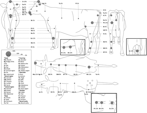
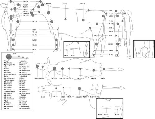
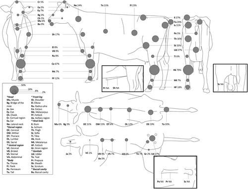
The anatomical distribution of CHMAs was illustrated using bovine silhouettes. For each of the 43 anatomical sites investigated, including the 5 sex-specific sites, a circle with an area proportional to the prevalence of the CHMAs was drawn.
The association between the presence of CHMAs and age group was assessed for each anatomical site using asymptotic or exact (if not, all expected counts ≥5) chi-square tests. A significance threshold of .001 was chosen to minimize the probability of committing a type I error. In the presence of a global association (P < .001), pairwise post hoc tests with Bonferroni adjustment were carried out. SAS software version 9.3 (SAS Institute, Inc, Cary, North Carolina) was used. Considering the descriptive nature of our study, no method to control for confounding factors or to manage missing data was used.
3 RESULTS
3.1 Study group
From July 1, 2018 to June 30, 2019, 610 cattle were admitted to the VTH. Of these, 528 fulfilled the inclusion criteria. Ninety-five of these cattle were excluded for failure to perform a dermatological examination within 48 hours of admission, mainly because of the absence of the observer (Supporting information 3). The remaining 433 cattle were included. Of those, 97.5% were female (422/433), and 89.8% were Holsteins (389/433; Supporting informations 4 and 5). The age of cattle in this study ranged from 0 days to 203.8 months old, with an average of 39.8 months (SD = 33.0). When the animals were divided into age groups, there were 85 cattle <3 months, 73 cattle 3 to 24 months, and 275 cattle >24 months old. The data entry error rate was estimated to be <0.3% (29/9900). The location of 36 CHMAs was recorded without being associated with a type of CHMA; these CHMAs were described as “undetermined” (Supporting information 6).
3.2 Prevalence, type, and anatomic location of CHMAs
3.2.1 All cattle (n = 433)
Of the 433 cattle in the study, 91.0% (394/433) had at least 1 identifiable skin/hair abnormality on physical examination, and 9.2% (40/433) presented with at least 1 buccal cavity abnormality. The most prevalent abnormalities were calluses on hocks (40.4%, 175/433) and on carpi (38.1%, 165/433), as well as alopecia on stifles (14.8%, 64/433; Supporting information 6). Nineteen CHMAs defined in the glossary (35%, 19/54) were never observed during the study (primary lesions, n = 4; secondary lesions, n = 2; primary or secondary lesions, n = 5; skin changes, n = 3; changes to hair coat, n = 4; type of discharge/exudate, n = 1).
3.2.2 Cattle <3 months (n = 85)
Of the 85 cattle <3 months, 65% (55/85) presented with at least 1 identifiable skin/hair abnormality on physical examination, and 7% (6/85) had at least 1 buccal cavity abnormality. The most frequently affected anatomical sites were the cornual region (19%, 16/85), muzzle (12%, 10/85), thighs (12%, 10/85), carpus (11%, 9/85), and the dorsal sacral region (11%, 9/85; Figure 1). The most common abnormalities were crusts (11%, 9/85) in the cornual region (disbudding sites), alopecia (9%, 8/85) on the thighs, and sores/wounds in the cornual region (7%, 6/85). The only male bovine with CHMAs in the prepuce, sheath, and scrotum was <3 months. The abnormalities observed were erythema, alopecia, and moist skin.

3.2.3 Cattle 3 to 24 months old (n = 73)
Of the 73 cattle aged 3 to 24 months, 90% (66/73) presented with at least 1 identifiable skin/hair abnormality on physical examination. The prevalence of abnormalities in the buccal cavity was 12% (9/73). The 5 anatomical sites most frequently affected in this age group were the lateral neck (19%, 14/73), dorsal cervical region (19%, 14/73), hocks (16%, 12/73), carpus (16%, 12/73), and muzzle (12%, 9/73; Figure 2). The most common abnormalities were crusts on the lateral neck (11%, 8/73), alopecia in the dorsal cervical region (10%, 7/73), crusts on the cheeks (8%, 6/73), and crusts on the convex pinnae (8%, 6/73). Among these 73 cattle, 35 (48%) presented with at least 1 crusted lesion.

3.2.4 Cattle >24 months (n = 275)
All 275 cattle >24 months were females. Almost all of them (99.3%, 273/275) had at least 1 identifiable skin/hair abnormality on physical examination. The prevalence of abnormalities in the buccal cavity was 9.1% (25/275). The 5 most frequently affected anatomical sites were the hocks (79.3%, 218/275), carpi (66.9%, 184/275), stifles (33.1%, 91/275), ischia (31.3%, 86/275), and the dorsal cervical region (30.9%, 85/275; Figure 3). Overall, the most common abnormalities were calluses on hocks (61.8%, 170/275), calluses on carpi (57.1%, 157/275), alopecia on hocks (23.3%, 64/275), alopecia on stifles (21.1%, 58/275), and thickened skin in the dorsal cervical region (18.2%, 50/275).

Among the 275 cattle >24 months, 23.3% (64/275) had at least 1 CHMA on at least 1 of their 2 flanks. The most frequently observed CHMAs on this site were alopecia, scars, and sores/wounds. 20.0% (55/275) of the animals presented with at least 1 CHMA on the tail or at its base. The most frequently observed CHMAs affecting this region were crusts and alopecia (29.1%, 16/55). 27.3% (75/275) of the animals presented with at least 1 CHMA localized to the udder (excluding the teats). The most common CHMAs observed in the udder were erythema, moist skin, crusts, and ulcers (Supporting information 6).
3.2.5 Comparisons between age groups
For many anatomical sites including the lateral neck, dorsal cervical region, flank, tail, shoulder, carpus, ilia, ischia, stifles, and hocks the prevalence of CHMAs in cattle >24 months was higher (P < .001) than that in cattle <3 months (Table 1). These increases were substantial, with 0 to 11% of cattle <3 months presenting with CHMAs on 1 of these sites (on the ilia and on the carpus respectively), and 16.7% to 66.9% of cattle >24 months presenting with CHMAs on 1 of these sites (on the shoulder and on the carpus respectively). For the cornual region, an inverse relationship was noted, with cattle <3 months being at higher risk of having CHMAs (19%) compared to cattle >24 months (5.1%) in this area. In female cattle, the probability of having an udder lesion was higher (P < .001) in the >24 months (27.3%, 75/275) than in the <3 months (3%, 2/77) groups. No association between CHMAs in male genitals and age category was tested because only 1 male had CHMAs.
| Site of CHMAs | Number (%) of animals with CHMAs | P-value | ||
|---|---|---|---|---|
| <3 months (n = 85) | 3-24 months (n = 73) | >24 months (n = 275) | ||
| Skin | ||||
| Head | ||||
| Muzzle | 10 (12) | 9 (12) | 15 (5.5) | .05 |
| Bridge of the nose | 3 (4) | 4 (6) | 9 (3.3) | .71a |
| Jaw | 1 (1) | 1 (1) | 6 (2.2) | .89a |
| Eye | 2 (2) | 11 (15) | 20 (7.3) | .01 |
| Cheek | 1 (1) | 6 (8) | 4 (1.5) | <.01a |
| Cornual region | 16 (19) A | 7 (10) AB | 14 (5.1) B | <.001a |
| Ear | 5 (6) | 9 (12) | 6 (2.2) | <.01a |
| Lateral neck | 2 (2) A | 14 (19) B | 66 (24.0) B | <.001 |
| Dorsal region | ||||
| Cervical | 8 (9) A | 14 (19) AB | 85 (30.9) B | <.001 |
| Wither | 7 (8) | 8 (11) | 42 (15.3) | .20 |
| Thoracic | 6 (7) | 4 (6) | 16 (5.8) | .92a |
| Lumbar | 5 (6) | 6 (8) | 32 (11.6) | .26 |
| Sacral | 9 (11) | 8 (11) | 53 (19.3) | .07 |
| Ventral region | ||||
| Cervical | 0 (0) | 2 (3) | 2 (.7) | .16a |
| Brisket | 1 (1) | 1 (1) | 25 (9.1) | <.01a |
| Sternal | 0 (0) | 1 (1) | 20 (7.3) | <.01a |
| Abdominal | 2 (2) | 9 (12) | 16 (5.8) | .03a |
| Body | ||||
| Thorax | 2 (2) | 6 (8) | 42 (15.3) | <.01 |
| Flank | 1 (1) A | 8 (11) AB | 64 (23.3) B | <.001 |
| Perineum | 4 (5) | 4 (6) | 13 (4.7) | 1.00a |
| Tail | 3 (4) A | 5 (7) AB | 55 (20.0) B | <.001 |
| Front leg | ||||
| Shoulder | 1 (1) A | 9 (12) AB | 46 (16.7) B | <.001 |
| Elbow | 3 (4) | 2 (3) | 23 (8.4) | .11a |
| Radius-ulna | 0 (0) | 2 (3) | 23 (8.4) | <.01a |
| Carpus | 9 (11) A | 12 (16) A | 184 (66.9) B | <.001 |
| Metacarpus | 2 (2) | 4 (6) | 19 (6.9) | .31a |
| Fetlock | 3 (4) | 5 (7) | 34 (12.4) | .04 |
| Axillary region | 0 (0) | 1 (1) | 0 (0) | .17 |
| Hind limb | ||||
| Ilia | 0 (0) A | 6 (8) A | 75 (27.3) B | <.001 |
| Ischia | 4 (5) A | 7 (10) A | 86 (31.3) B | <.001 |
| Thigh | 10 (12) AB | 7 (10) A | 81 (29.5) B | <.001 |
| Stifle | 4 (5) A | 4 (6) A | 91 (33.1) B | <.001 |
| Tibia | 3 (4) | 2 (3) | 15 (5.5) | .58a |
| Hock | 5 (6) A | 8 (11) A | 168 (61.1) B | <.001 |
| Metatarsus | 5 (6) | 2 (3) | 19 (6.9) | .43a |
| Fetlock | 3 (4) A | 5 (7) A | 49 (17.8) A | <.001 |
| Groin region | 1 (1) | 0 (0) | 6 (2.2) | .50a |
| Mucosa | ||||
| Buccal cavity | 6 (7) | 9 (12) | 25 (9.1) | .52 |
- Note: A and B: different letters indicate significant difference between age groups in the prevalence of cutaneous, hair and oral mucosal abnormalities (CHMAs) for the same anatomical site after Bonferroni adjustment for multiple comparisons (alpha = .001).
- a Exact chi-square test.
4 DISCUSSION
In this study, dairy cattle frequently had identifiable CHMAs at the time of admission to the VTH, and the prevalence of CHMAs at certain anatomic sites varied among the age groups.
Whereas studies on dermatosis in cattle have focused on a few specific conditions such as udder cleft dermatitis or environmental lesions on the hocks, carpus, and neck,6-12 the objective of this study was to determine how often CHMAs could be identified in dairy cattle upon physical examination. This study revealed new information on the prevalence and types of CHMAs affecting sites often not examined in dairy cattle. For example, the results highlighted the high prevalence of environmental and traumatic CHMAs on the stifles, ischia, and ilia of dairy cattle >24 months. This study specified the types of CHMAs affecting each anatomical site based on a detailed glossary, whereas published visual scores of hock and carpus environmental lesions in cattle only considered “hair loss,” “loss of skin integrity,” “swelling,” and “redness.”15, 16 For instance, this study-dedicated glossary distinguished “loss of skin integrity” from “erosion,” “ulcer” or “sore/wound,” and “redness” from “erythema” or “purpura.” These additional distinctions might guide the diagnostic approach.
Although this study did not investigate the etiologies of CHMAs, the substantial prevalence of specific types of CHMAs in cattle in some age groups suggests they are frequently affected by well-recognized bovine dermatoses.1 For instance, the high proportion of muzzle erosion in cattle <3 months is compatible with bovine papular stomatitis, whereas crusts around eyes, cheeks, convex pinnae, and lateral neck in cattle aged 3 to 24 months can suggest dermatophytosis. Finally, the presence of crusts at the base of the tail in cattle >24 months strongly suggests chorioptic mange. Other types of CHMAs likely result from specific interventions in dairy cattle. Thus, the high prevalence of crusts and wounds localized to the horn buds (cornual region) of cattle <3 months is compatible with early dehorning,17 whereas the alopecia, scars, and sores/wounds on the flanks of cattle >24 months are consistent with omentopexy/pyloropexy frequently performed in dairy cows.18
In this study, 27.3% (75/275) of cattle >24 months had at least 1 CHMA on the udder suggestive of an udder cleft dermatitis. Udder cleft dermatitis is a disease of the mammary fold, of the cranial attachment to the udder, or of a combination of these 2 anatomical locations. In Swedish and Dutch dairy herds, the reported prevalence ranges from 18.4%9 to 38%.12 Here, the prevalence was similar despite differences in study samples and visual scores used. The previous studies were conducted on Swedish and Dutch farms, where dairy cows were housed in free-stall barns. In this study, the study group consisted of Quebec dairy cows admitted to a VTH, the majority of which were housed in tie-stall barns.19 The study results differed from others regarding the prevalence of environmental hock lesions in dairy cows: previous studies identified a prevalence ranging from 19% to 64.4% for alopecia and 64% for swelling and ulcerations, while making no report of calluses in Canadian dairy herds.20, 21 Here, the prevalence of “loss of skin integrity” (combination of erosion, ulcer, and sore/wound; 10.5%) and “hair loss” (alopecia; 23.3%) were low to moderate, and the prevalence of calluses (defined as a thickened, rough, hyperkeratotic, alopecic plaque; 61.8%) was high. Even if dairy husbandry practices (inadequate tie-stall dimensions, flooring, or bedding) could favor the appearance of these environmental lesions,16 methodological differences limit the comparison of these results with the published literature.
An increase in the prevalence of CHMAs with age category was noted for many anatomical regions, including the dorsal cervical region, lateral neck, flanks, and limbs (mainly on bony prominences). This suggests that some CHMAs are chronic or traumatic, which is consistent with the types of many abnormalities observed on those sites, or that the underlying causes are more frequent in older cattle. The substantial variation in prevalence between age categories highlights the importance of considering the age of cattle when targeting interventions or comparing results between studies.
All dermatological examinations in this study were performed by a trained observer with a well-defined dermatological glossary, which strengthens the validity of the results. However, the study design did not evaluate the intra- and interobserver repeatability of the observations, which would be useful to better appraise the strengths and limitations of the evaluation. The conduct of the study over a 1-year period limited the influence of seasons on the results. Nevertheless, the external validity is limited, since only sick dairy cattle transported to a referral hospital were included, and they frequently had high economic, genetic, or sentimental value. Compared to the general population of Quebec dairy cattle, the study group could have been at higher risk of the types of CHMAs associated with transport or recumbency (frequent reasons for consultation).
In conclusion, this study reported a high prevalence of CHMAs in dairy cattle admitted to a Quebec VTH during a 1-year period and, based on a dermatological glossary adapted for cattle. These results are consistent with the types of CHMAs secondary to husbandry methods of dairy cattle in Quebec and with the current clinical descriptions of common dermatoses in cattle (udder cleft dermatitis and environmental lesions). With this high prevalence of CHMAs and suspected dermatoses in Quebec dairy herds, the authors hope to increase the awareness of researchers and bovine practitioners.
ACKNOWLEDGMENT
Funding provided by a Zoetis clinical research grant. This study was presented as a research abstract at the 2020 American College of Veterinary Internal Medicine (ACVIM) Forum On Demand and at the World Buiatrics Congress (WBC) 2022, Madrid, Spain. The authors thank Simon Dufour and Hélène Lardé for help with Access Software and Luke Parker from proofreading services for the English proofreading.
CONFLICT OF INTEREST DECLARATION
Authors declare no conflict of interest. Zoetis was not involved in the study design, data collection, data analysis, and writing of the manuscript.
OFF-LABEL ANTIMICROBIAL DECLARATION
Authors declare no off-label use of antimicrobials.
INSTITUTIONAL ANIMAL CARE AND USE COMMITTEE (IACUC) OR OTHER APPROVAL DECLARATION
Approved by the Comité d'éthique sur l'utilization des animaux [Ethics Committee for the Use of Animals], Université de Montréal (18-Rech-1957).
HUMAN ETHICS APPROVAL DECLARATION
Authors declare human ethics approval was not needed for this study.


