Computed tomography features of discospondylitis in dogs
Abstract
Background
Computed tomography (CT) findings of dogs with discospondylitis have not been widely described despite increased availability of this imaging modality.
Objectives
Describe the CT features of discospondylitis in a population of clinically affected dogs with discospondylitis diagnosed by magnetic resonance imaging (MRI).
Animals
Forty-one dogs (63 affected discs) with MRI-identified discospondylitis presented to a single referral hospital between 2012 and 2022.
Methods
Retrospective, single center, descriptive case series with analysis of MRI-identified discospondylitis sites and concomitant CT imaging. Computed tomographic features of MRI-affected sites including intervertebral disc space (IVDS), endplates, vertebral body, epidural space and paraspinal tissues were described.
Results
The most frequently found changes were: (1) endplate involvement (87.3%) most frequently bilateral (94.5%), with erosion (61.9%) and multifocal osteolysis (67.3%); (2) periosteal proliferation adjacent to the IVDS (73%) and spondylosis (66.7%); and (3) vertebral body involvement (66.7%) involving one-third of the vertebra (85.7%) with multifocal osteolysis (73.5%). Other less prevalent features included an abnormal IVDS (narrowed or collapsed), sclerosis of the adjacent vertebral body or endplates, presence of disseminated idiopathic skeletal hyperostosis or vacuum artifact.
Conclusions and Clinical Importance
We determined that bilateral endplate erosion and periosteal proliferation were very common in dogs with discospondylitis. Careful evaluation of CT in all 3 planes (dorsal, sagittal, transverse) is necessary to identify an affected IVDS. These described CT features can aid in the diagnosis of discospondylitis in dogs but equivocal cases might still require MRI.
Abbreviations
-
- CT
-
- computed tomography
-
- DISH
-
- disseminated idiopathic skeletal hyperostosis
-
- FNA
-
- fine needle aspirate
-
- IVDS
-
- intervertebral disc space
-
- MRI
-
- magnetic resonance imaging
1 INTRODUCTION
Discospondylitis describes infection of an intervertebral disc, associated cartilaginous vertebral endplates and vertebral bodies, with infection being centered in ≥1 intervertebral discs. Typically, dogs suffering from discospondylitis have considerable spinal hyperesthesia, accompanied by variable neurological dysfunction ranging from pain only to paraplegia and nonspecific clinical signs including lethargy, reluctance to move, pyrexia, anorexia and weight loss.1-4 Considering the variable and challenging clinical presentation, typically diagnosis is made based on imaging characteristics.5 Destructive lesions centered in ≥1 intervertebral discs can be identified either by plain radiography or advanced cross-sectional imaging.
Magnetic resonance imaging (MRI) is considered superior to other imaging techniques in the diagnosis of discospondylitis in dogs, cats and people, particularly in the early stages of the disease, because changes consistent with infection can be identified before bony destruction is evident.4-8 Computed tomography (CT) potentially can identify osseous lesions earlier in the disease process than can radiography.5 In addition to characteristic osseous lesions, CT can identify accompanying osseous abnormalities such as disseminated idiopathic skeletal hyperostosis (DISH) or vertebral malformations. Vertebral malformations recently have been proposed to be found frequently in association with discospondylitis in screw-tailed brachycephalic dogs.9 Despite increased availability of this imaging modality in veterinary medicine and several case reports incorporating CT in the diagnosis of discospondylitis,5, 10-15 no studies specifically have described CT imaging findings in a cohort of dogs with discospondylitis.
The aim of our retrospective study was to describe the CT features of discospondylitis in a population of dogs with MRI-diagnosed discospondylitis.
2 MATERIAL AND METHODS
2.1 Animals
Medical records of dogs diagnosed with discospondylitis between July 2012 and May 2022 at a single referral institution were reviewed.
Cases were included when presented with (1) clinical signs and history compatible with discospondylitis and with an MRI diagnosis of discospondylitis and (2) CT imaging of the vertebral column including the MRI-identified discospondylitis sites. Owners of all dogs presented to our referral center for spinal evaluation during the analyzed time period were offered both MRI and CT of the vertebral column. Compatible clinical signs included spinal hyperesthesia, pyrexia, lameness, and abnormalities on neurological examination compatible with a lesion found on advanced imaging. The MRI diagnosis was based on previously reported imaging characteristics of discospondylitis.4, 6 Characteristic MRI features included a combination of: (1) T2-weighted and short tau inversion recovery (STIR) hyperintense signal in the nucleus pulposus; (2) involvement of the adjacent vertebral endplates and T2-weighted and STIR hyperintense signal in neighboring soft tissue; (3) contrast enhancement of ≥1 of the following structures: nucleus pulposus, adjacent endplates or neighboring soft tissue.4, 6 The MRI diagnosis was obtained from clinical reports and all images were reassessed to confirm the imaging diagnosis, by 1 of the authors (SG). The diagnosis of discospondylitis was further confirmed by ≥1 of the following criteria: (1) ≥1 positive bacterial or fungal culture of urine, blood, or material from percutaneous fine needle aspiration (FNA) or surgical biopsy of affected intervertebral discs, epidural space, paraspinal tissues, associated abscesses, cerebrospinal fluid or infected lesions elsewhere in the body, (2) positive serologic tests for fungal agents, or (3) improvement of clinical signs after a course of antibacterial or antifungal drugs. Information obtained from the medical records included signalment, age, sex, weight, and duration of clinical signs.
2.2 Imaging and study design
Cross-sectional imaging was performed under general anesthesia. All dogs underwent CT using a 16-slice CT machine (Aquilion RXL; Toshiba Medical Systems Corporation, Tokyo, Japan) and MRI using a low field 0.25 Tesla (T) permanent magnet (Esaote VetMR Grande, Genova, Italy) or a high field 1.5 T magnet (Siemens MAGNETOM Sempra, Erlanger, Germany). The CT imaging conditions were standardized using a spinal protocol with the following acquisition parameters: pitch factor of 0.938 and helical pitch of 15.0, thickness of 0.5 to 1 mm (depending on the weight of the dog), modulation amperage (within 30-150 mA, depending on the weight of the dog), and tube peak voltage of 100 kV.
We evaluated all MRI studies to establish which IVDS were imaged and which had imaging features of discospondylitis (MRI-affected). Secondly, MRI-affected IVDS were assessed using concurrently acquired CT, as described below.
2.3 CT features
Non-contrast-enhanced CT examinations were retrospectively evaluated using commercially available imaging software (Horos, The Horos project, Purview, Annapolis, Maryland), and a bone reconstruction algorithm with a window width of 1500 to 2500 HU and window level of 300 to 600 HU on transverse, sagittal, and dorsal planes. Images were independently assessed by 2 board-certified neurologists (SG and ML). When initial agreement was not attained, features were revaluated and a consensus was reached.
The CT features evaluated were selected based on individual case reports of discospondylitis in dogs where CT had been utilized and on previously described radiographic features.5, 10-16 The imaging features of the affected intervertebral disc spaces (IVDS), adjacent endplates, vertebral bodies, contiguous epidural space and meninges and paraspinal tissues were evaluated.
To describe the IVDS between 2 endplates where the disc should be contained, subjective size was compared to the adjacent unaffected IVDS and categorized as normal, narrowed, collapsed or virtually enlarged.4, 8 Subjective size was classified as normal if equivalent in size with adjacent nonaffected IVDS, narrowed if smaller in size but still identifiable and collapsed if an IVDS was not visualized. A virtually enlarged IVDS was defined as an IVDS that appeared enlarged because of osteolysis of the endplates and vertebral body, with the intervertebral disc having been completely destroyed. The presence of a concomitant disc herniation or evidence of gas within the IVDS (termed vacuum artifact) also was recorded.16, 17
Periosteal proliferation adjacent to the IVDS was evaluated, as well as the presence of local spondylosis or local DISH at the infected site. Periosteal proliferation was described as new bone formation beyond the borders of the vertebral body periosteum, not crossing the intervertebral disc space and appearing in a plane perpendicular to the cortex as a brush border or palisade.18 Radiological features compatible with spondylosis were new bone formation mostly confined to the intervertebral disc junction fusing both involved vertebral bodies, whereas in DISH bone proliferation affected the ventral longitudinal ligament along the entire ventral plane of at least 4 contiguous vertebral bodies.19
Both endplates of the affected IVDS were analyzed by describing the changes as affecting 1 or both endplates and grading endplate changes as intact, eroded or destroyed. When present, the type of endplate osteolysis was classified as focal or multifocal, the latter being further subclassified as punctate if appropriate. The punctate subclassification was based on previous reports describing erosion resembling a lace design.8, 20 The presence of endplate sclerosis also was noted.4, 8
Both vertebral bodies of the affected IVDS were evaluated for the extent of involvement classified as affecting one-third, two-thirds, or the entire vertebral body. The presence and type of osteolysis was classified as focal, multifocal or punctate as previously described. The presence of vertebral body sclerosis was recorded. The affected vertebral bodies were evaluated for their morphology and spatial position, described as vertebral shortening, subluxation (including both dorsoventral and laterolateral subluxation), fracture, or congenital vertebral malformation.4, 6
The epidural space overlying the affected IVDS was evaluated by describing its involvement, the presence of presumed empyema, compression of neural structures (spinal cord, cauda equina, nerve root) or fluid collection or abscess. Paraspinal tissues surrounding the affected IVDS were analyzed by describing their involvement, presence of soft tissue opacity alterations, evidence of foreign body or fluid collection or suspected abscess.8 The vertebral region surveyed was noted.
3 RESULTS
3.1 Signalment
Fifty-seven dogs were found to have been diagnosed with discospondylitis, of which 44 had undergone CT at the time of diagnosis. All cases in which CT was not performed along with MRI were a consequence of refusal by the owners. Three cases were excluded for the following reasons: MRI not available for reassessment (1), diagnosis revaluated as indicative of osteomyelitis (1), and diagnosis revaluated as indicative of physitis (1). Forty-one dogs were included, and breed distribution included Labrador retriever (n = 10), mixed breed (8), Springer spaniel (5), German Shepherd Dog (3), Boxer (2), Flat-coated Retriever (2), American Bulldog (1), Border terrier (1), English Bulldog (1), Cavalier King Charles spaniel (1), Doberman (1), English Springer Spaniel (1), French Bulldog (1), Golden retriever (1), Lurcher (1), Rottweiler (1), and Staffordshire Bull Terrier (1). Sex distribution was 15 females and 26 males with a mean age of 83 months (median, 93; range, 7-151 months) and mean weight of 26.9 kg (median, 27; range, 8.4-45.6 kg). Mean duration of clinical signs was 8.9 weeks (median, 4; range, 0.3-48 weeks).
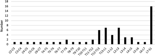
A total of 561 IVDS were reevaluated on MRI, with 63 having been identified as affected. Eight dogs had multifocal discospondylitis, with 4 having 2 affected discs, and individual dogs presenting with 7, 6, 5 and 4 affected discs. The distribution of infected sites is presented in Figure 1, with L7-S1 being the most commonly affected site 16/63 (25.4%).

A positive culture was obtained in 12/41 cases (28.2%), including Staphylococcus spp. (4/12), Streptococcus spp. (3/12), Brucella canis (2/12), and 1 occurrence each of Escherichia coli, Paecilomyces spp. and Serratia spp. Microbiological cultures were positive in ≥1 samples from samples of urine (n = 5), blood (4) including serological titers for Brucella canis (2), percutaneous FNA (2), surgical biopsy of affected intervertebral discs (2) or concomitant actively-infected lesions elsewhere in the body (1). All dogs experienced clinical improvement after antibacterial or antifungal medications.
3.2 Computed tomography findings
All 63 MRI-identified discospondylitis sites had available concurrently acquired CT images. Detailed descriptions of the CT features and their frequency are presented in Table 1.
| Region of interest | Assessed features for each region of interest | Number of occurrences | % (total sites or within altered assessed feature) |
|---|---|---|---|
| IVDS | Subjective size as compared to adjacent IVDS | ||
|
36/63 | 57.1 | |
|
27/63 | 42.9 | |
|
11/27 | 40.7 | |
|
11/27 | 40.7 | |
|
5/27 | 18.5 | |
| Concomitant disc herniation | 11/63 | 17.5 | |
| Gas within IVDS/vacuum artifact | 15/63 | 23.8 | |
| Periosteal proliferation adjacent to IVDS | 46/63 | 73.0 | |
| Ventral spondylosis | 42/63 | 66.7 | |
| Local DISH | 16/63 | 25.4 | |
| Endplates | 0 | 0.0 | |
| Endplate involvement | 55/63 | 87.3 | |
|
3/55 | 5.5 | |
|
52/55 | 94.5 | |
| Grading | |||
|
16/63 | 25.4 | |
|
39/63 | 61.9 | |
|
8/63 | 12.7 | |
| Osteolysis present | 55/63 | 87.3 | |
|
7/55 | 12.7 | |
|
37/55 | 67.3 | |
|
11/55 | 20.0 | |
| Sclerosis | 16/63 | 25.4 | |
| Vertebral body | Vertebral body involvement | 42/63 | 66.7 |
| Extent of abnormalities | |||
|
35/42 | 85.7 | |
|
6/42 | 14.3 | |
|
1/42 | 2.4 | |
| Osteolysis | 34/63 | 54.0 | |
|
8/34 | 23.5 | |
|
25/34 | 73.5 | |
|
1/34 | 2.9 | |
| Sclerosis | 23/63 | 36.5 | |
| Morphology and spatial position | 11/63 | 17.5 | |
|
10/11 | 90.9 | |
|
6/11 | 54.5 | |
|
2/11 | 18.2 | |
|
0/11 | 0.0 | |
| Epidural space | Involvement | 7/63 | 11.1 |
|
0/63 | 0.0 | |
|
2/63 | 3.2 | |
|
5/63 | 7.9 | |
|
0/63 | 0.0 | |
| Paraspinal tissues | Paraspinal involvement | 6/63 | 9.5 |
|
6/63 | 9.5 | |
|
0/63 | 0.0 |
All sites had at least 2 identifiable changes on CT. The most frequently found changes were: (1) endplate involvement (87.3%) affecting both sides in most cases (94.5%), endplate erosion (61.9%) and multifocal osteolysis (67.3%); (2) periosteal proliferation adjacent to the IVDS (73%) associated with spondylosis (66.7%); and (3) vertebral body involvement (66.7%) mostly of one-third of the vertebra (85.7%) with multifocal osteolysis (73.5%). Other features observed included an abnormal IVDS (42.9%) mostly characterized by narrowing (40.7%) or collapse (40.7%), sclerosis of the adjacent vertebral body (36.5%) or endplates (25.4%), presence of local DISH (25.4%) or local vacuum artifact (23.8%). Vertebral body morphology alterations (17.5%) consisted of both vertebral shortening and subluxation with no fractures found. Concomitant vertebral malformations were found in 2 dogs (an English Bulldog and French Bulldog).
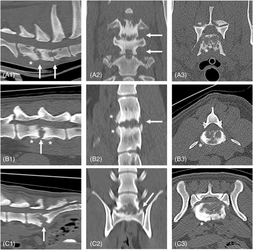
Both the epidural space and the paraspinal tissues were found to be involved in a minority of cases (11.1% and 9.5% of cases, respectively), with the presence of soft tissue opacity alterations or compressive lesions to the nerve roots, always found affecting 1 of the L7 nerves. Examples of some of the most common appearances of discospondylitis on CT found in our study are depicted in Figure 2.

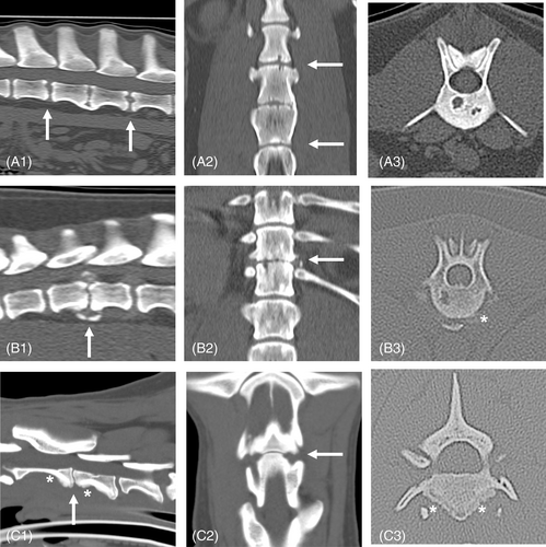
In sites where CT did not disclose endplate erosion, a detailed description of identifiable CT changes is provided in Supplemental information, highlighting other features of potential interest in equivocal cases. Intact endplates were found in 8 sites in 4 dogs: 1 case with multifocal discospondylitis, where 4 affected sites of 6 had intact endplates; 4 other cases had single site discospondylitis. The most commonly found CT change in these cases was periosteal proliferation (7/8). Examples of discospondylitis cases with less frequently identified features (cases with minimal or no endplate changes), are presented in Figure 3.

4 DISCUSSION
Magnetic resonance imaging is considered superior to other imaging modalities for the diagnosis of discospondylitis in both dogs and cats,4-6, 8 as well as in the counterpart spondylodiscitis (also termed vertebral osteomyelitis) in humans.7, 21 Magnetic resonance imaging offers several advantages including high contrast resolution, multiplanar imaging acquisition, high sensitivity to variations in structural water content, good visualization of the epidural space and degree of compression of neural structures, detailed depiction of spinal cord parenchyma and meninges, as well as visualization of bone marrow changes.4, 6 Despite these advantages, CT presents some advantages over MRI in the diagnosis of discospondylitis such as excellent depiction of bone, FNA guidance and enhanced utility in preoperative planning of spinal surgery in instances of subluxation or fractures. These capabilities are further improved when contrast studies are performed, particularly for observation of both the epidural space and paraspinal tissues. In our case series, plain CT was evaluated, and it is possible that contrast studies would have identified pathological changes in the epidural and paraspinal tissues in some cases. Other advantages of CT over MRI include increased availability, shorter acquisition times and occasionally not requiring general anesthesia (which could be an important consideration for systemically ill animals).
Classical imaging findings of discospondylitis on MRI include changes involving the IVDS, adjacent vertebral endplates and neighboring soft tissue with signal changes that are most commonly T2-weighted and STIR hyperintense signals along with contrast enhancement of ≥1 of these structures, commonly the intervertebral disc and the neighboring soft tissue.4, 6 These MRI features are considered virtually pathognomonic for infection,22 with intervertebral discs being only rarely affected by neoplasia.23, 24 Our findings supported this notion, with endplate involvement being the most prevalent finding (87.3%), in most cases with both endplates involved (94.5% of cases) along with endplate erosion (61.9%). Other differential diagnoses should be considered for endplate changes, including reactive changes, fatty infiltration of the body and endplates, vertebral osteochondrosis or Schmorl's nodes.25 These changes have been described previously on MRI but not CT. The MRI signal patterns of these changes can overlap with those of discospondylitis. In a previous case series describing endplate changes in dogs, most were deemed secondary to discospondylitis. Cases such as those depicted in Figure 3 and described in Supplemental information with subtle or no endplate changes can be problematic, and discospondylitis may not be considered. We consider the presence of bilateral endplate changes (i.e., osteolytic lesions even if subtle) as a warning sign of a possible discospondylitis. Furthermore, in the presence of clinical signs compatible with spondylodiscitis in humans, in particular when pain is of short duration, repeat advanced imaging (e.g., MRI) is recommended if early imaging is nondiagnostic but the clinical suspicion persists.26
Characteristics of discospondylitis on plain radiography have been well established including endplate erosion, endplate sclerosis, vertebral body osteolysis, IVDS morphology changes, osseous proliferation adjacent to the IVDS, spondylosis, soft tissue opacity alterations, as well as signs of vertebral fracture, subluxation or shortening.5, 27-29 Considering that CT is also an ionizing radiation modality, findings of discospondylitis on CT should, in principle, share similar features to those of plain radiography. Our findings confirm this hypothesis to be the case. Nonetheless, the excellent depiction of bone on CT permitted a more detailed description of endplate and vertebral body osteolysis (pattern and extension) as well as enhanced evaluation of sclerosis and more detailed information regarding vertebral body morphology. Osteolysis could be subclassified in categories and interestingly, although punctate osteolysis has been reported as a feature of discospondylitis in cats and horses,8, 20, 30 we found in the dogs of our study that osteolytic foci coalesced into larger regions of bone destruction in most cases (Figure 2C3). The possibility of multiplanar reconstructions and lack of superimposition of structures on images permitted identification of mild changes unlikely to be identifiable on plain radiography, such as cases in which only mild or no endplate erosion was identifiable (Figure 3 and Supplemental information).
Computed tomography also has growing importance in the identification of osseous proliferation, ventral spondylosis and DISH.19, 31 Periosteal proliferation was the second most common finding in our study (after endplate changes). Periosteal reactions are nonspecific findings that can occur in many situations, such as a response to injury, local inflammatory or infectious processes, neoplasia (local or distant) or metabolic conditions.18 Although its presence is nonspecific, when accompanied by clinical signs compatible with discospondylitis, the finding of vertebral periosteal proliferation along with endplate changes should be considered indicative of potential discospondylitis. Ventral spondylosis and DISH are frequently incidental findings more common in older dogs,19 with spondylosis being commonly associated with intervertebral disc protrusion.31 A causal relationship between spondylosis and discospondylitis is possible; but the most common locations of asymptomatic ventral spondylosis in previous studies (i.e., cranial lumbar, midthoracic portion of the vertebral column or L7-S1) also are some of the most commonly reported discospondylitis sites.3, 31
The presence of vertebral malformations at infected sites also was investigated in our study and was found in only 2 cases. A recent abstract proposed that vertebral malformations frequently were found in association with discospondylitis in screw-tailed brachycephalic dogs.9 We did not find such an association in our study population. However, few breeds typically affected by vertebral malformations (e.g., Pug, French Bulldog, English Bulldog) were represented. Other features observed for which CT has the potential of being superior to other imaging modalities included the presence of local vacuum artifact (23.8%) and vertebral body morphological changes. Vacuum phenomenon within the IVDS previously was reported in dogs with discospondylitis,16 but it is considered more common with intervertebral disc extrusion.17 Despite not being pathognomonic for discospondylitis, this phenomenon could be a helpful diagnostic imaging sign in conjunction with other more typically encountered features in the diagnosis of discospondylitis (Supplemental information).
The identification of fractures and luxations of the vertebral column is easier on CT as compared to plain radiography or MRI.32 In accordance with previous literature in which subluxation has been reported in 7% to 20% cases, subluxation was present in 9.5% of our cases. We did not encounter evidence of fractures in our population. Nonetheless, fracture remains a possible consequence of discospondylitis with a few instances of fractures being reported in dogs diagnosed with discospondylitis on radiographs or MRI.4, 6, 33
In equivocal cases, we found that evaluation of the endplates and a search for periosteal changes should be performed in all 3 planes. The importance of doing so was exemplified by cases where sagittal reconstructions did not clearly identify endplate changes, but dorsal or transverse planes allowed detection of faint osteolytic areas (Figure 3A1,B1). In cases without endplate erosion, other factors such as periosteal proliferation or the presence of a vacuum artifact could be warning signs of possible underlying discospondylitis in patients with compatible clinical signs. The clinical suspicion that CT could lead to the identification of osseous lesions earlier than radiography5 is yet to be confirmed, but the aforementioned identification of subtle endplate and periosteal changes supports this conjecture.
Our study had some limitations. Data were collected retrospectively and diagnosis of discospondylitis relied on clinical features and MRI evidence of a suspected infectious process affecting the IVDS, vertebral endplates or both. Diagnosis by MRI was superior to diagnosis by CT for discospondylitis in dogs in our study. Identification of an infectious agent was possible in <33% of the cases, which is a common problem in the diagnosis of discospondylitis in companion animals.3, 34 In our case series, approximately 24% (13/54) of the dogs with MRI-diagnosed discospondylitis were excluded because CT was not performed concomitantly with MRI. Thus, conclusions about signalment, duration of clinical signs, weight, distribution of infected sites of discospondylitis, and percentage of cases with positive microbiological culture cannot be directly compared with previous studies.3 However, we believe that the CT features we observed are representative of discospondylitis cases in dogs, having included approximately 76% of the total population of dogs presented to our referral hospital within a 10-year time period.
5 CONCLUSIONS
Our study highlights the CT features of discospondylitis in dogs. Common features included an abnormally-sized IVDS, with erosion of both endplates and multifocal osteolysis, periosteal proliferation adjacent to the affected site, and vertebral body changes most typically seen as multifocal osteolysis. We also identified cases in which only subtle changes were identifiable, emphasizing that even subtle bilateral endplate changes along with periosteal proliferation should alert the clinician of the possibility of underlying discospondylitis. Although these CT features can aid in the diagnosis of discospondylitis in dogs, equivocal cases might still require MRI, which remains the investigation method of choice in the diagnosis of discospondylitis in dogs.
ACKNOWLEDGMENT
No funding was received for this study.
CONFLICT OF INTEREST DECLARATION
Authors declare no conflict of interest.
OFF-LABEL ANTIMICROBIAL DECLARATION
Authors declare no off-label use of antimicrobials.
INSTITUTIONAL ANIMAL CARE AND USE COMMITTEE (IACUC) OR OTHER APPROVAL DECLARATION
Authors declare no IACUC or other approval was needed.
HUMAN ETHICS APPROVAL DECLARATION
Authors declare human ethics approval was not needed for this study.


