Noninvasive hydrostatic reduction of an ileocecocolic intussusception in a puppy
Abstract
A 5-week-old male intact Golden Retriever puppy was presented for a history of vomiting and diarrhea with hematochezia. Ultrasound findings confirmed the presence of an ileocecocolic intussusception. Surgical correction was declined because of financial concerns. Based on a pediatric procedure used in humans, an ultrasound-guided hydrostatic reduction (USGHR) was performed. This procedure consisted in injecting saline rectally under controlled pressure to mechanically reduce the intussusception. Reduction of the intussusception and evaluation of potential complications were concurrently evaluated by ultrasound during the procedure. No recurrence was observed the next day and the puppy was discharged. Follow-up indicated that the dog was still doing well 6 months later. This case report describes a new technique in veterinary medicine allowing successful nonsurgical reduction of an ileocecocolic intussusception in a dog. This procedure is innovative, simple, and substantially decreases the cost and minimizes morbidity potentially associated with surgical management.
Abbreviation
-
- USGHR
-
- ultrasound-guided hydrostatic reduction
1 INTRODUCTION
Intussusception is an invagination of 1 segment of the gastrointestinal tract into the lumen of another segment. The invaginated segment is the intussusceptum and the enveloping segment is called the intussuscepiens. Clinical signs are consistent with partial or complete bowel obstruction and include acute diarrhea, vomiting and abdominal pain.1, 2 In dogs, the etiology usually is unknown, but foreign bodies, intestinal parasites, gastroenteritis, neoplasia, previous abdominal surgery or any condition affecting intestinal motility are potential triggering factors.1 Ileocolic intussusception is the most common type in small animals.2, 3 A tentative diagnosis can be made clinically by palpation of an abdominal mass but definitive diagnosis usually requires ultrasound examination with identification of a characteristic target-like structure involving the intestines.4 Spontaneous reduction has been described, but surgical correction is commonly required.3, 5
In human medicine, a nonsurgical procedure called ultrasound-guided hydrostatic reduction (USGHR) has been used to correct ileocolic intussusception. It consists of filling the colon with saline using gravity-controlled hydrostatic pressure.6-9 This technique was developed in infants and children as a safe, noninvasive and efficient procedure.10, 11
This case report describes a similar nonsurgical procedure to correct ileocecocolic intussusception that has not yet been described in small animals.
2 CASE DESCRIPTION
A 5-week-old Golden Retriever intact male puppy was first presented to the referring veterinarian for evaluation of vomiting, diarrhea, hematochezia and inappetence over the past 2 days.
On admission, physical examination showed only mild lethargy. A fecal parvovirus snap test (Idexx SNAP parvo) was negative. Abdominal radiographs were recommended but declined by the owner. The puppy then was discharged with symptomatic treatment consisting of maropitant 1 mg/kg SC, metronidazole 10 mg/kg PO q12h and fenbendazole 50 mg/kg PO q24h for 5 days.
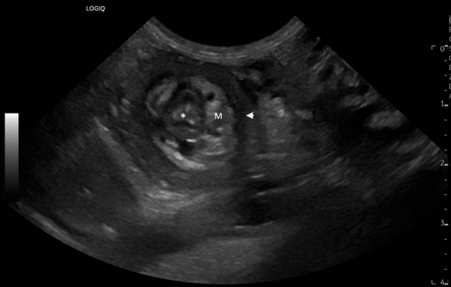
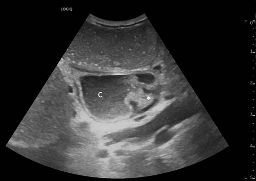
The dog did not improve and was referred to the emergency department 4 days after initial presentation. On admission, the puppy was lethargic, had mild abdominal distension and was mildly to moderately dehydrated. Abdominal ultrasound examination (GE LOGIQ E10) identified an ileocecocolic intussusception (Figure 1), mechanical ileus, generalized steatitis and mild abdominal effusion. The ileum was completely invaginated into the proximal colon. Blood flow was still visualized within the intussusceptum on Doppler interrogation.
Surgical management was recommended but declined by the owner because of financial constraints. Consequently, the possibility of attempting the ultrasound-guided reduction was discussed and accepted by the owner.
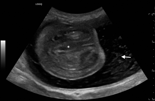
The dog was placed in dorsal recumbency for the procedure. The perineal region was clipped. The dog was sedated with butorphanol (0.4 mg/kg IV) and midazolam (0.2 mg/kg IV). A well-lubricated (18F) Foley catheter was inserted into the rectum and the balloon inflated with 10 mL of air to provide a tight closure. A bag of prewarmed 0.9% sodium chloride was suspended 100 cm above the level of the patient. The Foley catheter then was connected to the bag of 0.9% saline and the colon was allowed to fill by gravity. The retrograde flow of saline and reduction of intussusception were continuously monitored by ultrasound (Figure 2).
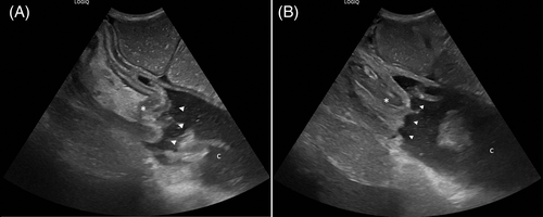
For the first attempt, the bag of saline was set 100 cm above the patient (corresponding to a 100 cm H2O pressure) until the colon was fully filled. Once the colon was full and maximum pressure was achieved (determined by a cessation of saline flow), the colon was kept under maximal pressure for 2 minutes with ultrasound monitoring (Figure 3A,B). At the first attempt, reduction was only partially achieved. A second reduction was performed 15 minutes later. For the second attempt, the bag of saline was placed approximately 120 cm above the patient. After 2 minutes at maximal pressure, the bag was elevated to 135 cm above the patient for an additional minute until complete reduction was observed by ultrasound (Figure 4).
An abdominal ultrasound examination performed 15 minutes after the second procedure indicated partial recurrence of the intussusception. A new reduction was attempted using the saline bag set at a height of 135 cm above the patient. Additional sedation using a 2 mg/kg IV propofol bolus was performed during the third attempt. After approximately 1 minute at a pressure of 135 cm H2O, the intussusception fully resolved again. A repeat abdominal ultrasound examination performed 2 hours after the reduction did not show any recurrence.
The dog was monitored in the hospital overnight to identify any potential complications and was discharged the next day without any sign of recurrence on ultrasound examination. A phone follow-up with the owner 2 weeks later and again >6 months later indicated that the dog was doing well with no recurrence of gastrointestinal signs.
3 DISCUSSION
In the current report, we describe the use of USGHR as an alternative to a surgery for treatment of an ileocolic intussusception in a puppy.
Intussusception causes signs of partial or complete bowel obstruction.2, 12 The invaginated bowel may experience ischemia, which can cause necrosis.12 In both small animals and children, the site most commonly involved is the ileocecal area.2, 3
Successful USGHR initially was performed in human medicine with the objective of providing an alternative to hydrostatic reduction using barium enema and fluoroscopy.7 This technique is used widely in pediatric medicine and is associated with decreased duration of hospitalization and decreased mortality. Morbidity associated with anesthesia and surgery also is decreased.13 In addition, the use of ultrasound instead of fluoroscopic guidance in children eliminates exposure to radiation and allows for wider availability and easier set-up of the procedure.12, 14 Because ultrasonography has become an effective modality to diagnose intussusception, saline enema is considered a valuable nonsurgical alternative to barium or pneumatic enemas in the management of intussusception in children.10, 12, 14, 15
Affected human patients are placed in lateral or dorsal recumbency and a Foley catheter is inserted into the rectum with the balloon inflated using air or saline.10, 11, 16-19 The catheter size used ranges from 10 to 24 F.11, 16, 19 In our case, a tight seal was achieved with the help of an assistant tightening the end of the catheter. This technique is not perfect, and saline leakage around the catheter may occur during the procedure leading to a decrease in pressure within the colon. A purse-string suture could be considered as an alternative to minimize saline leakage.
To limit potential complications and uncontrolled increases in pressure, saline is infused into the colon by gravity. The pressure is directly correlated with the height of the saline bag, which is usually set between 100 and 150 cm above the patient.10, 11, 16, 18, 19 The optimal pressure necessary to reduce intussusception in dogs is not known and we followed the guidelines used in human pediatric medicine. In human medicine, up to 3 or 4 attempts are usually attempted.11, 16, 18, 19 There is no clear consensus regarding the duration of each attempt and the duration of the interval between reductions. Previous studies recommended approximately 15 to 20 minutes reduction and an interval of several hours between 2 enemas.11, 18 Another study did not exceed 5 minutes of reduction time with an interval of <3 minutes between attempts.15 A final study maintained pressure for 30 seconds after filling and repeated the saline enema after a 1-minute interval if the intussusception was not reduced.19 We performed 3 attempts with a maximum of 3 minutes (at maximal pressure) in duration. Between 2 reductions, an interval of 15 minutes was used. This protocol allowed successful reduction with no recurrence at the third attempt. In human medicine, success rate with ultrasound-guided hydrostatic reduction is >80% in children (81.5%-96.8%).10, 11, 16-19 In children, a recent prospective study showed that the success rate of USGHR (96.8%) was significantly higher than that of pneumatic reduction (83.9%).17
Compared to surgical management, USGHR does not allow assessment of the bowel integrity and possible tissue necrosis. In the case of suspected intestinal tissue necrosis, surgical management is recommended and intestinal resection and anastomosis should be performed if deemed necessary.3 For this reason, USGHR carries a risk of bowel perforation if a large volume of saline is instilled under pressure. In the human pediatric population, the reported risk of perforation remains very low at <1%.11, 17 Abdominal ultrasonography or fluoroscopy performed during the procedure allows for monitoring of increasing volume of free abdominal fluid or gas which could be suggestive of bowel perforation.
In veterinary medicine, intussusceptions commonly are treated by surgical reduction. Several intestinal surgical procedures are used to reduce intussusceptions, laparotomy with manual reduction and intestinal resection and anastomosis being the most common procedures. Enteroplication may be performed to prevent future recurrences.3
Only a few studies have explored nonsurgical management of intussusception in small animals. In 27 experimentally-induced ileocolic intussusception dogs, laparoscopic-assisted pneumatic reduction by insufflation of carbon dioxide through the rectum was successful in 26/27 (96%) of cases and 1 perforation occurred.20 Incomplete reduction was first observed in 3/26 dogs. Successful reduction finally was obtained by using a higher carbon dioxide pressure and grasping forceps in these dogs. This method allowed real time monitoring of the reduction and assessment complications, but required general anesthesia as well as specialized equipment and expertise.20 A recent report described nonsurgical management of rectal prolapses leading to colocolic intussusception in 2 puppies.21 Saline enema and manual rectal reduction of the prolapses were used in combination in both dogs to obtain complete reduction. Real time reduction was not confirmed or visualized. The role of saline enema in the successful reduction of the intussusception was not evaluated, and the authors did not know whether spontaneous reduction, rectal manipulation alone or a combination of manipulations and saline enema led to complete reduction.
Spontaneous reduction also has been described in children and dogs.5, 22 Spontaneous reduction in children was reported in up to 17% of cases over a 6-year period in 1 study.22 In dogs, spontaneous reduction appears to be less common. In a case series, 5 of 63 dogs had spontaneous reduction of their intussusceptions.5 Median duration of clinical signs appeared to be shorter for dogs with spontaneous reduction of intussusception (median, 2 days; range, 1-3 days) than in those in which surgery was performed (median, 8.63 days; range, 3-20 days). In human medicine, there is no consensus on whether duration of clinical signs is associated with successful USGHR. Some studies suggested that a longer duration of clinical signs is associated with unsuccessful USGHR and may be considered as an important predictive factor for outcome of nonsurgical management of intussusception.11, 17, 23 However, other reports showed that the duration of clinical signs did not influence the reduction of intussusception using enemas.17, 24 In dogs, duration of clinical signs does not seem to be a reliable predictor of intestinal viability.3 In our case, the patient experienced clinical signs for at least 5 to 6 days before reduction, suggesting saline enema could be efficient several days after the onset of clinical signs.
Our case report describes a new noninvasive procedure to correct ileocecocolic intussusception in dogs. This technique does not require specialized training and provides veterinarians with an alternative less expensive and less invasive option compared to conventional surgery. We suggest using a large Foley catheter so as to quickly infuse a large amount of saline into the colon and obtain good occlusion of the rectum. Manual compression of the anus or a purse string suture might be necessary to completely occlude the rectum. We recommend starting the infusion with the saline bag approximately 100 cm above the patient and to increase the height (and therefore pressure) if necessary to obtain reduction of the intussusception. A maximum pressure of 135 cm H2O was sufficient in our case. According to the human medical literature, a pressure of 150 cm H2O should be avoided. Several attempts may be made and a maximum of 3 to 5 attempts is considered reasonable. Abdominal ultrasound monitoring is recommended throughout the procedure to identify any evident leakage from the colon and to visualize reduction of the invagination. Additional studies are necessary to assess efficiency, safety and repeatability of this procedure in small animals.
ACKNOWLEDGMENT
No funding was received for this study. The authors thank the technicians from the internal medicine service for their help during this procedure.
CONFLICT OF INTEREST DECLARATION
Authors declare no conflict of interest.
OFF-LABEL ANTIMICROBIAL DECLARATION
Authors declare no off-label use of antimicrobials.
INSTITUTIONAL ANIMAL CARE AND USE COMMITTEE (IACUC) OR OTHER APPROVAL DECLARATION
Authors declare no IACUC or other approval was needed.
HUMAN ETHICS APPROVAL DECLARATION
Authors declare human ethics approval was not needed for this study.


