Elimination of fibrin polymer formation or crosslinking, but not fibrinogen deficiency, is protective against diet-induced obesity and associated pathologies
Abstract
Background
Obesity predisposes individuals to metabolic syndrome, which increases the risk of cardiovascular diseases, non-alcoholic fatty liver disease (NAFLD), and type 2 diabetes. A pathological manifestation of obesity is the activation of the coagulation system. In turn, extravascular fibrin(ogen) deposits accumulate in adipose tissues and liver. These deposits promote adiposity and downstream sequelae by driving pro-inflammatory macrophage function through binding the leukocyte integrin receptor αMβ2.
Objectives
An unresolved question is whether conversion of soluble fibrinogen to a crosslinked fibrin matrix is required to exacerbate obesity-driven diseases.
Methods
Here, fibrinogen-deficient/depleted mice (Fib- or treated with siRNA against fibrinogen [siFga]), mice expressing fibrinogen that cannot polymerize to fibrin (FibAEK), and mice deficient in the fibrin crosslinking transglutaminase factor XIII (FXIII–) were challenged with a high-fat diet (HFD) and compared to mice expressing a mutant form of fibrinogen lacking the αMβ2-binding domain (Fib𝛾390-396A).
Results and Conclusions
Consistent with prior studies, Fib𝛾390-396A mice were significantly protected from increased adiposity, NAFLD, hypercholesterolemia, and diabetes while Fib- and siFga-treated mice gained as much weight and developed obesity-associated pathologies identical to wildtype mice. FibAEK and FXIII– mice displayed an intermediate phenotype with partial protection from some obesity-associated pathologies. Results here indicate that fibrin(ogen) lacking αMβ2 binding function offers substantial protection from obesity and associated disease that is partially recapitulated by preventing fibrin polymer formation or crosslinking of the wildtype molecule, but not by reduction or complete elimination of fibrinogen. Finally, these findings support the concept that fibrin polymerization and crosslinking are required for the full implementation of fibrin-driven inflammation in obesity.
ESSENTIALS
- Fibrin(ogen) deposits in tissues promote obesity and associated metabolic sequalae.
- Fibrinogen deficiency or depletion does not protect against high-fat diet–induced obesity.
- Selectively eliminating fibrin polymerization and crosslinking partially protects against obesity-related pathologies.
1 INTRODUCTION
Obesity, defined as body mass index of >30 kg/m2, is a global health issue and affects more than 40% of the US population.1 Obesity is characterized by the pathological accumulation of fatty acids in tissues that alters healthy metabolic function (e.g., glucose homeostasis). As such, obesity is a risk factor for metabolic syndrome, a constellation of pathological conditions that increases the risk for non-alcoholic fatty liver disease (NAFLD), type 2 diabetes, insulin resistance, and cardiovascular diseases.2-4 Enlarged visceral adipose tissues, such as omental and mesenteric white adipose tissue (WAT) in humans and epididymal WAT (eWAT) in rodents, is associated with increased macrophage-driven inflammation that exacerbates insulin resistance and subsequently metabolism of glucose, cholesterol, and lipids.5
A mechanistic underpinning for many of the obesity-associated sequelae is that obesity promotes a procoagulant and anti-fibrinolytic state that ultimately increases the risk of cardiovascular events. Circulating levels of procoagulant proteins such as von Willebrand factor, factor VII, and fibrinogen are commonly elevated in obese individuals.6-9 Further, elevated levels of biomarkers of coagulation activity, including thrombin–antithrombin complexes, thrombin f1 + 2, and D-dimer are routinely observed in obese individuals.10, 11 Importantly, both initial risk and severe morbidity and mortality outcomes for vaso-occlusive diseases, including coronary artery disease and stroke, are positively correlated with the degree of obesity in patients.12, 13 Collectively, these observations support a causal link between obesity and an increased risk for thromboembolic disease.
Elevated coagulation system activity is not just a consequence of obesity but is also a contributing factor to the severity of obesity, metabolic syndrome, and associated sequelae. Extravascular fibrin(ogen) deposits accumulate in the adipose tissue and the liver of obese individuals.14 Fibrin(ogen) co-localizes with pro-inflammatory macrophages within crown-like structures of obese white adipose tissue and areas surrounding steatosis in the liver.14 Genetically modified Fibγ390-396A mice, which express fibrinogen with a mutated integrin αMβ2 binding motif, were previously shown to be protected from developing high fat diet (HFD)-induced obesity, insulin resistance, and NAFLD.14 These studies supported a mechanism of action whereby fibrin-driven macrophage-mediated metabolic inflammation promotes HFD-induced obesity and associated pathologies, such as NAFLD and insulin resistance.
Fibrinogen contains a cryptic binding site for the integrin receptor αMβ2, expressed by macrophages and neutrophils, such that only immobilized fibrinogen or fibrin matrix, and not soluble fibrinogen, engages the receptor.15, 16 An unresolved question is whether the αMβ2−binding motif requires fibrin polymerization and crosslinking for the full implementation of fibrin(ogen)-mediated HFD-induced obesity and associated pathologies. In this study, the contribution of fibrinogen itself in promoting HFD-driven obesity was explored using Fibγ390-396A mice and mice that are genetically deficient or pharmacologically depleted in fibrinogen (Fib- and siRNA targeting fibrinogen [siFga]-treated, respectively).17-19 In addition, to differentiate between distinct fibrin(ogen) molecular forms, we utilized mice expressing fibrinogen locked in the monomeric form (FibAEK) and mice deficient in the fibrin crosslinking transglutaminase factor XIII (FXIII–).20, 21
2 MATERIALS AND METHODS
2.1 Mice
Animal experiments and protocols were approved by the Animal Care and Use Committee of the University of North Carolina at Chapel Hill. Fibγ390-396A, Fib–, FibAEK, and FXIII– mice were all previously described.18-21 Age-matched littermates of male mice between the ages of 6 and 10 weeks were used. For siFga experiments, male C57BL/6J mice were injected with 2 mg/kg of lipid nanoparticles containing siRNA against luciferase (siLuc) or fibrinogen Aα-chain (siFga) 1 week before the start of the HFD challenge and subsequently every 10–11 days to minimize fluctuations in circulating fibrinogen levels.17 Mice were fed ad libitum low-fat diet (LFD, 10–13% fat; Laboratory Autoclavable Rodent Diet 5010, LabDiet or D12450, Research Diets) or HFD (60% fat; catalog D12492, Research Diets). The total body weights of mice and the weight of food consumed were determined weekly. At the end of the observation period, mice were anesthetized before plasma and organs were harvested, weighed, and processed for further analysis.
2.2 RNA isolation, cDNA synthesis, and real-time quantitative polymerase chain reaction
Total RNA was isolated from tissues using Trizol (Ambion) according to the manufacturer’s protocol. RNA (1 μg) was used for cDNA synthesis with a High-Capacity cDNA Reverse Transcription kit (Applied Biosystems). Levels of Tnfa, Ccl2, Adgre1, Pparg, Cidea, Cd36, Hmgcr, Cyp7a1, Cyp27a1, Ucp1, and B2m were determined using TaqMan gene expression assays (Applied Biosystems) on an ABI StepOne Plus sequence detection system (Applied Biosystems). The expression of each gene was normalized relative to B2m expression levels using the Pfaffl method.22
2.3 Analysis of diabetes and insulin resistance
Mice were fasted for at least 6 h prior to analysis of circulating glucose levels, as previously described.14 For glucose tolerance tests (GTT) performed on week 16 of diet, dextrose was administered by i.p. injection (1 g/kg). For insulin tolerance tests (ITT) performed on mice on week 18, human recombinant insulin (Humulin; Eli Lilly) was administered by i.p. injection (1.5 U/kg). Blood glucose readings were taken at 15-minute intervals.
2.4 Analysis of NAFLD and hypercholesterolemia
Hepatic triglyceride content was determined as described previously.14 Briefly, frozen liver tissue was homogenized and analyzed spectrophotometrically for triglycerides using commercially available reagents (Pointe Scientific). Plasma alanine transferase (ALT) activity was determined using a commercially available reagent (Thermo Fisher Scientific). ELISA was used to determine the circulating fibrinogen levels (Immunology Consultants Laboratory). Circulating levels of total, high-density lipoprotein (HDL), and free cholesterol were measured using commercially available kits (Wako). The left lateral lobe of the liver was used for histological analysis. Formalin-fixed tissue sections (5 μm) were stained with hematoxylin and eosin (H&E) stain as well as fibrin(ogen) antibody (DAKO). Extravascular deposition of fibrin was analyzed using automated capillary western blotting as previously described.14
2.5 Statistics
All analyses were performed using Prism 9. Comparisons of multiple groups were performed using two-way analysis of variance and Fisher’s least significant difference test. Results were considered significant when p < .05.
3 RESULTS
3.1 Obesity, adiposity, and white adipose tissue inflammation
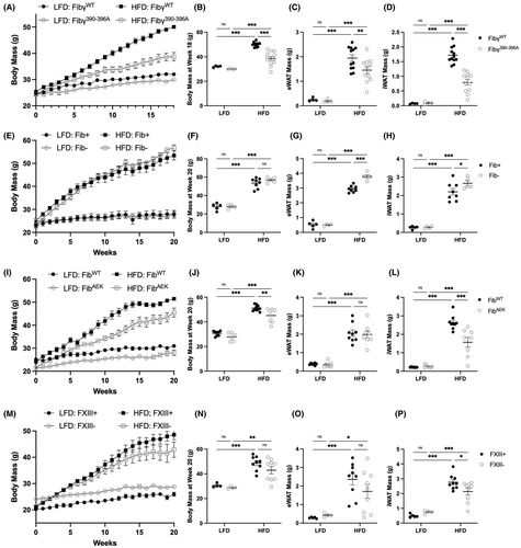
To determine if fibrinogen, fibrin, and crosslinked fibrin matrix each contribute to the development of HFD-induced obesity, Fib–, FibAEK, and FXIII– mice were fed a LFD or HFD diet for 20 weeks and compared to Fibγ390-396A mice. Consistent with previous reports, Fibγ390-396A mice were protected from an HFD-driven increase in total body mass (Figure 1A,B) and had smaller eWAT and subcutaneous inguinal WAT (iWAT) mass after 20 weeks on diet (Figure 1C,D) relative to HFD-fed FibγWT mice.14 To examine the impact of fibrinogen deficiency on HFD-induced obesity, Fib– mice were challenged with HFD. Surprisingly, Fib– mice gained as much weight as Fib+ mice on HFD and had larger eWAT and iWAT mass than Fib+ mice (Figure 1E-H). Complementary studies with pharmacological knockdown of fibrinogen were also performed. Here, by administering a lipid nanoparticle containing siRNA directed against the fibrinogen Aα-chain (siFga), circulating fibrinogen was reduced to ~10% compared to control mice treated with siLuc. Over the course of 20 weeks, siFga-treated mice gained as much weight as siLuc-treated mice on HFD and displayed an equivalent increase in eWAT and iWAT mass (Figure S1A-E in supporting information). To examine the impact of fibrin polymerization, FibAEK mice were challenged with HFD; FibAEK mice displayed modest but significant protection from HFD-induced obesity, with a significant reduction in total body weight and iWAT mass at 20 weeks, but comparable eWAT mass relative to HFD-challenged FibWT mice (Figure 1I-L). Finally, to examine the impact of fibrin crosslinking, FXIII– mice were challenged with HFD; FXIII– mice gained as much weight as FXIII+ mice on HFD (Figure 1M,N). HFD-fed FXIII mice had an eWAT mass comparable to FXIII+ mice, but the mass of the iWAT was significantly lower in FXIII– mice relative to FXIII+ mice after 20 weeks on a HFD (Figure 1O,P), similar to previous reports.14, 23
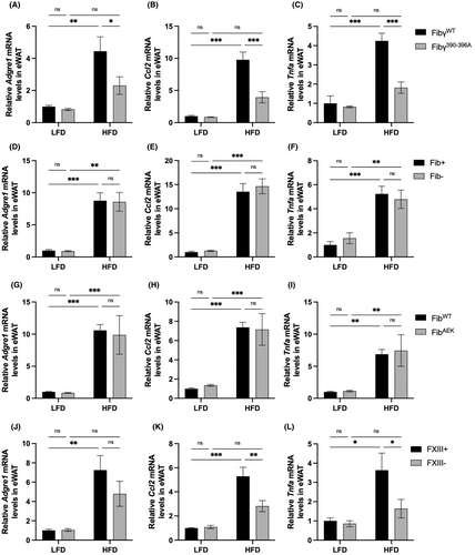
The development of metabolic syndrome is strongly associated with increased local inflammation in the eWAT.5 To examine local inflammatory changes, mRNA levels of proinflammatory genes Adgre1 (F4/80 receptor), Ccl2 (monocyte chemoattractant protein [MCP]-1), and Tnfa (tumor necrosis factor α) were measured. Comparative studies were performed in Fibγ390-396A, Fib–, FibAEK, and FXIII– mice challenged with HFD to determine whether distinct molecular forms of fibrin(ogen) alter the local expression of macrophage-associated inflammatory mediators within the eWAT. As expected, levels of Adgre1, Ccl2, and Tnfa were low, with no genotype-dependent differences in LFD-fed mice across each of the genotypes analyzed (Figure 2A-L). Adgre1, Ccl2, and Tnfa levels were significantly decreased in eWAT of HFD-fed Fibγ390-396A mice compared to HFD-fed FibγWT mice (Figure 2A-C). However, there were no significant genotype-dependent differences in the expression of Adgre1, Ccl2, or Tnfa in HFD-fed Fib– and FibAEK mice relative to their respective controls. Ccl2 and Tnfa, but not Adgre1 levels were significantly decreased in eWAT of HFD-fed FXIII– relative to HFD-fed FXIII+ mice (Figure 2D-L). Overall, protection from HFD-induced eWAT adiposity was associated with reduced local markers of macrophage-driven inflammation in eWAT.
3.2 NAFLD, hypercholesterolemia, and diabetes
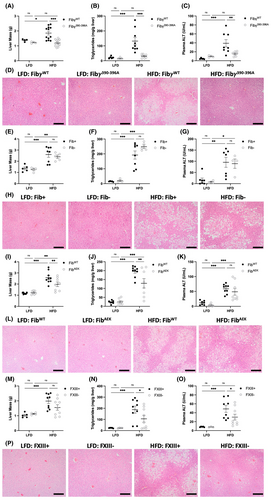
NAFLD is a spectrum disorder that begins as elevated lipid accumulation within hepatocytes (steatosis) that can progress to hepatic inflammation and non-alcoholic steatohepatitis (i.e., NASH).4 Consistent with previous findings, HFD-fed Fibγ390-396A mice had smaller liver mass as well as reduced hepatic triglyceride accumulation and circulating ALT activity relative to HFD-challenged FibγWT mice, indicative of reduced hepatic steatosis and hepatocellular damage, respectively (Figure 3A-C). Histological analysis of liver tissues also revealed evidence of fatty liver disease. Liver sections of HFD-challenged FibγWT mice displayed the accumulation of lipid within the hepatocytes as evidenced by the appearance of clear macrovesicular (large) and microvesicular (small) droplets within the cells. These droplets were either reduced or absent in sections from HFD-challenged Fibγ390-396A mice and LFD-fed mice of each genotype (Figure 3D). In contrast, elimination of fibrinogen, either genetically or pharmacologically, did not reduce HFD-induced NAFLD development. No differences in liver mass, hepatic triglyceride accumulation, circulating ALT activity, or liver tissue histopathology were observed comparing HFD-fed Fib+ and Fib– mice or HFD-fed siLuc-treated and siFga-treated mice (Figure 3E-H, Figure S1F-I). The severity of NAFLD in HFD-fed FibAEK mice appeared intermediate to wildtype (WT) and Fibγ390-396A mice. Liver mass and hepatic triglyceride accumulation were significantly reduced in HFD-fed FibAEK mice compared to FibWT mice (Figure 3I,J). However, ALT activity induced by HFD only trended lower in FibAEK mice relative to FibWT animals (Figure 3K). The liver histology was also consistent with reduced hepatosteatosis in HFD-fed FibAEK mice(Figure 3L). Intriguingly, FXIII– mice were protected from HFD-driven increase in hepatic triglyceride accumulation and circulating ALT activity (Figure 3M-O). Moreover, liver histology revealed less apparent hepatosteatosis in HFD-fed FXIII– mice compared to FXIII+ mice (Figure 3P).
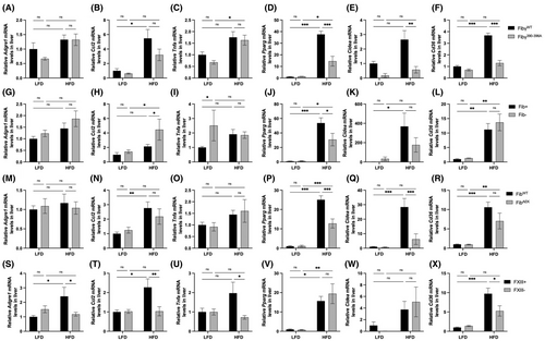
Hepatic fibrin content was measured to determine whether NAFLD severity was associated with changes in the level of extravascular fibrin deposits in the liver. However, there were no genotype-dependent changes in hepatic fibrin(ogen) deposition in the liver measured immunohistochemically or biochemically using capillary western blotting in HFD-fed Fibγ390-396A, FibAEK, and FXIII– mice compared to their respective control cohorts (Figure S2 in supporting information). Local hepatic mRNA levels were measured for proinflammatory mediators, such as Adgre1, Ccl2, and Tnfa, as well as markers of lipid metabolism including the nuclear receptor Pparg, the fatty acid sensor Cidea, and fatty acid transporter Cd36. As expected, the HFD-driven increases in mRNA levels of Pparg, Cidea, and Cd36 observed in FibγWT mice were reduced in livers from HFD-fed Fibγ390-396A mice (Figure 4A-F). In contrast, hepatic mRNA levels of Adgre1, Tnfa, Cidea, and Cd36 were not significantly different between Fib+ and Fib– mice (Figure 4G-L). In HFD-fed FibAEK mice, inflammatory mRNA levels were similar to FibWT mice. However, there was protection from HFD-driven increases in Pparg and Cidea levels in HFD-fed FibAEK mice compared to HFD-fed FibWT mice. Additionally, Cd36 levels trended lower in HFD-fed FibAEK mice relative to HFD-fed FibWT mice (Figure 4M-R). For FXIII– mice, HFD-induced hepatic mRNA levels of Adgre1, Ccl2, Tnfa, and Cd36, but not Pparg and Cidea, were lower relative to HFD-fed FXIII+ mice (Figure 4S-X). Collectively, studies of HFD-driven changes in liver weight, hepatic triglyceride accumulation, circulating ALT activity, liver histology, and hepatic gene expression suggest that prevention of fibrin polymerization and crosslinking help to suppress the development of NAFLD in a manner that mimics, but does not fully recapitulate, elimination of the fibrin(ogen)–αMβ2 binding motif.
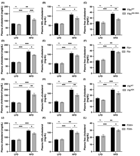
To evaluate the contribution of fibrin(ogen) in HFD-induced hypercholesterolemia, total cholesterol, HDL cholesterol, and free cholesterol levels in plasma isolated from LFD- and HFD-challenged mice were measured. As expected, cholesterol was significantly increased in all mice following the HFD challenge (Figure 5A,D,G,J). HFD-fed Fibγ390-396A mice had significantly reduced levels of total, HDL, and free cholesterol compared to HFD-challenged FibγWT mice (Figure 5A-C). Fib– mice were modestly protected from HFD-driven increase in total circulating cholesterol, but not in HDL– and free cholesterol compared to Fib+ mice (Figure 5D-F). Both FibAEK and FXIII– mice displayed modest protection against HFD-induced hypercholesterolemia, with significantly reduced levels of total, HDL, and free cholesterol relative to HFD-challenged control mice (Figure 5G-L). The changes in cholesterol observed in Fibγ390-396A, FibAEK, and FXIII– cohorts were not linked to changes in hepatic gene expression of key cholesterol regulatory molecules. Specifically, no genotype-dependent differences were observed in the hepatic mRNA levels of the cholesterol biosynthesis rate-limiting enzyme HMG-CoA reductase (Hmgcr) (Figure S3 in supporting information). In addition, hepatic gene expression of CYP7A1 (Cyp7a1) and CYP27A1 (Cyp27a1), key enzymes regulating the classical and alternative pathways of bile acid synthesis, respectively, were analyzed. For Cyp7a1, no differences were observed in HFD-fed Fibγ390-396A, Fib–, and FibAEK mice compared to their respective controls, whereas HFD-fed FXIII– mice had modestly higher hepatic levels than FXIII+ mice (Figure S3). For Cyp27a1, no differences in HFD-fed control and Fibγ390-396A, FibAEK, and FXIII– were observed whereas HFD-fed Fib– mice had modestly lower hepatic levels than Fib+ mice (Figure S3). These findings suggest that although fibrin(ogen) is a determinant of circulating cholesterol, the impact of fibrinogen occurs through mechanisms independent of altered hepatic cholesterol metabolism.
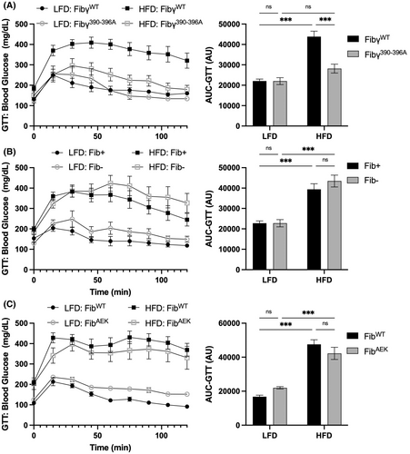
The contribution of fibrin(ogen) to the development of HFD-driven diabetes and insulin resistance was also examined. As expected, control mice in each HFD group displayed a significant prolongation in blood glucose clearance relative to LFD control animals in glucose tolerance tests (Figure 6, Figure S1J). Notably, only HFD-challenged Fibγ390-396A mice, but not Fib–, siFga-treated and FibAEK mice, exhibited a significantly improved glucose clearance relative to the HFD-challenged control mice (Figure 6A-C, Figure S1J). A previous study demonstrated that FXIII– also respond similar to WT mice following HFD challenge in a glucose tolerance test.23 Similarly, the HFD-challenged Fibγ390-396A mice, but not HFD-challenged Fib–, siFga-treated, FibAEK, and FXIII– mice, exhibited a significant improvement in response in an insulin tolerance test relative to their respective control groups (Figures S1K and S4 in supporting information), suggesting that changes in glucose homeostasis are particularly sensitive to elimination of the fibrin(ogen)–αMβ2 binding motif, but not perturbation of fibrin polymerization and crosslinking.
3.3 Brown adipose tissue and thermogenesis
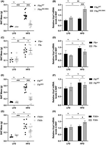
The contribution of fibrin(ogen) to HFD-driven reactive changes in brown adipose tissue (BAT) were also examined. In addition, mRNA levels of the regulator gene of facultative thermogenesis, uncoupling protein 1 (Ucp1), was determined. The HFD-driven increase in the BAT mass of HFD-challenged FibγWT mice was reduced in HFD-challenged Fibγ390-396A mice (Figure 7A). The mRNA level of Ucp1 was elevated in HFD-fed Fibγ390-396A mice relative to FibγWT mice (Figure 7B). In HFD-challenged Fib– and siFga-treated mice, there were no differences in BAT mass (Figure 7C, Figure S1L) nor Ucp1 mRNA levels (Figure 7D). As with Fibγ390-396A mice, the BAT mass of HFD-challenged FibAEK mice was reduced relative to HFD-challenged FibWT mice (Figure 7E). However, Ucp1 mRNA levels were equivalent between the two genotypes (Figure 7F). HFD-fed FXIII– mice had smaller BAT mass as well as modest increase in Ucp1 expression relative to FXIII+ mice (Figure 7G-H).
4 DISCUSSION
The key objective of this study was to determine the molecular forms of fibrin(ogen) that contribute to the development of HFD-induced obesity and associated metabolic sequelae in mice. The present analysis was motivated by previous observations that the fibrinogen integrin αMβ2 binding motif is a potent driver of metabolic inflammation, leading to increased adiposity, fatty liver disease, and diabetes.14 An unresolved question has been whether the different molecular forms of fibrinogen (i.e., immobilized monomer, fibrin matrix, crosslinked fibrin matrix) confer distinct or graded effects on disease processes that have been linked to the αMβ2 binding motif. In this report, we show that fibrin polymerization significantly promotes the development of HFD-induced adiposity, NAFLD, and hypercholesterolemia while FXIII significantly promotes NAFLD and hypercholesterolemia. Moreover, contrary to our initial expectations, fibrinogen deficiency did not confer protection against HFD-driven obesity or any of the associated metabolic pathologies.
The binding site for the αMβ2 integrin on fibrinogen is cryptic such that it is inaccessible in soluble fibrinogen, but becomes exposed by immobilization on a surface or by conversion to an insoluble fibrin matrix.15, 16 FibAEK mice express fibrinogen that cannot be converted to fibrin (i.e., locked in the monomeric form) due to a mutation in the α-chain thrombin cleavage site.20 Notably, the αMβ2 binding motif in fibrinogenAEK remains otherwise unperturbed. Here, we found that FibAEK mice were partially protected from an increase in HFD-driven adiposity and NAFLD, although Fibγ390-3906A mice had a greater degree of protection. Interestingly, FibAEK mice were partially protected from development of hepatic steatosis and iWAT adiposity, but not eWAT adiposity. Enlarged visceral adipose, such as the eWAT depot, is most associated with an increased chronic inflammation and subsequently exacerbated metabolic dysregulation.5 Consistently, gene expression of inflammatory markers in eWAT of HFD-fed FibAEK mice were similar to that of FibWT mice. The observed intermediate phenotype of HFD-fed FibAEK mice relative to that of HFD-fed WT mice and Fibγ390-396A mice is consistent with the concept that whereas fibrinogen can support αMβ2-driven activity on obesity and associated sequalae, fibrin polymerization is required for the maximum function of the γ-chain αMβ2 binding motif.
The observation that FibAEK mice display some protection from HFD-induced obesity and metabolic sequelae is also consistent with previous studies examining mice administered dabigatran, a direct thrombin inhibitor.14 In both prophylactic and therapeutic approaches, dabigatran-treated mice displayed protection from HFD-induced obesity and metabolic syndrome. Although it is difficult to make direct comparisons across studies, the protection conferred by the FibAEK mutation appears to be less pronounced than that provided by dabigatran. One possible explanation for this difference in phenotype could be that thrombin levels are lower in FibAEK mice compared to FibWT mice. However, analysis of plasma thrombin–antithrombin complexes (TATs), a marker of thrombin generation, revealed no genotype-dependent differences between control mice and Fib–, FibAEK, and FXIII– mice on HFD (data not shown). Another explanation is that dabigatran would inhibit the activation of all downstream thrombin targets, including protease-activated receptor (PAR)-1 and FXIII.23, 24 Notably, PAR-1−/− mice on Western diet have been shown to gain as much weight as WT mice but are completely protected from NAFLD.24 Similarly, as documented here and elsewhere, HFD-challenged FXIII– mice are protected from several aspects of metabolic syndrome.23 Studies examining the obesity phenotype of double or triple mutant mice could be designed (e.g., FibAEK/PAR-1−/− or FibAEK/FXIII– mice) to both better approximate the impact of thrombin inhibition and detail the impact of loss of multiple thrombin targets. Such studies will be the focus of future reports delineating the contribution of the coagulation system to obesity and metabolic syndrome.
Consistent with a previous study, FXIII– mice were not protected from HFD-driven total body weight gain, but had a modest, albeit statistically significant, reduction in iWAT mass and a trend in reduced eWAT mass.23 FXIII– mice were previously shown to be protected from insulin resistance and hyperinsulinemia. In contrast, we observe that HFD-fed FXIII– mice were not significantly protected from insulin resistance. The difference in ITT results may be explained by the different dosage and source of insulin used (0.5 U/kg of Novolin vs. 1.5 U/kg of Humulin); the higher insulin concentration used in our experiments may have masked the subtle differences in insulin sensitivity. It is possible that local inflammation within other organs may perturb metabolic function and contribute to the development of obesity and obesity-associated pathologies. For example, in a preliminary analysis we found that gene expression of inflammatory markers trended toward reduction in the pancreas of HFD-fed Fibγ390-396A compared to FibγWT mice (data not shown). The complex network of contributions from various organ systems to fibrin(ogen)-mediated regulation of glucose homeostasis will require extensive detailed analyses beyond those performed here.
We showed for the first time that the development of NAFLD and hypercholesterolemia is attenuated in HFD-challenged FXIII– mice. A key question is the mechanism by which FXIII exacerbates obesity and metabolic sequelae. One possibility is FXIII contributing to fibronectin assembly and collagen accumulation in WAT that alters adipocyte proliferation and macrophage activation/infiltration.23 However, studies specifically analyzing fibronectin as an experimental variable (e.g., imposing fibronectin deficiency or a functional mutation) in the context of FXIII deficiency remain to be performed. We postulate that the differences in the obesity phenotype (e.g., adiposity, NAFLD, hypercholesterolemia) observed in HFD-fed FXIII– mice are primarily due to a loss of extravascular fibrin crosslinking in adipose and/or liver tissue and that changes in fibronectin are secondary. Such a model is supported by recent studies suggesting that (1) extravascular fibrin deposition and FXIII-mediated crosslinking both influence the severity of liver injury following other challenges and (2) transglutaminase-crosslinked fibrin versus non-crosslinked fibrin exerts unique effects on macrophage inflammatory function.25-28 Definitive analyses remain to be performed on whether FXIII alters the obesity phenotype through mechanisms linked to fibrin, fibronectin, or both.
An unexpected finding was the observation that neither genetic fibrinogen deficiency nor pharmacological knockdown of fibrinogen to ~10% of normal was protective against HFD-induced obesity and associated metabolic sequelae. In contrast, elimination of the fibrinogen–αMβ2 integrin binding motif was highly effective in preventing HFD-induced adiposity, NAFLD, diabetes, and hypercholesterolemia. Distinct responses, and thus unique pathogenic mechanisms, between Fib– mice and Fibγ390-396A have been reported in other disease models, including acetaminophen-induced liver injury.29 Collectively, these findings suggest that the impact of fibrin matrices on HFD-induced obesity can be multifactorial. In the native form, the capacity of fibrin to drive inflammatory activity through the integrin αMβ2-binding motif appears to be a dominant mechanism to exacerbate obesity and metabolic syndrome. However, when fibrin is stripped of its αMβ2-proinflammatory function, as with fibrinogen γ390-396A, the presence of “non-inflammatory” fibrin appears to serve a protective role in suppressing HFD-driven adiposity and associated metabolic sequelae. It could be that fibrin deposits, in the absence of its αMβ2-proinflammatory functions, act as a physical barrier that restrains adipocyte hypertrophy and hyperplasia, such that fibrin deficiency promotes unregulated eWAT and iWAT expansion, as observed in Figure 1G,H. Consistently, adipocytes and adipocyte precursors express intercellular adhesion molecule 1 and integrin αVβ3, known fibrinogen receptors, that could regulate preadipocyte proliferation, maturation, and hypertrophy.30, 31 Alternatively, fibrinogen deficiency or depletion may lead to compensatory increases in other extracellular matrix (ECM)-associated proteins such as fibronectin. As noted, fibronectin is a FXIII substrate that can support adipocyte differentiation and hypertrophy.32 Compensatory increases in fibronectin protein and function have been identified in mice with reduced fibrinogen. For example, α-granules of platelets isolated from Fib– mice have significantly higher fibronectin than those of Fib+ mice, resulting in similar in vitro platelet aggregation function following PAR activation.33, 34 Moreover, in models of thrombosis, plasma fibronectin appears to compensate for the loss of fibrinogen to promote the formation of platelet-rich thrombi.35 Whether similar compensatory changes occur as a consequence of fibrinogen deficiency remains an open question.
Here, we show that fibrin polymerization and potentially crosslinking are required for full fibrin(ogen)–αMβ2 integrin function in driving the development of HFD-induced adiposity, NAFLD, and hypercholesterolemia in mice. However, the beneficial effects of fibrin(ogen) mutations on HFD-driven obesity and metabolic sequelae are not conferred by partial or full elimination of fibrinogen itself. The findings presented here suggest that selective reduction of fibrin-driven inflammation, through modulation of fibrin polymerization and β2 integrin binding, could be a novel therapeutic strategy for the treatment of obesity and associated pathologies, particularly for NAFLD and hypercholesterolemia.
AUTHOR CONTRIBUTIONS
W.S.H., K.C.K., Y.N.P., Y.N., Z.W. Y.Y., and M.J.F. designed the research, performed experiments, and analyzed the data; L.J.J. and J.L. provided valuable reagents; C.J.K., A.S.W., and J.P.L. provided critical guidance on experimental procedures and helped write the manuscript. W.S.H. and M.J.F wrote the manuscript. All authors read and approved the final manuscript.
ACKNOWLEDGMENTS
The authors thank Sara Abrahams, Alyssa Dandridge, and Colton Strong for their technical assistance. This work was supported by the Canadian Institutes of Health Research (MFE181897 to W.S.H.) and National Institutes of Health grants R01DK112778 to M.J.F., R01CA211098 to M.J.F., and R01HL160046 to M.J.K., A.S.W., and C.J.K. The content is solely the responsibility of the authors and does not necessarily represent the official views of the National Institutes of Health.
CONFLICTS OF INTEREST
C.J.K. is a director and shareholder of NanoVation Therapeutics, Inc., which is developing RNA-based therapies. C.J.K., L.J.J., and J.L. have filed intellectual property on RNA-based therapies, with the intention of commercializing these inventions. The remaining authors declare no competing financial interests.


