Electromagnetic field for supramaximal muscle stimulation: A retrospective study of safety, efficacy, and patient satisfaction in Brazil
Abstract
Background
Currently, even individuals who do physical activity regularly have some degree of dissatisfaction with their own bodies. The electromagnetic field for supramaximal muscle contraction has been the subject of research. High-intensity supramaximal muscle stimulation (HI-SMS) is a non-invasive technology used to strengthen, firm, and tone the abdominal muscles, arms, buttocks, and thighs and has been indicated for aesthetic purposes.
Aims
The present study aimed to examine the safety and efficacy of HI-SMS used in the abdominal muscles of patients through the analysis of clinical evaluation, biochemical serum profile, and patient satisfaction with the procedure.
Patients/Methods
This is retrospective non-randomized and non-controlled study collected in a private clinic; all data from healthy participants (n = 25), aged between 18 and 55 years, were compiled and analyzed. All received eight 30 min sessions of electromagnetic field ONIX HI-SMS (intensity of the 90%–100%) located in abdominal, twice a week with intervals of 2–3 days.
Results
The results show that BMI, fat thickness, and waist circumference improved the body contour after the treatment. There was no statistical difference in the data referring to the values of AST, ALT, ALP, creatinine, cholesterol, LDL-C, VLDL-C, HDL-C, glycemia, LDH, CK, and IL-6. However, there was a reduction of “non-esterified” free fatty acids when compared to baseline. This treatment provided high levels of tolerance, comfort, and high level of satisfaction.
Conclusions
Thus, it can be suggested that the treatment with HI-SMS in abdominal muscles proves to be a safe technology with potential for non-invasive therapy for aesthetic purposes.
1 INTRODUCTION
Currently, even individuals who do physical activity regularly present some degree of dissatisfaction with their own bodies.1 Surgical and non-invasive body shaping procedures are effective, but in most cases, they require patients with well-defined localized fat volumes for successful and safe treatment.2-4 The most common non-invasive fat reduction procedures in aesthetic medicine include cryolipolysis, radiofrequency, or laser therapy, among others; however, all these modalities are designed to address only adipose tissue and/or skin. It is noteworthy that none of the procedures focus on stimulating the underlying musculature, which is highly responsible for the toned and aesthetically pleasing abdominal appearance.3-5
Subcutaneous fat is an important factor that affects the patient's body contour since it comprises approximately 25% of human body composition. However, muscle tissue comprises an even larger portion of the human body composition (42% male/36% female), and depending on the individual's characteristics, the patient's muscle condition can play an equal or even greater role in defining the appearance of aesthetics in general. Even so, physical exercise is currently the only method generally available for the natural strengthening of muscles.6 In addition to physical exercise, electrical and electromagnetic stimulation have been used for muscle training. Electromagnetic muscle stimulation appears to dominate over electrical stimulation since it induces twice the peak torque, penetrates deeper into tissue, and is not associated with any pain or risk of burns.7-9 Although electromagnetic muscle stimulation has been used for decades in physical therapy10 and urological applications,11 it is a relatively new technology in the field of aesthetics.
Recently, there was the introduction in Brazil of a new device that uses a high-intensity electromagnetic field for supramaximal muscle stimulation (HI-SMS), with frequencies that induce supramaximal tonic muscle contractions. Safety studies regarding systemic biochemical effects with a similar device were performed on a swine model;12 however, no studies using this type of device were found in humans.
The present study aimed to examine the safety and efficacy of HI-SMS technology for toning the abdominal muscles of patients through the analysis of clinical evaluation, biochemical serum profile, and patient satisfaction with the procedure.
2 PATIENTS AND METHODS
2.1 Type of study and ethical considerations
The present article is a retrospective, non-randomized, and non-controlled evaluation, collected in a private clinic located in São Paulo, Brazil, and approved by the research ethics committee of Universidade Brasil, under protocol number CAEE 59334922.0.0000.5494. The collection of data contained in the anamnesis was carried out between June 2021 and June 2022. Data records were accessed regarding anthropometry measurements, bioimpedance, perimetry, measurements of abdominal fat thickness and abdominal rectus muscle by diagnostic ultrasound imaging, biochemical assessment, self-esteem assessments, and patient satisfaction with the focused electromagnetic field treatment in the abdominal region.
2.2 Evaluation and intervention
For the present study, the medical records of patients who met the inclusion and exclusion criteria were selected. The inclusion criteria adopted were as follows: healthy participants, aged between 18 and 55 years, who underwent clinical and laboratory tests before and after treatment with HI-SMS.
Exclusion criteria were overweight (BMI above 29), pregnancy, cardiac pacemaker, implanted electronic devices, metallic implants, heart disease, and any medical conditions that contraindicate the use of the electromagnetic field.9 Underlying diseases such as uncontrolled systemic arterial hypertension, hepatic, renal, hematologic (coagulopathies and/or purpura), inflammatory, rheumatologic, and diabetes diseases were also considered as exclusion criteria.
Based on the inclusion criteria, data were collected from 25 subjects treated with 8 sessions of HI-SMS lasting 30 min in the abdominal region, twice a week with intervals of 2–3 days. All participants were assessed before and immediately after the last treatment session.
The device used has a circular coil built into the applicator that generates a variable magnetic field that can reach magnetic flux densities of 2.5 Tesla, ÔNIX HI-SMS (Fismatek, São Paulo, Brazil).
The applicator was positioned over the navel and secured by a Velcro belt, under the rectus abdominis, external, and internal oblique muscles. During each session, the initial stimulation intensity was set according to the patient's tolerance threshold, and then, the field intensity was gradually increased to 90%–100% to induce vigorous supramaximal muscle contractions. During treatment, participants were instructed to maintain their activities of daily living.
2.3 Anthropometric and diagnostic ultrasound assessment and Digital Photographs
Weight, height, BMI (kg/height m2) data were measured by the InBody770™ bioimpedance device (Ottoboni, Rio de Janeiro, RJ, Brazil). For circumference measurements (perimetry), a flexible tape measure was used in the region to be treated: abdomen (reference point, umbilicus), upper abdomen (5 cm above the umbilicus), and lower abdomen (5 cm below the umbilicus). Furthermore, assessment values of the thickness of the abdominal subcutaneous adipose tissue were collected by diagnostic ultrasound (BodyMatrix™ PRO BX 2000, Araraquara, Brazil), with a linear transducer with a frequency of 2.5 MHz. The Body View™ software was used to calculate the percentage of fat. In addition, digital photographs of all participants, anterior and lateral views were taken.
2.4 Biochemical evaluation data
To measure the biochemical profile, as well as assess fat/muscle metabolism, data on alanine aminotransferase (ALT), aspartate aminotransferase (AST), alkaline phosphatase (ALP), urea, and creatinine (Crea) were obtained as safety parameters for liver and kidney function. Blood parameters involved in the stability of lipid metabolism and muscle metabolism were also analyzed, including the following: total cholesterol (TC), VLDL cholesterol, LDL cholesterol, HDL cholesterol, free fatty acids (FFA), glucose (Glu), creatinine kinase (CK), lactate dehydrogenase (LDH) and interleukin 6 (IL-6), respectively.
2.5 Patient comfort and satisfaction
Sensory baseline data on the degree of tolerance and on the degree of satisfaction with treatment using the five-point Likert scale were evaluated. Assessment of the treatment tolerance degree: Numerical equivalence × Tolerance degree where 1 = Intolerable; 2 = Tolerable; 3 = Indifferent; 4 = Comfortable; 5 = Very comfortable. Evaluation of the degree of satisfaction with the treatment: Numerical equivalence × Degree of satisfaction: 1 = Very dissatisfied; 2 = Dissatisfied; 3 = Not sure; 4 = Satisfied; 5 = Very satisfied. All vestments were evaluated before the start of treatment and at the end of treatment. Data were also collected regarding the claims of treatment indication.
2.6 Statistical analysis
Data were analyzed and presented in the form of tables and graphs, with values expressed as mean and standard error of the mean. The Shapiro–Wilk's normality test was used for all variables. In case of normal distribution and with homogeneous variance, Student's t test was used. For non-normal distributions, the Mann–Whitney test was used. The significance level adopted was 5% (p ≤ 0.05). All analyses were performed using the GraphPad Prism 6.0 (GraphPad Software, San Diego CA, USA).
3 RESULTS
3.1 Subject demographics, Baseline characteristics, and Digital Photographs
At the end of the analysis of the medical records, 5 men and 20 women were selected, totaling 25 participants (N = 25). The age of the patients ranged from 20 to 47 years with a mean of 34 ± 7.7 years. Overall, patients did not significantly change their lifestyle or food intake.
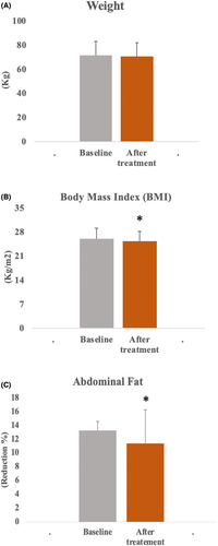
Regarding body weight, no statistical difference was observed, with an initial value of 71.5 ± 11.4 and a final value of 70.5 ± 11.3 (Figure 1A); however, BMI measurements were significantly reduced (p < 0.05), with the initial value being 26.1 ± 3.1 kg/m2 and the final value being 23.4 ± 3.9 kg/m2 (Figure 1B).

Regarding the thickness of fat evaluated by diagnostic ultrasound, the analyzed data showed a statistically significant reduction in percentage (p < 0.01), with initial means and standard deviation of 13.3 ± 1.3 and final of 11.4 ± 4.9 (Figure 1C).
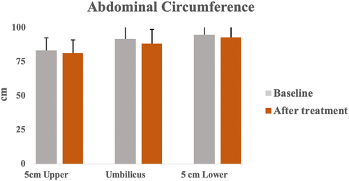
Regarding the analysis of waist circumference, a significant reduction was observed at the end of treatment (p < 0.01) in all measurement levels collected. The mean and standard deviation of the mean 5 cm above the umbilicus presented initial data of 83.2 ± 9.4 and final data of 81.2 ± 9.6; on the umbilicus, initial data of 90.8 ± 10 and final of 88.2 ± 10.1 and 5 cm below the umbilicus, initial of 94.8 ± 9.0 and final of 92.7 ± 8.7 (Figure 2).

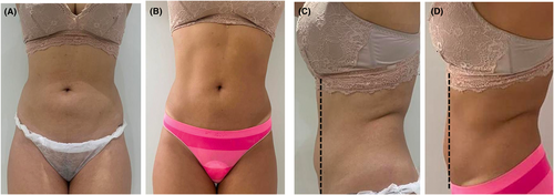
Figure 3 demonstrates, through digital photographs, anterior view (A and B) and side views (C and D), the visual results obtained after 8 sessions of treatment with HI-SMS when compared to the beginning of treatment.

3.2 Biochemical analyses
The safety parameters for the liver and kidney functions analyzed did not show statistical differences between the beginning and end of treatment (Table 1).
| Biomarkers | Baseline | After treatment | |
|---|---|---|---|
| ALT (U/L) | 21.48 ± 8.8 | 20.38 ± 15.4 | NS |
| AST (U/L) | 20.90 ± 6.5 | 23.62 ± 14.5 | NS |
| ALP (U/L) | 68.00 ± 25.3 | 67.00 ± 24.3 | NS |
| Urea (U/L) | 31.81 ± 7.3 | 32.29 ± 10.0 | NS |
| Crea (mg/dl) | 0.81 ± 0.1 | 0.80 ± 0.1 | NS |
- Note: Values are mean ± standard deviation *p < 0.05, paired t test and NS not significant.
- Abbreviations: ALT, Alanine aminotransferase; AST, Aspartate aminotransferase; ALP, Alkaline phosphatase; Crea, Urea and Creatinine.
Parameters related to fat metabolism were evaluated at the beginning and end of treatment. No statistical difference was observed in the values of total cholesterol (TC), LDL cholesterol, VLDL cholesterol, HDL cholesterol, as well as in the glycemic indexes. However, the values related to “non-esterified” FFA showed a statistically significant reduction (p < 0.05) immediately after treatment (Table 2).
| Biomarkers | Baseline | After treatment | |
|---|---|---|---|
| TC (mg/dl) | 185 ± 39.6 | 182.24 ± 38.3 | NS |
| LDL-C (mg/dl) | 114.29 ± 35.7 | 111.57 ± 38.1 | NS |
| VLDL-C (mg/dl) | 21.90 ± 9.6 | 20.90 ± 7.4 | NS |
| HDL-C (mg/dl) | 49.76 ± 15.1 | 49.76 ± 15.3 | NS |
| FFA (mmol/L) | 86.81 ± 10.3 | 87.71 ± 10.1 | * |
| Glu (mg/dl) | 86.81 ± 10.3 | 87.71 ± 10.1 | NS |
| LDH (U/L) | 171.29 ± 28.7 | 176.95 ± 37.4 | NS |
| CK (U/L) | 141.86 ± 86.6 | 178.40 ± 165.7 | NS |
| IL-6 (pg/ml) | 2.56 ± 0.94 | 2.56 ± 0.86 | NS |
- Note: Values are mean ± standard deviation *p < 0.05, paired t test and NS not significant.
- Abbreviations: CK, Creatinine kinase; FFA, Free fatty acids; Glu, Glucose; IL6, Interleukin 6; LDH, Lactate dehydrogenase; LDL, Low-density lipoproteins cholesterol; TC, Total cholesterol; VLDL, Very-low-density lipoproteins cholesterol.
3.3 Patient Comfort and Satisfaction
Patient satisfaction questionnaires showed a relatively high rate of patient satisfaction with the treatment outcomes. In total, 19 of 25 subjects (70%) reported being satisfied or strongly satisfied with the results; 4 patients (20%) were unsure about their satisfaction, and 2 patients (10%) expressed dissatisfaction.
Regarding the tolerance and comfort rates with the treatment, 75% (n = 13) of patients found the treatments tolerable to very comfortable (little or no discomfort); 30% (n = 6) considered it uncomfortable and 5% (n = 1) considered the treatment uncomfortable. Most patients tolerated stimulation intensities ranging between 90% and 100% at the end of the first session or during the second session, depending on individual sensitivity. No adverse events occurred. The only adverse effect observed was in some cases, fatigue and mild muscle soreness 1 day after the first treatment session; in all cases, the pain resolved spontaneously within the next 24 h. Regarding the additional questioning, whether the participant would recommend the treatment to a friend or relative, 23 of 25 individuals stated that yes, they would indicate the therapy, and only 2 were in doubt and chose not to.
4 DISCUSSION
The present study was intended to retrospectively evaluate the effects of HI-SMS used on the abdominal muscles of healthy patients. The main results show that 8 treatment sessions promoted a reduction in thickness, abdominal circumference, and FFA. Still, a high degree of tolerance and satisfaction with the treatment was observed.
The results of the present study showed that treatment with HI-SMS was able to reduce BMI, fat thickness, abdominal circumference, and body contour. The treatment did not change any biochemical parameters related to lipid profile, glycemic index, liver and kidney function, muscle metabolism and did not promote significant adverse effects, suggesting treatment safety. Furthermore, the treatment met the patients' expectations regarding tolerance and satisfaction with the treatment, and 92% of the treated individuals would recommend the treatment to a friend or relative. Thus, the findings of the study are indicative of the evolution of HI-SMS in the field of aesthetics. However, it is of fundamental importance that prospective studies with a greater number of volunteers using this technology are developed.
Electromagnetic fields are composed of electric and magnetic fields that produce biochemical and cellular phenomena promoted by the electromagnetic force that includes electricity and magnetism.13 As previously mentioned, previous studies using equivalent technologies have shown effects in improving body contour.9, 14-16 In the present study, the effects of a device that provides an alternating magnetic field with intensities of up to 2.5 T and frequencies of up to 150 Hz, which induce electrical currents in the underlying tissue were evaluated. Thus, the current study is the first study to evaluate the effects of abdominal muscle toning using this specific device.
It is known that electromagnetic pulses are delivered at a high-frequency rate, inhibiting muscle relaxation, which results in a phenomenon known as supramaximal or tetanic contractions, not reproducible by voluntary muscle contraction. The study by Kent et al.17 investigated the effects of high-intensity focused electromagnetic technology for induction of changes in abdominal subcutaneous fat and abdominal muscles. The authors found reduction in subcutaneous fat and simultaneous 14.8% thickening of the rectus abdominis muscle, suggesting that the investigated device is effective for abdominal body sculpting, however,the mechanisms of action are still not clear. In the present study, a reduction in the percentage of fat, fat thickness, and circumference of the abdominal region was also observed, as well as an improvement in body contour after the use of HI-SMS in the region of the abdominal muscles. Positive data were not related to weight loss and corroborate other studies.14-16
It is known that the mature adipocyte is composed of a large lipid droplet containing triglycerides (TAG) occupying most of the cell. These TAGs are mobilized according to the body's energy demand and need to be cleaved through a mechanism called lipolysis. During lipolysis, intracellular TAG undergoes hydrolysis by the action of lipases. The efflux of FFA and glycerol from adipose cells is followed by the transport of these metabolites in the bloodstream to other tissues, mainly the liver, skeletal muscle, and heart.18
The results of the present study can be justified by the study by Weiss et al.12 carried out on a swine model. The authors hypothesize that induced supramaximal contractions may lead to an increase in metabolic activity in the region of stimulation and subsequent breakdown of lipids into FFA and glycerol as seen during intensive resistance training. Halaas et al.19 carried out a study, also on a swine model, which evaluated whether the metabolic stress caused by the high muscle energy demand during the application of the electromagnetic field for 30 min in the animal's abdominal region after a single treatment session could induce apoptosis. The authors collected tissue samples from the treated site through biopsy and observed through liquid chromatography and spectrophotometry, in addition to other markers, that FFA levels around adipocytes increased immediately after treatment and remained high 8 h after the session when compared to the control sample.19 Both studies supported the hypothesis that treatment with electromagnetic contraction can induce programmed cell death, since in supramaximal contractions, lipid breakdown can lead to FFA extrusion into the intracellular and extracellular space,19 and studies indicate that when the amount of FFA exceeds a certain level in the intracellular space, this can lead to adipocyte dysfunction and stress-induced apoptosis of the endoplasmic reticulum.19, 20 These data suggest a potential relationship between FFA released after muscle contractions and fat cell apoptosis, but this hypothesis was challenged by the study carried out by Zachary et al.21 who used post-abdominoplasty excision human tissue samples after a single treated session and compared two different types of electromagnetic field muscle stimulation systems and a cryolipolysis system. This hypothesis regarding FFA stress-induced adipocyte apoptosis caused by supramaximal contractions requires further research, and its validation was not the aim of the present study.
In addition to the results shown above, the present study also aimed to assess the safety of the electromagnetic field through biochemical analyses using plasma markers. In the experimental study proposed by Weiss et al.12 the serum levels of safety biomarkers related to liver and kidney function on fat and muscle metabolism were also evaluated before the beginning of the session, 1 h and 8 h after treatment. The animals were treated for 30 min in the abdominal region. In the present retrospective clinical study, we verified the stability of serum biomarkers involved with hepatic, renal and muscle fat metabolism. Data from the following biomarkers were evaluated: alanine aminotransferase (ALT), aspartate aminotransferase (AST), alkaline phosphatase (ALP), urea and creatinine (Crea) related to liver and kidney function, respectively; total cholesterol (TC), low-density lipoproteins cholesterol (LDL), very-low-density lipoproteins cholesterol (VLDL); FFA indicators related to the metabolism of fats and glucose (Glu). The muscle damage biomarkers creatinine kinase (CK) and lactate dehydrogenase (LDH), and acute inflammation (IL-6) were also evaluated. All mean and standard deviation values found in the present study, measured before and immediately after the eighth treatment session, did not show statistically significant clinical changes.
However, interestingly, lower values of circulating FFA were observed in the present study, corroborating the results of thickness, circumference, and improved body contour. It is likely that there is an increase in paracellular FFA in adipose tissue due to the high rate of lipolysis and a decrease in plasma due to muscle energy demand, simultaneously. Studies carried out with humans have shown a decrease in circulating FFA levels that occur physiologically during intense physical activity and prolonged fasting,18, 22, 23 which corroborates the findings of the present study. It is possible to consider that these findings are correlated, because in the same way that it occurs during intense physical exercise, the muscular contraction induced by the magnetic field can increase the extracellular uptake of FFA and lower its concentration in the blood plasma.22, 23 No studies were found that evaluated these serum biomarkers in humans using similar electromagnetic field devices.
Secondary assessments included patient satisfaction assessments and therapy comfort assessments. In the present study, the results showed very high patient satisfaction 30 days after the end of treatment. No patient had an adverse event or side effect other than a slight sensation of muscle fatigue that resolved spontaneously.
The present study has limitations, both because it is a retrospective study, as well as the small number of the sample, and the use of animal studies for confrontation can also be considered as a limitation since the observed results may not be fully transferable to humans.
It should be noted that, based on the fact that treatment with HI-SMS is relatively new, prospective studies are essential to help us obtain a better understanding of the ideal treatment parameters (frequency of stimulation, intensity, number of sessions, interval between sessions, need for maintenance between sessions, etc.) and also the ideal patient profile (BMI, subcutaneous fat thickness). However, the present non-comparative retrospective analysis of data from medical records of patients treated with HI-SMS provides us with important information about the behavior of serum parameters related to the safety of the treatment.
5 CONCLUSION
The results of the present study demonstrate that HI-SMS exerts positive effects on the reduction of circumference measurements and adipose tissue thickness at the treated site, having non-invasive therapeutic potential for aesthetic purposes. It is noteworthy that the use of HI-SMS did not produce abnormalities in the biochemical parameters evaluated: AST, ALT, ALP, creatinine, lipid profile, glycemic index, liver, and kidney function; biomarkers of muscle damage LDH and CK; and acute inflammation IL-6, proving to be a safe treatment. Furthermore, the treatment met the patients' expectations regarding tolerance and satisfaction with the treatment, and these findings are indicative of the evolution of the field of aesthetics.
With these results, it is possible to suggest that the treatment with HI-SMS in abdominal muscles proves to be a safe technology with potential for non-invasive therapy for aesthetic purposes; however, prospective studies with a greater number of volunteers should be developed.
AUTHOR CONTRIBUTIONS
E.S., M.M. performed the research. M.M, P.F., and R.P. participated in the data acquisition. ES. L.A. CC, and S.G. analyzed the data. E.S. and L.A. wrote the paper.
FUNDING INFORMATION
No funding was provided for the present study.
CONFLICT OF INTEREST
Dr. Sant'Ana, Dr. Meleck, and Dr. Ferrari are speakers for Fismatek Industries. The other authors have no conflict of interest to disclose.
ETHICAL APPROVAL
The following study was conducted in accordance with the principles of the Declaration of Ethics Committee of Universidade Brasil.
Open Research
DATA AVAILABILITY STATEMENT
The data that support the findings of this study are available on request from the corresponding author. The data are not publicly available due to privacy or ethical restrictions.


