Blue light-emitting diodes for the treatment of localized vitiligo: A retrospective study
Abstract
Background
Vitiligo is an autoimmune dermatological disease characterized by hypopigmented macules.
Treatments include topical agents, phototherapy, and laser therapies. Different lasers should be individually chosen regarding location, extent, activity of the disease.
Aims
This article aims to demonstrate how blue LED is effective and safe, as its wavelength is very close to the UV spectrum (415 nm vs. 400 nm), but, unlike UV therapy, blue LED have not shown any long-term cancerogenic side effects.
Patients/Methods
We treated 30 patients affected by vitiligo localized on different anatomical areas with blue light-emitting diodes.
Results
Complete repigmentation occurred in 75.33% of treated patients (22 out of 30 patients, 14 males, and 8 females). Partial repigmentation occurred in the remaining patients.
Conclusions
Blue LED light may be a safe and well-tolerated way to induce repigmentation in patients affected by vitiligo.
1 INTRODUCTION
Vitiligo is a depigmenting skin condition that affects 0.5%–2% of the global population of all ethnic groups and skin types, with no distinctions. The condition is defined by the selective loss of melanocytes, resulting in non-scaly, chalky-white macules. Vitiligo is considered a cosmetic issue, even though its symptoms can be psychologically distressing and significantly impact everyday life.
It is now firmly recognized as an autoimmune disease, with hereditary and environmental components, as well as metabolic issues, oxidative stress, and cell detachment.1
The diagnosis of vitiligo is clinical and usually straightforward, based on the presence of acquired, amelanotic macules with clear edges in a typical distribution: periorificial, lips and distal extremity tips, penis, segmental, and frictional areas. Generally, laboratory tests are not required, but histological samples show a total loss of melanin pigment and a lack of melanocytes. Lymphocytes may occasionally be seen at the advancing edge of the lesions.2
Phototherapy (ultraviolet A and B), topical and systemic immunosuppressants (corticosteroids, calcineurin inhibitors), may assist in slowing the progression of vitiligo, stabilize depigmented lesions, and stimulate repigmentation.3, 4 As most therapies are time-demanding and require long-term follow-up, management may need an individualized therapeutic strategy in which patients should always be consulted. Cosmetic camouflage advice from a cosmetician or a professional nurse should be supplied to people with vitiligo that affects exposed regions.1
Another recent approach involves using lasers (excimer, CO2, Er:YAG, and non-ablative resurfacing laser), even combined with topical treatments.5, 6
Among all these treatments, the most frequent technique of treating vitiligo is ultraviolet (UV) light therapy (320–400 nm); however, UV light therapy needs a prolonged treatment time, and continuous exposure to UV radiation causes adverse effects. On the contrary, blue light with a spectrum of about 415 nm is not cancerogenic, and it was seen to be efficient in the treatment of vitiligo combined with other substances since they promote melanin synthesis in activated cells via the CREB/MITF/TYR pathway by stimulating adenylyl cyclase.7
Blue light causes an increase of MITF (the master gene of pigmentation) without increasing cell proliferation. The efficacy of this light may also be linked to the role of Opsin-3 (OPN3), a melanocyte photoreceptor and visible light sensor that is highly expressed in human epidermal melanocytes (HEMs): It senses blue light in melanocytes and mediates melanogenesis.8
OPN3 is the crucial sensor on melanocytes, and it is responsible for hyperpigmentation induced by the shorter wavelengths of visible light.9
Moreover, photobiomodulation, the process of absorbing red/near-infrared light energy, boosts mitochondrial ATP generation, cell signaling, and growth factor synthesis while reducing oxidative stress.10 This may have a positive effect on vitiligo, increasing melanocytes turnover, without induce the formation of Reactive Oxygen Species (ROS).
2 MATERIALS AND METHODS
During winter 2020/2021, a not randomized retrospective study included 30 patients with localized, both segmental and non-segmental, and stable vitiligo (20 males and 10 females) aged from 7 to 45 years old, treated at the Magna Graecia university hospital dermatologic center in Catanzaro, Italy. Patients who had any of the following characteristics were excluded from the study: prior treatment with laser therapies, gold-containing medications, recent exfoliation procedures, surgical procedures, prior skin conditions (including keloids), hypersensitivity to light (visible and near-infrared), medications that make users more sensitive to light, anticoagulant and immunosuppressive treatments, gestation or breastfeeding, a personal or family history of skin cancer, patients whose vitiligo was not stable, where stability means that older lesions have not progressed during the last 2 years, no more lesions appeared at the same timeframe, lack of a contemporary Koebner phenomena, either historical or experimental.
All patients (or their legal tutor) completed informed consent forms admitting the risks of the treatment and giving permission for photographs to be taken and shown. The local ethics committee authorized the study.
Concerning children, we enrolled a 7-year-old child, that had a stable plaque of segmental vitiligo. As reported in literature, in fact, segmental vitiligo stabilizes in few months.11
A blue LED (light-emitting diode—provenience China) device at 417 ± 10 nm was used with a fluence of 120 J/cm2, and power intensity of 60 mW/cm2 ± 20%, for the time of 9 min; treatments were performed twice a week for 10 consecutive weeks.
Patients presented different locations of vitiligo: acral, foot (5 patients) and hands (3 patients), face (16 patients), and shank (6 patients).
Before the first session, clinical photography was conducted, and it was done again 10 weeks later and 3 months after the final session. The same shooting choices, a twin flash, and the same illumination were employed, as well as the same camera (Nikon 5600d, Nikon Corporation) and settings.
The patients were given a Visual Analogue Scale (VAS) from 1 to 10 during the ten-week follow-up to estimate their satisfaction.
2.1 Primary outcome measures
- VASI score improvement of 0%–10%, minimally improved
- VASI score improvement of 10%–25%, improved
- VASI score improvement of 25%–50%, much improved
- VASI score improvement of 50%–100%, very much improved.
2.2 Secondary outcome measures
Safety and tolerability were assessed considering the adverse reactions, clinically evaluated after the session treatment, and collecting data on late adverse events reported by patients.
2.3 Post-procedure care
We expected to have no severe adverse events but at most episodes of skin rash, burning and pain after treatment.
After the procedure, no treatment was intended to be administered, except for people who would have adverse events: topic gentamicin to avoid risk of overinfection and emollient cream, while paracetamol 1 g in case of pain.
2.4 Statistical analysis
Mean, SD, and percentages were calculated with IBM® SPSS® Statistics 26.0.
3 RESULTS
The study involved 30 individuals: 20 (66.6%) men and 10 (33.3%) women. Mean age was 30.36 ± 14.63. The participants' skin type ranged from type I (n = 1, 6.6%), to type II (n = 17, 56.6%) and type III (n = 12, 40%) on the Fitzpatrick scale. Patient's characteristics are reported in Table 1.
| Patient | Sex | Age | Skin fitzpatrick phototype | VASI score | Localization |
|---|---|---|---|---|---|
| 1 | M | 7 | 2 | 2% | Foot |
| 2 | M | 45 | 3 | 3375% | Face |
| 3 | M | 33 | 3 | 1% | Hand |
| 4 | M | 29 | 2 | 4.50% | Shank |
| 5 | M | 23 | 3 | 2.25% | Face |
| 6 | M | 19 | 2 | 3375% | Shank |
| 7 | M | 35 | 2 | 1.50% | Foot |
| 8 | M | 35 | 2 | 2.25% | Face |
| 9 | M | 17 | 2 | 3375% | Shank |
| 10 | M | 44 | 3 | 4.05% | Shank |
| 11 | M | 41 | 1 | 4.05% | Face |
| 12 | M | 22 | 2 | 1.50% | Hand |
| 13 | M | 39 | 2 | 3375% | Face |
| 14 | M | 45 | 3 | 2.25% | Face |
| 15 | M | 36 | 2 | 2.25% | Face |
| 16 | M | 43 | 2 | 3375% | Face |
| 17 | M | 27 | 3 | 3375% | Face |
| 18 | M | 43 | 3 | 6.75% | Shank |
| 19 | M | 38 | 2 | 1.80% | Foot |
| 20 | M | 20 | 2 | 2.25% | Face |
| 21 | F | 20 | 2 | 2.25% | Shank |
| 22 | F | 34 | 3 | 3375% | Face |
| 23 | F | 37 | 2 | 1125% | Face |
| 24 | F | 40 | 3 | 1.80% | Foot |
| 25 | F | 23 | 3 | 2.25% | Face |
| 26 | F | 25 | 2 | 1125% | Face |
| 27 | F | 30 | 3 | 3375% | Face |
| 28 | F | 34 | 2 | 1% | Foot |
| 29 | F | 29 | 2 | 1125% | Face |
| 30 | F | 18 | 3 | 0.75% | Hand |
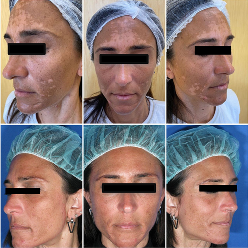
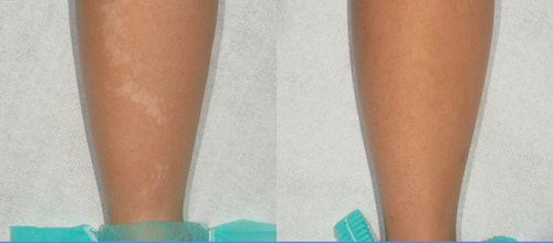
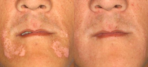
Blue light proved to be well tolerated by patients. In fact, nobody had severe adverse events. Only three patients reported hyperpigmentation on the treated area that resolved in a few weeks (one patient, 3.33%) and mild erythema (two patients, 6.66%) that was treated with emollient cream and gentamicin cream, that was treated with emollient cream and gentamicin cream, and the power of LED was reduced to 45 mW/cm2 at the following session, even though the erythema healed before doing next treatment. All patients concluded the treatment and experienced a repigmentation rate in different percentages, so we did not register any drop out. Complete repigmentation occurred in 75.33% (22 patients, 14 males, and 8 females), while partial repigmentation occurred in the remaining patients (Figures 1-3). In particular, two patients improved their VASI score by 90% (vitiligo localized on shank and foot), two patients by 75% (localized on foot and hand), and the other four patients, respectively, by 67% (hand), 87% (face), 83% (shank), and 77% (face) as shown in Table 2.


| Patient | VASI score after 10 session treatment | VASI score 3 months after the final session | VAS score | VASI score improvement |
|---|---|---|---|---|
| 1 | 0.50% | 0.50% | 7 | 75% |
| 2 | 0% | 0% | 10 | 100% |
| 3 | 0.25% | 0.25% | 7 | 75% |
| 4 | 0% | 0% | 9 | 100% |
| 5 | 0% | 0% | 7 | 100% |
| 6 | 0% | 0% | 7 | 100% |
| 7 | 0% | 0% | 9 | 100% |
| 8 | 0% | 0% | 8 | 100% |
| 9 | 0% | 0% | 8 | 100% |
| 10 | 0.45% | 0.45% | 8 | 90% |
| 11 | 0% | 0% | 10 | 100% |
| 12 | 0.50% | 0.50% | 6 | 67% |
| 13 | 0% | 0% | 9 | 100% |
| 14 | 0% | 0% | 9 | 100% |
| 15 | 0% | 0% | 9 | 100% |
| 16 | 0.45% | 0.45% | 7 | 87% |
| 17 | 0% | 0% | 8 | 100% |
| 18 | 1125% | 1125% | 8 | 83% |
| 19 | 0% | 0% | 10 | 100% |
| 20 | 0% | 0% | 9 | 100% |
| 21 | 0% | 0% | 8 | 100% |
| 22 | 0% | 0% | 9 | 100% |
| 23 | 0% | 0% | 8 | 100% |
| 24 | 0% | 0% | 8 | 100% |
| 25 | 0% | 0% | 9 | 100% |
| 26 | 0% | 0% | 8 | 100% |
| 27 | 1125% | 1125% | 6 | 77% |
| 28 | 0.10% | 0.10% | 7 | 90% |
| 29 | 0% | 0% | 8 | 100% |
| 30 | 0% | 0% | 8 | 100% |
Partial repigmentation occurred in subjects with a higher VASI score due to a larger surface area or greater depigmentation than the other patients.
Moreover, all patients were satisfied with the treatment, with an average VAS score of 8133.
Table 2 shows that the results did not change in the 3 months following the final session, so we can assert that outcomes remain constant throughout time, without recurrence.
Outcomes seem to be not linked to the age of the patients and the Fitzpatrick phototype.
4 DISCUSSION
Vitiligo is a frequent disease of the skin, classified as autoimmune. It causes depigmentation of different body areas. It affects people of all races, ages, and latitudes with no differences, while it is slightly higher in females than males. Treatment of vitiligo must be safe and effective but also quick and long-lasting. All these criteria are not always present in treatments now available. The most used therapy is ultraviolet (UV) therapy, especially UVA (whose spectrum is between 320 and 400 nm) and narrowband UVB (280–320 nm).12
PUVA (UV with Psoralen) is known to cause repigmentation but must be performed for a long time, with at least 100–200 sessions spaced at least a day apart, 2–3 times a week.
Although varying degrees of repigmentation has been accomplished, long-term repigmentation is challenging. Because the precise dosage is required for safe therapy, the UVA system's spectrum power distribution must be determined, and the UVA dose must be adjusted correspondingly.13
PUVA shows various side effects such as itch, erythema, xerosis, eye toxicity, and photoaging. Moreover, nonmelanoma skin cancer, particularly squamous cell carcinoma, is linked to high cumulative oral PUVA exposure (SCC).14
The primary control of UV-induced melanogenesis in melanocytes is mediated by keratinocyte production of cytokines (ROS pathways) and hormones.
Blue LED, on the contrary, uses different pathways to induce repigmentation and, for this reason, does not lead to skin cancers: especially in dark skin-type melanocytes, it causes the development of a multimeric tyrosinase/tyrosinase-related protein complex, which leads to persistent tyrosinase activity and finally acts on MITF, the master gene of melanogenesis that leads to increment melanin in cells.8
Blue LED, moreover, has good efficacy on vitiligo, probably owing to its wavelength being extremely near to UV rays (415 nm versus 400 nm), yet it is not cancerogenic, unlike UV therapy.
Other promising therapies for vitiligo are JAK inhibitors.15 CD8 T lymphocytes destroy melanocytes to cause vitiligo, and IFN- plays a crucial role in the pathophysiology of the illness. JAK inhibitors may be effective in treating vitiligo because IFN- signaling uses the JAK–STAT pathway.16 For instance, tofacitinib therapy for a patient with global vitiligo led to nearly complete repigmentation of the hands, forearms, and face over 5 months, and ruxolitinib led to facial repigmentation after 20 weeks.17, 18 In both cases, if the drug is suspended, vitiligo returns.
Moreover, these systemic drugs are not free of side effects and require prolonged hematochemical monitoring. Ruxolitinib may also be used topically twice daily19: Rothstein et al. reported a VASI improvement in 23% of treated patients.20 Compared to our outcomes, all patients (100%) had a VASI improvement, so topical treatment with JAK inhibitors maybe less encouraging if compared to the systemic JAK inhibitors, and blue LED treatment.
Promising early results have been shown with anti-IL-15 drugs in vitiligo, but further studies with larger casuistic should be done to ensure that these drugs may be safely used in clinical practice.21
For all these reasons, the blue LED treatment proved to be safe and effective, without systemic adverse events. Furthermore, it does not interfere with other drugs. Only few mild temporary local reactions have been reported, and few limited precautions need to be taken in order to carry out this treatment safely.
Results do not appear to be correlated with patient age or Fitzpatrick phototype, but future studies could help to deepen this aspect.
Limitations of the study are small sample, follow-up of only 3 months and treatment of specific patients with localized and not full-body vitiligo. In fact, higher VASI score participants experienced partial repigmentation because they had a bigger surface area or more depigmentation than the other patients and that is why more studies are needed to know whether this treatment has the same efficacy on larger affected areas.
5 CONCLUSIONS
This study shows the first results of a new possible vitiligo treatment with blue LED. This treatment has been demonstrated to be a safe and well-tolerated way to induce repigmentation in patients of all ages and different skin type and shows promising results in fewer sessions when compared to other phototherapies. All patients improved by over 50% compared to baseline, and everyone was satisfied with the treatment. Mild adverse events were temporary and localized.
In conclusion, although the outcomes are encouraging, further studies are needed to confirm clinical results, long-term efficacy and validate our hypothesis.
AUTHOR CONTRIBUTIONS
G.L., C.D.R., G.C., and M.S. performed the research. S.N. and LB. designed the research study. G.L. analyzed the data. L.B. wrote the paper.
CONFLICT OF INTEREST
None declared for all the authors.
ETHICAL APPROVAL
Authors declare human ethics approval was not needed for this study.
Open Research
DATA AVAILABILITY STATEMENT
The data that support the findings of this study are available from the corresponding author upon reasonable request.


