Gastrointestinal: Melanotic schwannoma of the pancreas associated with Carney complex: A cause of acute neoplastic symptom
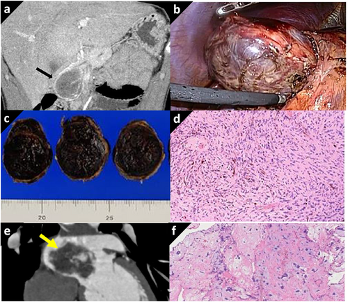
An 18-year-old man who had no familial/past history presented with acute severe epigastralgia. Computed tomography revealed a 45-mm low-density pancreatic head tumor with rim enhancement (Fig. 1a). The tumor grew approximately 5 mm/week. Thirteen days after symptom onset, laparoscopic excision was performed (Fig. 1b). The resected specimen was a 55 × 46 × 33 mm black-brown colored expansive tumor, with hemorrhage and necrosis (Fig. 1c). It was histologically identified as a melanotic schwannoma with intracytoplasmic melanin granules (Fig. 1d). Fourteen months after the pancreatectomy, the patient was readmitted to the hospital due to right hemiplegia. Imaging revealed a lacunar infarction owing to a tumor embolism from the left atrial myxoma (Fig. 1e). Surgical resection of the myxoma and atrial septal defect repair were performed to avoid further infarction (Fig. 1d). Anticoagulant therapy was initiated, and he recovered without any symptoms. Although no genetic tests have been performed, he has been identified as a patient with Carney complex based on the clinical course and has been undergoing regular systemic examinations. Thus far, no symptoms of tumor recurrence and endocrine disorders were noted 60 months after pancreatectomy.

Schwannomas rarely arise from the pancreatic nerve-sheath. Since its imaging findings are similar to cystic lesions, discrimination from other pancreatic cystic neoplasms is crucial for its diagnosis. Melanotic schwannoma composed of schwannoma cells with melanin pigment is a variant of schwannoma; however, it has some distinctive clinical features. Firstly, more than 50% of the patients is associated with Carney complex, and secondly, it is potentially malignant. Carney complex is multiple neoplastic syndrome featuring endocrine, connective tissue, neural, and cardiac tumors, associated with skin pigmentation and endocrine disorders. Half are hereditary autosomal dominant conditions associated with a PRKAR1A gene mutation on chromosome 17q24.2. As melanotic schwannoma with Carney complex have potential for metastasis or local spread, eradication of tumor is necessary. Also, clinicians should be aware of this disease because this may cause an acute neoplastic condition in the youth.


