Gastrointestinal: Gastric heterotopic pancreas has potential of malignancy requiring appropriate resection
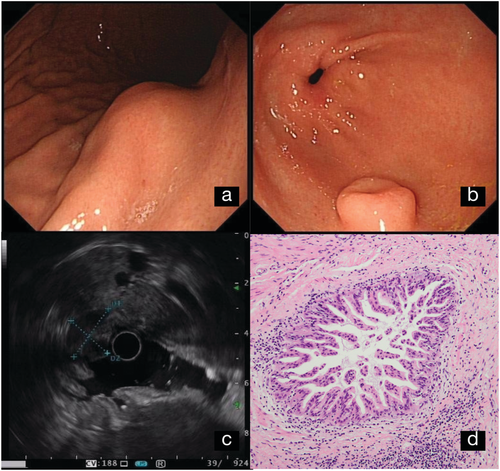
A 48-year-old woman, without any symptoms, was found to have two subepithelial lesions in the stomach during a gastroscopy examination. Hematological examination and biochemical tests were all within normal limits. On examination, a 3 × 2.5-cm smooth protrusion lesion was found in the post wall of the gastric body (Fig. 1a), while another 1-cm subepithelial tumor in the antrum was characterized with a central umbilication (Fig. 1b). CT revealed irregular wall thickening containing an enhancing mass in the gastric body but missed the antrum lesion. On endosonographic scanning, the body mass displayed hypoechoic and heterogenous echogenicity, which originated from the muscular layer of the stomach with indistinct margin (Fig. 1c). However, the gastric antral lesion was found arising from the submucosa layer and exhibited heterogeneous, mixed echoic echogenicity.

Laparoscopic resection of gastric subepithelial tumors was performed, without any complications. The two resection specimens were confirmed ectopically located pancreatic tissues through pathology observation. Importantly, there was high-grade pancreatic intraepithelial neoplasia in the gastric body ectopic pancreas, with irregularly arranged epithelium cells (Fig. 1d).
Traditional concept holds that asymptomatic gastric heterotopic pancreas can be followed expectantly. However, those lesions also have potential of malignancy, which need appropriate resection.


