Gastrointestinal: A case of a monster enterolith in a Crohn's disease patient
Enterolithiasis is considered uncommon, although its true prevalence remains unknown. The diagnosis is often made incidentally, during cross-sectional imaging. Complications include acute or subacute small bowel obstruction, perforation, or even hemorrhage. These events are rare. A gallstone ileus is the main and most frequent differential diagnosis. Enteroliths are believed to form through a precipitation and sedimentation of enteric contents in the context of luminal stasis, commonly associated with conditions such as small bowel diverticulosis or other alterations of luminal anatomy. Crohn's disease has been implicated in occasional case reports. Some of these reports refer to the enterolith size as large or giant, but none comes even close to the undisputed champion presented here.
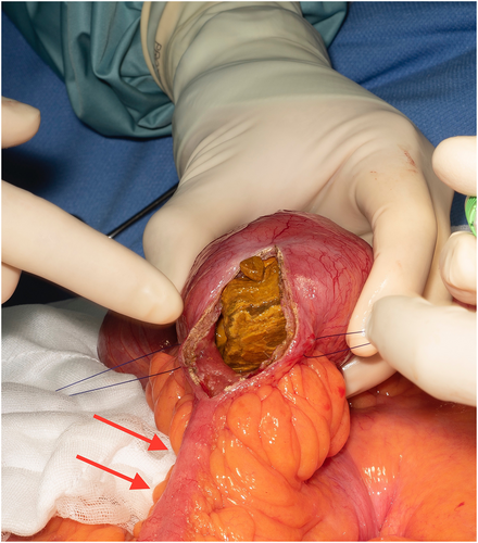
This case involves a 50-year-old Caucasian man with a background of long-standing small bowel Crohn's disease. He had no history of prior surgeries. Previous treatment involved immunomodulator monotherapy. This was ceased at the patient's discretion several years ago. Despite ongoing symptoms of intermittent abdominal discomfort, he continued to decline both treatment and further investigations. His symptoms have eventually progressed to those of suspected intermittent bowel obstruction. Investigations revealed normal inflammatory markers as well as normal ileo-colonoscopy. Magnetic resonance enterography reported on a laminated calculus measuring up to 80 mm in diameter in mid-ileum with further adjacent smaller calculi (Fig. 1). Distally, there was a short segment of significant luminal narrowing with full-thickness enhancement compatible with chronic stricture as well as a marked proximal small bowel dilatation of up to 75 mm. There was no evidence of small bowel diverticulosis or cholecystolithiasis. Reluctantly, he proceeded to elective laparotomy. This confirmed the magnetic resonance enterography findings. A small bowel enterotomy was performed, and the enterolith was delivered (Fig. 2). A small bowel resection followed to remove the strictured segment. This was histologically confirmed as severe fibrotic stricture without evidence of active inflammation or malignancy.


