Hepatobiliary and Pancreatic: Sub-Glisson's capsule hepatic multiple nodular formations
An 81-year-old man was evaluated in our hospital for upper abdominal pain and dyspepsia. At the time of his presentation, hematological examinations were within the normal limits, except for C-reactive protein (5.18 mg/dL) and hemoglobin (92.00 g/L). There was no alteration in liver function tests. Gastro-duodenoscopy was within normal limits.
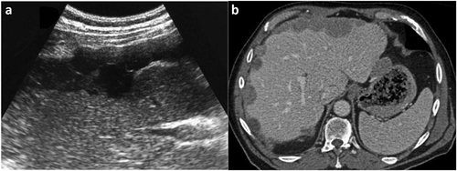
He underwent abdominal ultrasound (Fig. 1a) and contrast-enhanced multi-detector computed tomography (Fig. 1b), which revealed a heterogeneous hypo-echoic and hypo-dense sub-Glisson's capsule hepatic multiple nodular formations (range diameter from 0.9 to 3.1 cm) and the presence of hepatic hilar lymph nodes. The patient then performed serological and tumor markers; both types of tests resulted negative. After multidisciplinary discussion, the patient underwent ultrasound guided liver core needle biopsy.

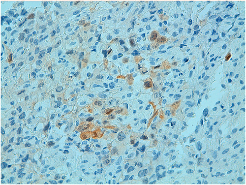
Hepatic histopathological evaluation demonstrated a malignant mesothelioma, consisting in predominantly epithelioid poorly differentiated as tumor cells or also known as ‘primary intrahepatic mesothelioma’ (Fig. 2). Given the intrahepatic multi-nodular form of malignant mesothelioma in our patient, he undergone to chemotherapy.

Primary intrahepatic mesotheliomas are exceedingly rare tumors, and their incidence is not well defined. Clinical manifestation is present only in advanced forms and has non‑specific signs; this makes them difficult to diagnose. Intrahepatic malignant mesotheliomas develop from mesothelial cell layers covering Glisson's capsula and subsequently invades the hepatic parenchyma.
Initial evaluation includes tumor markers for possible differential diagnosis of primary and secondary liver neoplasms. Consequently, imaging studies (ultrasound, multi-detector computed tomography, or magnetic resonance imaging) are to be performed as they allow us to evaluate the morphological characteristics and the intrahepatic and extrahepatic extension of the pathology. Generally, primary intrahepatic mesothelioma appears as a solitary tumor mass that is localized into the liver parenchyma. To our knowledge, an initial presentation of primary intrahepatic mesotheliomas with sub-Glisson's capsule hepatic multiple nodular formations, as in our patient, has not been described previously in the literature. Biopsy is mandatory to establish the correct nature diagnosis. Surgery is indicated in single hepatic nodule, all other case are treated by chemotherapy.


