CAD–CAM natural restorations—Reproducing nature using a digital workflow
Abstract
Objectives
Natural restorations combine digital workflow and shell technique to create CAD/CAM restorations with the form and texture of natural teeth. This case report describes an interdisciplinary digital workflow combined with CAD/CAM natural restorations to achieve the naturalness of an anterior rehabilitation. Clinical considerations: A 38-year-old patient attended to the office with esthetic issues. An interdisciplinary treatment plan was conducted, which included periodontal surgery to recreate the gingival contour, associated with bleaching and manufacturing CAD/CAM ceramic veneers to return an optimal teeth surface texture and shape.
Conclusions
The design and manufacturing of CAD/CAM natural restorations using a digital workflow allowed a predictable result and overcame the limitations of conventional shell technique.
Clinical Significance
Conventional shell technique is used to create restorations with the form and texture of natural teeth. This article presents a combination of the conventional shell technique with a digital workflow, facilitating the design and manufacturing of CAD/CAM natural restorations.
1 INTRODUCTION
The main proposal of restoring anterior teeth is to restore the smile's natural appearance. Despite the favorable esthetic outcomes achieved with ceramic restorations, mimicking nature may be challenging, since the natural tooth is composed of vertical and horizontal concavities. Anatomic details and surface texture directly influence value, color saturation, and light reflection of natural dentition.1
The reproduction of a natural surface texture using conventional techniques can be challenging, thus relying on the dental technician's ability. To overcome this limitation,2-4 Kano et al.1 described the anatomic shell technique, in which composite resin shells are fabricated from Jan Hajtó models to copy the texture and morphology of the natural dentition. Based on this, CAD/CAM restorations with natural morphology are fabricated, ensuring the predictability of the esthetic outcome.
A complete digital workflow brings benefits for manufacturing CAD/CAM restorations.5-8 Digital libraries containing different sizes and shapes of natural teeth make it possible to virtually select a natural smile and to perform an anatomic shell technique. Furthermore, based on principles of smile design, tooth form is chosen and evaluated.6, 9, 10 The concept of “natural restorations” involve the combination of a digital workflow and a shell technique to create CAD/CAM restorations with natural form and texture. This case report describes an interdisciplinary periodontal/restorative digital workflow to achieve anterior CAD/CAM restorations with natural results.
2 CASE REPORT
A 38-year-old patient attended the dental clinic due to esthetic issues. His chief complaint was the presence of spaces between anterior teeth and the teeth misalignment, which was not solved by a previous orthodontic treatment with fixed appliances. The patient was not willing to a second orthodontic treatment and was looking for a treatment option. The detailed medical and dental history revealed no abnormality or contra-indication for dental treatment. The clinical examination showed a disharmony between dental and periodontal tissues, which was characterized by gingival recession, presence of diastemas, unbalanced gingival levels, and unfavorable dental proportions. The damaged tooth surface and texture, especially at the incisal edges, suggested a process of biocorrosion.11 The patient presented excellent oral hygiene and the absence of occlusal disorders or parafunctional habits.
2.1 Digital Smile Design protocol
An interdisciplinary treatment planning was conducted to restore the harmony between hard and soft tissues, thus improving facial esthetics and smile. The treatment began with a facially driven esthetic analysis based on objective and subjective criteria. For this purpose, a Digital Smile Design (DSD) protocol was performed using the DSD App (Digital Smile Design, Madrid, Spain).12 In order to record the initial situation, digital documentation, such as photos and intra-oral scans digitally, was taken (Figure 1).

DSD Photographic protocol was used for documentation.13 First, a smile frame with ideal dental proportions was drawn on facial photos and overlapped with intraoral scans. The desired smile was simulated digitally in order to provide the clinician and the patient an overview of treatment possibilities.
2.2 Digital planning
Based on these proportions, a digital wax-up was designed from a digital library containing different shapes and sizes of natural teeth. The “copy and paste” technique made it possible to replicate anatomical details and surface texture of a natural dentition into the digital wax-up.
Afterward, the relation between a smile and facial expressions was analyzed by overlaying the digital wax-up onto the photographs (Figure 2). Then, a motivational mock-up was printed and proved clinically. As soon as the patient accepted the expected result, multidisciplinary treatment planning, including periodontal and restorative treatment, was elaborated. The treatment included periodontal surgery to recreate the gingival contour, dental bleaching, and fabrication of CAD/CAM ceramic veneers to restore teeth surface texture and shape optimally.

2.3 Periodontal surgery
Cone-beam computed tomography (CBCT) scans were requested. The surgical planning involves matching the intra-oral scanning (STL files) to the CBCT (DICOM) scan files. Surgical planning was done (Nemo Studio, Biotech Dental, Madrid, Spain) and transferred to a digitally designed surgical guide. It was possible to assess the relation between periodontal and dental tissue in cross-sectional views. The need for gingival or bone reduction at the anterior dentition was analyzed, taking the biological width as a reference. Based on these measurements, a crown lengthening guide was designed and printed to guide gingival and bone resection (Figure 3).

2.4 Restorative treatment
Photos and intra-oral scans showing the brand-new periodontal condition were taken after a healing period of 2 months to guide the esthetic rehabilitation. Based on the previously designed digital wax-up, shells (post-operative mock-up) were printed and tried in clinically (Figure 4).

Restorative procedures began by manufacturing resin guides to orient teeth preparation. The initial digital wax-up was used as reference to determine the need for tooth substance removal. Preparation guides were designed and printed (Figure 5A). The tooth reduction was performed according to the first preparation guide. Labial and incisal reduction were performed using additional guides (Figure 5B,C).

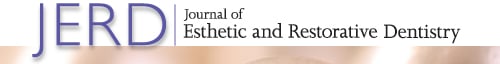
The monolithic CAD/CAM restorations were designed (InLab, 18.0, Dentsply Sirona, Bensheim, Germany) and milled (MCXL, Dentsply Sirona, Bensheim, Germany) with a lithium disilicate material (Celtra Duo LT, shade BL3, Dentsply Sirona, Bensheim, Germany) (Figure 6). The restorations were characterized (Stain & Glaze kit, Dentsply Sirona) for a natural appearance. Internal surfaces were etched with hydrofluoridric acid and cementation was conducted using an adhesive protocol (Variolink Esthetic, Neutral, Ivoclar Vivadent, Schaan, Liechtenstein). After tooth preparation, the intraoral situation was scanned, followed by shade selection. The digital models obtained in this phase were superimposed on the digital wax-up to guide the definitive restorations (Figure 7). Figure 8 summarizes the complete digital workflow for this case. Notice the similarity between the wax-up, the mock-up, the designed restorations, and the final restorations.


3 DISCUSSION
One of the main aspects of a successful esthetic restoration is to restore the natural aspect of a smile. However, reproducing tooth texture in dental restorations is challenging, time-consuming, and demands an experienced technician. Previously, Kano et al.1 described the resin-shell technique to create a natural morphological appearance in final restorations. The original protocol included the recreation of teeth anatomical details based on Jan Hajtó's models, associated with the adaptation of resin composite shells in the mouth prior to the tooth preparation.1, 2
The availability of a digital library containing natural teeth enabled the creation of a natural surface and texture. The “smile donator” concept and the “copy-and-paste” technique are applied to incorporate the anatomical details of a natural tooth into the digital wax-up.
After obtaining the digital wax-up, a motivational mock-up is printed for the clinical try-in. These digital tools help achieve esthetic results with natural morphology. Treatment was performed based on esthetic concepts, in which ideal dental proportions and dentofacial symmetry were considered for the final result.6, 9, 10 Briefly, digital workflow consisted of the digitalization of patient's documentation acquired by facial and intraoral scanners, photographs, and CBCT scans. All files were overlapped and analyzed together to define the treatment plan. DSD was conducted based on facial photographs and overlapped intra-oral scans to guide the digital wax-up.
Further advantages of a digital workflow include the implementation of interdisciplinary treatment planning.4, 12 To ensure a harmonious relationship between dental and gingival tissues, the described case report benefited from periodontal management before the restorative treatment.
A crown lengthening surgical guide allowed to transfer of linear measurements among teeth, bone, and soft tissues during the surgical procedure. This procedure allowed to determine the quantity of bone or soft tissue that must be removed to achieve the planned result. Likewise, preparation guides also helped visualize the extension of teeth preparation according to the thickness of the future CAD/CAM restorations. In addition, the accuracy of the outcome was ensured by the 3D-printed preparation guides.
DISCLOSURE
Christian Coachman is founder of the Digital Smile Design Group. Ângelo Rafael Toste Coelho Segundo is director of the Digital Smile Design (DSD) Lab.
ACKNOWLEDGMENT
Open Access funding enabled and organized by Projekt DEAL.
Open Research
DATA AVAILABILITY STATEMENT
The data that support the findings of this study are available from the corresponding author upon reasonable request.