Correction to Non-canonical Raf-1/p70S6K signalling in non–small-cell lung cancer
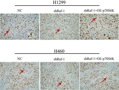
FIGURE 5D.
D. Immunohistochemical staining with Ki-67 (left) showing NSCLC tumour cell proliferation in each group (×400). Large brown Ki-67 staining (arrow) indicates proliferating cells. TUNEL staining (middle and right) demonstrating apoptotic cells in each group (×400); positive cells are indicated by arrows.


