Cross-talk between EGF and BMP9 signalling pathways regulates the osteogenic differentiation of mesenchymal stem cells
Abstract
Mesenchymal stem cells (MSCs) are multipotent progenitors, which give rise to several lineages, including bone, cartilage and fat. Epidermal growth factor (EGF) stimulates cell growth, proliferation and differentiation. EGF acts by binding with high affinity to epidermal growth factor receptor (EGFR) on the cell surface and stimulating the intrinsic protein tyrosine kinase activity of its receptor, which initiates a signal transduction cascade causing a variety of biochemical changes within the cell and regulating cell proliferation and differentiation. We have identified BMP9 as one of the most osteogenic BMPs in MSCs. In this study, we investigate if EGF signalling cross-talks with BMP9 and regulates BMP9-induced osteogenic differentiation. We find that EGF potentiates BMP9-induced early and late osteogenic markers of MSCs in vitro, which can be effectively blunted by EGFR inhibitors Gefitinib and Erlotinib or receptor tyrosine kinase inhibitors AG-1478 and AG-494 in a dose- and time-dependent manner. Furthermore, EGF significantly augments BMP9-induced bone formation in the cultured mouse foetal limb explants. In vivo stem cell implantation experiment reveals that exogenous expression of EGF in MSCs can effectively potentiate BMP9-induced ectopic bone formation, yielding larger and more mature bone masses. Interestingly, we find that, while EGF can induce BMP9 expression in MSCs, EGFR expression is directly up-regulated by BMP9 through Smad1/5/8 signalling pathway. Thus, the cross-talk between EGF and BMP9 signalling pathways in MSCs may underline their important roles in regulating osteogenic differentiation. Harnessing the synergy between BMP9 and EGF should be beneficial for enhancing osteogenesis in regenerative medicine.
Introduction
Mesenchymal stem cells (MSCs) are multipotent progenitors and differentiate into osteogenic, chondrogenic, adipogenic and other lineages 1-5. Although MSCs have been isolated from numerous tissues, one of the major sources in adults is the bone marrow stromal cells. Osteogenesis is a sequential cascade that recapitulates most of the cellular events occurring during embryonic skeletal development 6. Bone is one of a few organs that retain the potential for regeneration and is the only tissue that can undergo continual remodelling throughout life. Bone morphogenetic proteins (BMPs) play an important role in development 4, 7, 8, stem cell proliferation and osteogenic differentiation 9, 10. Bone morphogenetic proteins belong to the TGFβ [transforming growth factor (TGF)] superfamily and consist of at least 14 members in humans 4, 7, 8, 11, 12. We previously analysed the osteogenic potential of the 14 types of BMPs and found that BMP9 is one of the most potent BMPs in inducing osteogenic differentiation of MSCs both in vitro and in vivo by regulating several important downstream targets during BMP9-induced osteoblast differentiation of MSCs 8, 13-21.
BMP9 (also known as growth differentiation factor 2, or GDF-2), originally identified in the developing mouse liver 22, may also play a role in regulating cholinergic phenotype 23, hepatic glucose and lipid metabolism 24, adipogenesis 25 and angiogenesis 26, 27. Bone morphogenetic proteins initiate their signalling activity by binding to the heterodimeric complex of BMP type I and type II receptors 12. We have recently demonstrated that BMP type I receptors ALK1 and ALK2 are essential for BMP9-induced osteogenic signalling in MSCs 28. The activated receptor kinases phosphorylate Smads 1, 5 and/or 8, which in turn, regulate downstream targets in concert with co-activators during BMP9-induced osteoblast differentiation of MSCs 8, 13-20. BMP9 is one of the least studied BMPs and its functional role in skeletal development remains to be fully understood.
It has been reported that epidermal growth factor (EGF) signalling may play an important role in endochondral bone formation and bone remodelling 29-31. Epidermal growth factor is a key molecule in the regulation of cell growth and differentiation 30. Earlier studies indicated that EGF administration at physiological doses induces distinct effects on endosteal and periosteal bone formation in a dose- and time-dependent manner 32, 33, although it was also reported that EGF exhibited biphasic effects on bone nodule formation in isolated rat calvaria cells in vitro 34. Epidermal growth factor receptor (EGFR or ERBB1) is a transmembrane glycoprotein with intrinsic tyrosine kinase activity and activated by a family of seven peptide growth factors including EGF 31. It is conceivable that the osteoinductive activity of BMP9 may be further regulated by cross-talking with other growth factors, such as EGF.
In this study, we investigate if EGF signalling cross-talks with BMP9 and regulates BMP9-induced osteogenic differentiation of MSCs. We show that EGF potentiates BMP9-induced early and late osteogenic markers of MSCs in vitro, which can be effectively blunted by EGFR inhibitors Gefitinib and Erlotinib or protein tyrosine kinase inhibitors AG-1478 and AG-494 in a dose-dependent manner. Furthermore, EGF significantly augments BMP9-induced endochondral bone formation in the cultured mouse foetal limb explants, which can be blocked by EGFR inhibitors. In vivo stem implantation experiments reveal that exogenous expression of EGF in MSCs effectively potentiates BMP9-induced ectopic bone formation, yielding larger and more mature trabecular bone masses. Mechanistically, EGF is shown to induce BMP9 expression in MSCs, whereas EGFR expression is directly up-regulated by BMP9 through Smad1/5/8 signalling pathway. Thus, the regulatory circuitry of EGF and BMP9 signalling pathways in MSCs may underline their important roles in regulating osteogenic differentiation. Harnessing the synergy between BMP9 and EGF may be beneficial for enhancing osteogenesis in regenerative medicine.
Materials and methods
Cell culture and chemicals
HEK293, C2C12 and C3H10T1/2 cells were from ATCC (Manassas, VA, USA). The reversibly immortalized mouse embryonic fibroblasts (iMEFs) were previously established 35. Cell lines were maintained in the conditions as described 13, 15, 19, 36. Recombinant human EGF (rhEGF) was purchased from Sigma-Aldrich (St. Louis, MO, USA). Epidermal growth factor receptor/tyrosine kinase inhibitors, including Gefitinib (aka, Iressa or ZD1839), Erlotinib (aka, Tarceva, CP358, OSI-774, or NSC718781), AG494 and AG1478 were purchased from Cayman Chemical (Ann Arbor, MI, USA) and EMD Chemicals (Gibbstown, NJ, USA). Unless indicated otherwise, all chemicals were purchased from Sigma-Aldrich (St. Louis, MO, USA) or Fisher Scientific (Pittsburgh, PA, USA).
Recombinant adenoviruses expressing BMP9, EGF, RFP and GFP
Recombinant adenoviruses were generated using AdEasy technology as described 13, 14, 25, 37, 38. The coding regions of human BMP9 and EGF were PCR amplified and cloned into an adenoviral shuttle vector and subsequently used to generate recombinant adenoviruses in HEK293 cells. The resulting adenoviruses were designated as AdBMP9 and AdEGF. AdBMP9 also expresses GFP, whereas AdEGF expresses RFP as a marker for monitoring infection efficiency. Analogous adenovirus expressing only monomeric RFP (AdRFP) or GFP (AdGFP) was used as controls 18, 19, 37-45.
RNA isolation and semi-quantitative RT-PCR
Total RNA was isolated using TRIzol RNA Isolation Reagents (Invitrogen, Grand Island, NY, USA) and used to generate cDNA templates by RT reaction with hexamer and M-MuLV Reverse Transcriptase (New England Biolabs, Ipswich, MA, USA). The cDNA products were diluted 5- to 10-fold and used as PCR templates. Semi-quantitative RT-PCR was carried out as described 19, 21, 25, 41, 43, 46-48. PCR primers (Table S1) were designed using the Primer3 program to amplify the genes of interest (~150–180 bp). A touchdown cycling program was as follows: 94°C for 2 min. for one cycle; 92°C for 20 sec., 68°C for 30 sec. and 72°C for 12 cycles decreasing 1°C per cycle; and then at 92°C for 20 sec., 57°C for 30 sec. and 72°C for 20 sec. for 23–28 cycles, depending on the abundance of a given gene. PCR products were resolved on 1.5% agarose gels. All samples were normalized by the expression level of GAPDH.
Alkaline phosphatase (ALP) assay
Alkaline phosphatase activity was quantitatively assessed using a modified Great Escape SEAP Chemiluminescence assay (BD Clontech, Mountain View, CA, USA) and/or histochemical staining assay (using a mixture of 0.1 mg/ml napthol AS-MX phosphate and 0.6 mg/ml Fast Blue BB salt) as described 13, 14, 16-19, 25, 36, 41, 42, 45. For the chemiluminescence assays, each assay condition was performed in triplicate and the results were repeated in at least three independent experiments. Alkaline phosphatase activity was normalized by total cellular protein concentrations among the samples (using the BCA protein assay).
Alizarin Red S staining
C3H10T1/2 and iMEF cells were seeded in 12- or 24-well cell culture plates and infected with AdBMP9, AdEGF, AdGFP or AdBMP9+AdEGF. Infected cells were cultured in the presence of ascorbic acid (50 μg/ml) and β-glycerophosphate (10 mM). At 14 days after infection, mineralized matrix nodules were stained for calcium precipitation by means of Alizarin Red S staining, as described previously 13, 14, 16-19, 25, 36, 41. Cells were fixed with 0.05% (v/v) glutaraldehyde at room temperature for 10 min. After being washed with distilled water, fixed cells were incubated with 0.4% Alizarin Red S (Sigma-Aldrich) for 5 min., followed by extensive washing with distilled water. The staining of calcium mineral deposits was recorded under bright field microscopy.
Foetal limb explant culture
The forelimbs (i.e. humerus containing elbow joint) of mouse embryos (E18.5) were dissected under sterile conditions and incubated in DMEM (Invitrogen) containing 0.5% bovine serum albumin, 50 μg/ml ascorbic acid, 1 mM β-glycerophosphate and 100 μg/ml penicillin–streptomycin solution at 37°C in humidified air with 5% CO2 for up to 14 days as described 21, 42, 45. Limb explants were directly infected with AdGFP or AdBMP9 24 hrs after dissection in the presence of EGF and/or Gefitinib. The medium was changed every 2–3 days. Cultured tissues were observed in different time-points under microscope to confirm the survival of cells and the expression of fluorescence markers. New bone formation was visualized by adding calcein (100 mM) to the medium.
Immunohistochemical staining
Cultured cells were treated, fixed with 10% formalin and washed with PBS. The fixed cells were permeabilized with 1% NP-40 and blocked with 10% goat serum, followed by incubation with an anti-osteocalcin (OCN), or osteopontin (OPN) antibody (Santa Cruz Biotechnology, Santa Cruz, CA, USA) for 1 hr. After being washed, cells were incubated with biotin-labelled secondary antibody for 30 min., followed by incubating cells with streptavidin–HRP conjugate for 20 min. at room temperature. The presence of the expected protein was visualized by 3,3′-Diaminobenzidine (DAB) staining and examined under a microscope. Stains with control IgG were used as negative controls.
Stem cell implantation and μCT analysis
Subconfluent iMEFs were infected with AdBMP9/AdRFP or AdBMP9/AdEGF. At 16 hrs after infection, cells were harvested, and resuspended in PBS for subcutaneous injection (5 × 10⁶/injection) into the flanks of athymic nude (nu/nu) mice (five animals per group, 4–6 week old, female, Harlan Sprague Dawley, Indianapolis, IN, USA). At 4 weeks after implantation, animals were killed. The retrieved specimens were fixed and imaged using the μCT component of a GE Triumph (GE Healthcare, Piscataway, NJ, USA) trimodality preclinical imaging system. All image data analysis was performed with Amira 5.3 (Visage Imaging Inc., San Diego, CA, USA), and 3D volumetric data and bone mean density heat maps were obtained as previously described 21, 28, 35, 42, 45.
Haematoxylin and eosin, trichrome and alcian blue staining
The retrieved tissues were fixed, decalcified in Cal-Ex II Fixative/Decalcifier (Fisher Scientific) overnight and embedded in paraffin. Serial sections of the embedded specimens were stained with haematoxylin and eosin. Masson Trichrome and Alcian Blue stains were performed as previously described 14, 17-19, 25, 28, 36, 41, 42, 45.
ChIP analysis
Subconfluent iMEFs were infected with AdGFP or AdBMP9. At 30 hrs after infection, cells were cross-linked and subjected to ChIP analysis as previously described 18, 19, 41. Smad1/5/8 antibody (Santa Cruz Biotechnology) or control IgG was used to pull down the protein-DNA complexes. The presence of Egfr promoter sequence was detected by PCR amplification using two pairs of primers corresponding to mouse Egfr promoter region.
Statistical analysis
All quantitative experiments were performed in triplicate and/or repeated three times. Data were expressed as mean ± SD. Statistical significances between vehicle treatments versus drug treatments were determined by one-way anova and the Student's t-test. A value of P < 0.05 was considered statistically significant.
Results
EGF enhances BMP9-induced osteogenic differentiation of MSCs
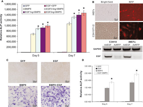
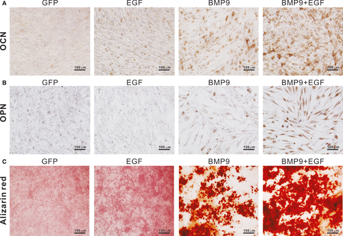
We previously identified that BMP9 is one of the most osteogenic BMPs in MSCs 8, 13, 14, 19, 20. Although we have identified several early targets that are important mediators of BMP9-induced osteogenic signalling 15-18, 20, 21, much remain to be learnt about how BMP9 cross-talks with other signalling pathways. Here, we investigated the possible cross-talk between BMP9 and EGF signalling pathways. Using a well-characterized MSC iMEFs, we found that rhEGF enhanced BMP9-induced early osteogenic marker ALP activity in a dose-dependent fashion, although rhEGF alone did not induce any detectable ALP activity (Fig. 1A). To further confirm these results, we constructed a recombinant adenovirus that expresses human EGF and demonstrated that this vector was able to transduce MSCs, such as iMEFs and C3H10T1/2 with high efficiency (Fig. 1B). Co-expression of BMP9 and EGF in MSCs led to a significant increase in ALP activity qualitatively (Fig. 1C) and quantitatively (Fig. 1D). Furthermore, exogenous expression of EGF was shown to enhance BMP9-induced expression of the late osteogenic markers osteocalcin (OCN) (Fig. 2A) and OPN (Fig. 2B). BMP9-induced matrix mineralization was significantly enhanced by co-expression of EGF in MSCs (Fig. 2C). These in vitro results strongly suggest that EGF signalling may synergize with BMP9-induced osteogenic signalling, although EGF signalling alone is seemingly insufficient to initiate osteogenic differentiation of MSCs.
BMP9-induced osteogenic differentiation can be effectively blocked by EGFR inhibitors
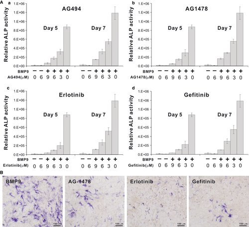
Epidermal growth factor initiates downstream events by activating its tyrosine kinase receptor EGFR (aka HER1). We sought to determine if commonly EGFR inhibitors would exert any effect on BMP9-induced osteogenic differentiation of MSCs. We tested four EGFR inhibitors, AG494, AG1478, Erlotinib and Gefitinib, and found all of them inhibited BMP9-induced ALP activity in a dose-dependent manner (Fig. 3A). Among the four tested inhibitors, Erlotinib and Gefitinib are clinically used as anticancer drugs, which were shown to be slightly more effective than AG494 and AG1478 in terms of inhibiting BMP9-induced ALP activity (Fig. 3A and B). Thus, these in vitro findings suggest that EGFR signalling may play an important role in BMP9-regulated osteogenic differentiation, although these inhibitors may also target other tyrosine kinase.
BMP9-regulated osteogenesis of mouse foetal limbs is promoted by EGF, but inhibited by EGFR inhibitors
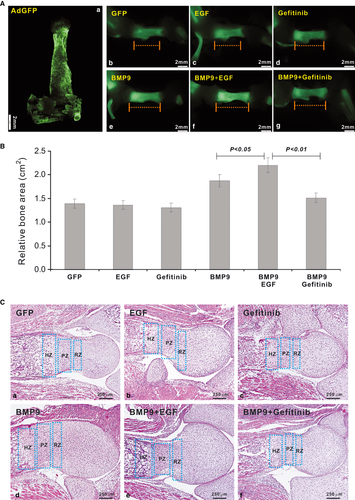
We sought to analyse the effect of EGF and BMP9 on developing bone using previously described ex vivo foetal limb culture assay 21, 42. We isolated mouse humerus containing elbow joint (with some soft tissues attached) from E18.5 embryos, which could be effectively transduced by adenoviral vectors (Fig. 4A, panel a). The isolated limbs were infected with AdBMP9 or AdGFP with or without EGF or Gefitinib. New bone formation was visualized by the incorporation of fluorescent dye calcein (Figure S1A). We found that the BMP9 group and BMP9+EGF group exhibited the highest fluorescence intensity, indicating active new bone formation (Fig. 4A, panels b–g). The BMP9+EGF group exhibited the largest fluorescence-positive area and was higher than that of BMP9 alone group (P < 0.05), while Gefitinib significantly inhibited BMP9-promoted bone formation (P < 0.01; Fig. 4B). Consistent with the in vitro results, EGF stimulation alone did not significantly promote osteogenesis, whereas Gefitinib might slightly inhibit osteogenesis, but the effect was not statistically significant (Fig. 4B). These results were further confirmed by histological analysis. The height and different chondrocyte zones at humerus growth plate did not exhibit any significant differences in EGF or Gefitinib treatment group compared with that of GFP control (Fig. 4C, panels a versus b & c). However, BMP9 or BMP9+EGF treatment led to a significant expansion of the humerus growth plate and endochondral bone formation (Fig. 4C, panels d & e). Notably, a combination of BMP9 and EGF led to a marked increase in cell numbers at the proliferating zone (Fig. 4C, panel e, Figure S1B). Conversely, the BMP9-induced growth plate expansion was blunted by EGFR inhibitor Gefitinib (Fig. 4C, panels d versus f). These in vitro results suggest that EGF may potentiate BMP9-induced endochondral ossification by increasing proliferative chondrocytes at the growth plate.
EGF enhances BMP9-induced ectopic bone formation and matrix maturation in stem cell implantation assay
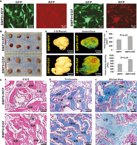
We further carried out stem cell implantation assays to determine the EGF effect on BMP9-induced bone formation in vivo. Subconfluent iMEFs were effectively co-infected with BMP9, RFP and/or EGF for 16 hrs (Fig. 5A). The infected cells were harvested to inject into athymic mice subcutaneously. Bony masses were found in BMP9+RFP and BMP9+EGF (Fig. 5B, panel a)-transduced cell groups at 4 weeks. No masses were formed in the cells transduced with RFP or EGF alone. The gross masses retrieved from BMP9+EGF group were larger than that from the BMP9 only group, which was confirmed by μCT scanning (Fig. 5B, panels a & b). The 3D isosurface analysis of the μCT scanning data revealed that the total bone volume in BMP9+EGF group was larger than that of BMP9 only group (Fig. 5B, panel c). Furthermore, a combination treatment of BMP9 and EGF promoted the mineralization of bone masses as demonstrated by μCT analysis of relative bone mean density in Hounsfield unit (Fig. 5B, panel c). Histological analysis of the retrieved ectopic bone masses indicated that MSCs treated with both BMP9 and EGF led to higher numbers of trabeculae and more mature bone matrix than that of the BMP9 alone group (Fig. 5C, panel a). These results were further confirmed by Masson's Trichrome staining of the retrieved bone masses, as more mature osteoid matrices were found in the specimens treated with both BMP9 and EGF (Fig. 5C, panel b). Conversely, alcian blue staining indicated that iMEFs co-infected with AdBMP9 and AdEGF exhibited slightly less cartilage matrix staining than those infected with AdBMP9 only (Fig. 5C, panel c). Thus, these in vivo results demonstrate that EGF can accelerate BMP9-induced osteoid matrix mineralization.
BMP9 cross-talks with EGF signalling pathway in regulating osteogenic differentiation of MSCs
Our in vitro and in vivo results show that EGF can significantly enhance BMP9-induced osteogenic differentiation of MSCs, suggesting that EGF may function in parallel and/or upstream of BMP9 signalling. The fact that EGFR inhibitors can block BMP9-induced osteogenic activity indicates that BMP9 could act upstream of EGFR function. We sought to determine the mechanistic aspects of BMP9 and EGF signalling in MSCs. Under unstimulated conditions, the pre-osteoblast progenitors (such as C3H10T1/2, iMEFs and C2C12 cells) expressed very low or undetectable levels of EGF and EGFR (Fig. 6A). When MSC cells (e.g. C3H10T1/2 and iMEFs) were stimulated with EGF, we found that BMP9 expression was apparently up-regulated (Fig. 6B), suggesting that EGF may act at least in part upstream of BMP9 signalling in MSCs. Consistent with this possibility, EGF was shown to enhance BMP9-induced phosphorylation of Smad1/5/8 (Figure S2). Furthermore, BMP9 stimulation in MSCs led to a significant increase in EGFR expression both at mRNA level (Fig. 6C, panel a) and protein level (Fig. 6C, panel b), while we did not observed any detectable changes in EGF expression (data not shown).
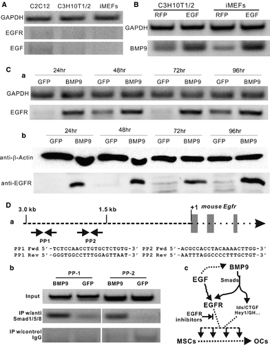
These results suggest that EGFR may be directly regulated by BMP9 signalling. We sought to determine if BMP9 regulates EGFR expression through Smad pathway using the chromatin immunoprecipitation (ChIP) assay. We analysed the possible presence of BMP R-Smad binding site(s) in the 3.0 kb Egfr promoter region using two pairs of PCR primers (Fig. 6D, panel a). For ChIP analysis, iMEFs were infected with AdGFP or AdBMP9 and cross-linked. Genomic DNA–protein complexes were sonicated and immunoprecipitated with anti-Smad1/5/8 antibody. The retrieved genomic DNA was subjected to PCR amplification using the two pairs of primers, PP1 and PP2. Although the genomic DNA input was comparable among the samples, primer pairs PP1 and PP2 were shown to amplify the expected fragments in BMP9-stimulated cells, but not in GFP control cells or in the samples precipitated by the control IgG (Fig. 6D, panel b). Thus, these results strongly suggest that EGF may be a direct downstream target of BMP9 signalling in MSCs.
Taken together, our results suggest a following mode of action about the cross-talk between BMP9 and EGF signalling pathways. Upon BMP9 stimulation of MSCs, several important downstream targets including EGFR are up-regulated. Epidermal growth factor signalling synergizes with other BMP9 targets and leads to efficient osteogenesis. BMP9-induced osteogenic signalling can be blocked by EGFR inhibitors. Intriguingly, EGF itself may up-regulate the expression of BMP9 through a yet-to-be-determined mechanism (Fig. 6D, panel c).
Discussion
We previously identified BMP9 as one of the most osteogenic BMPs both in vitro and in vivo 8, 13-20. As one of the least studied BMPs, BMP9 may exert its signalling activity by regulating a distinct set of downstream mediators in MSCs. We have recently identified several downstream targets in MSCs upon BMP9 stimulation 8, 15-18, 20. As for any major signalling molecules, BMP9 may fulfil its biological functions by cross-talking with other pathways. In this study, we show that EGF potentiates BMP9-induced early and late osteogenic markers of MSCs, which can be effectively inhibited by EGFR inhibitors Gefitinib and Erlotinib or protein tyrosine kinase inhibitors AG-1478 and AG-494. Epidermal growth factor significantly augments BMP9-induced endochondral bone formation in the cultured mouse foetal limb explants, which can be blocked by EGFR inhibitors. In vivo stem cell implantation experiments reveal that exogenous expression of EGF in MSCs effectively potentiates BMP9-induced ectopic bone formation, yielding larger and more mature trabecular bone masses. Mechanistically, EGF is shown to induce BMP9 expression in MSCs, whereas EGFR expression is directly up-regulated by BMP9 through Smad1/5/8 signalling pathway. Thus, the regulatory circuitry of EGF and BMP9 signalling pathways in MSCs may underline their important roles in regulating osteogenic differentiation.
Our results indicate that EGF itself exhibits negligible capability to induce osteogenic differentiation of MSCs. In fact, it was reported that the activation of EGF signalling may promote the proliferation and expansion of the pre-osteoblast progenitor cells at the expense of lineage-specific differentiation 30, 49, 50. However, it was also shown that EGF stimulation enhances osteogenic differentiation of human MSCs in the presence of chemical cues 51. Although no satisfactory explanation is offered about the conflicting results, one possibility is that the EGF effect on MSC cells may be dependent on the proliferation versus differentiation status of the pre-osteoblast progenitor cells. Another possible explanation is that the biological outcome of the EGF-stimulated MSCs may be significantly affected by the potency/efficacy of the differentiation cue. We have demonstrated that osteogenic BMPs (including BMP9)–induced osteogenesis is a well-coordinated process of the proliferative expansion and terminal differentiation of pre-osteoblast progenitor cells 4, 8, 16, 20, 52. Our previous studies have demonstrated that BMP9 is one of the most osteogenic factors 4, 8, 13, 14, 20. Thus, our current studies indicate that the EGF-stimulated expansion of progenitor cells can be efficiently driven to osteogenic lineage and terminal differentiation by BMP9. Consistent with our findings is an earlier study, which showed that overexpression of Egf in transgenic mice using the β-actin promoter results in growth retardation and over-proliferation of osteoblasts 53.
It has been reported that EGFR signalling plays an important role in growth plate development, endochondral bone formation and longitudinal bone growth 29, 31, 54. The EGFR family of receptor tyrosine kinases includes EGFR/ErbB1, HER2/ErbB2, HER3/ErbB3 and HER4/ErbB 31, 55. Epidermal growth factor receptor binds several ligands including EGF, TGF-α, β-cellulin, epiregulin and amphiregulin. Upon ligand binding, EGFR dimerizes and undergoes auto- or transphosphorylation on tyrosine residues in the intracellular domain, leading to the activation of several important intracellular signalling pathways, such as Ras-Raf-MAP-kinase and PI-3 Kinase-Akt. ErbB2 is the preferred partner for EGFR, and the signals mediated by EGFR/ErbB2 account for most of EGFR's biological activities 31, 55. Egfr-deficient mice exhibit impaired endochondral ossification, probably secondary to a defect in hypertrophic chondrocyte maturation and osteoblastic cell proliferation, and delayed primary endochondral ossification because of defective osteoclast recruitment 54, 56. More recently, it was shown that in vivo administration of Gefitinib or mice with cartilage-specific Egfr inactivation leads to defective endochondral ossification and hypertrophic cartilage enlargement as a result of suppressed osteoclastogenesis 57, although it is not clear if these effects resulted from the blockade of TGFα signalling. Nonetheless, it has been shown that the Egfr dominant–negative mutant mice exhibited a remarkable decrease in tibial trabecular bone mass with abnormalities in trabecular number and thickness, whereas mice with a constitutively active Egfr allele displayed increases in trabecular and cortical bone content 58. Thus, our findings are supported by these prior studies on EGFR functions in bone and skeletal development.
Finally, it is noteworthy that aberrant activations of EGFR signalling are frequently observed in many types of advanced stage cancers and bone metastases 59, 60. Various EGFR tyrosine kinase inhibitors, such as Gefitinib and Erlotinib, have shown promising results in clinical and/or preclinical anticancer studies 59-61. Further investigations may be required to determine if the use of the EGFR inhibitors could affect the bone density in cancer patients, leading to pathological fractures. Furthermore, it is also important to investigate if the use of these inhibitors to treat bone metastases could worsen osteolytic lesions.
In summary, we have demonstrated that EGF potentiates BMP9-induced early and late osteogenic markers of MSCs, which can be effectively blunted by EGFR inhibitors. EGF also augments BMP9-induced endochondral bone formation in the cultured mouse foetal limb explants. Exogenous expression of EGF in MSCs effectively potentiates BMP9-induced ectopic bone formation. Mechanistically, EGF is shown to induce BMP9 expression in MSCs, whereas EGFR expression is directly up-regulated by BMP9 through Smad1/5/8 signalling pathway. Thus, the cross-talk between EGF and BMP9 pathways in MSCs may play an important role in regulating osteogenic differentiation. Harnessing the synergy between BMP9 and EGF should be beneficial for enhancing osteogenesis in regenerative medicine.
Acknowledgements
We thank Dr. Chad Hanley of the Department of Radiology at The University of Chicago for his assistance and advice on μCT scanning and imaging analysis. This study was supported in part by research grants from the Orthopaedic Research and Education Foundation (RCH and HHL), the National Institutes of Health (RCH, TCH and HHL), and the Natural Science Foundation of China (#81100309 to YB, #81001197 to YS, #30973062 and 81172545 to QL, and #81272172 to GN).
Conflicts of interest
All authors state that they have no conflicts of interest.


