Osteology of the posterior vertebral column and caudal skeleton of marine amphibious gobies (mudskippers) (Teleostei: Gobioidei)
Ghanbarifardi and Gut contributed equally to this work.
Abstract
Mudskippers are amphibious gobies (Teleostei: Gobioidei, Oxudercinae) that have served as models for the specialised physiology and behaviour of fishes out of water. In this study, a comparative analysis of the posterior vertebral column and the caudal skeleton of ten mudskipper species was conducted on the basis of X-ray imaging. The species considered were Apocryptes bato, Apocryptodon madurensis, Boleophthalmus dussumieri, Oxuderces dentatus, Periophthalmodon freycineti, Pn. schlosseri, Periophthalmus novemradiatus, Ps. waltoni, Pseudapocryptes borneensis, and Scartelaos tenuis. For the osteological description the new term 'modified caudal vertebra' is used for all those vertebrae that display visibly modified neural and/or haemal spines compared to the spines of a 'usual' caudal vertebra, but are not involved in the support of caudal rays. The results reveal that the most terrestrial forms (Pn. freycineti, Pn. schlosseri, Ps. novemradiatus, Ps. waltoni) possess distinct traits that are seldom found in the other species. Among these features are (a) the existence of at least two modified caudal vertebrae (also present in S. tenuis), (b) a particularly close, dovetailing association between the neural spines of the preural vertebrae two and three (restricted to Ps. novemradiatus and Ps. waltoni), and (c) thickening and shortening of the ventralmost principal caudal rays (also present in B. dussumieri and S. tenuis). These findings support the idea that the posterior caudal vertebrae and caudal skeleton of the mentioned species are modified to enhance locomotion on land. Moreover, a relationship between character development and degree of terrestrial adaptation is probable, as all three traits are most pronounced in Ps. waltoni, which correlates with its strikingly high level of adaptation to amphibious life. A further aspect of this study is that the newly recognized skeletal structures have good fossilization potential and could therefore facilitate recognition of fossil species of mudskippers, which are currently unknown.
1 INTRODUCTION
Mudskippers are small to moderately sized amphibious fishes that belong to the Gobiidae sensu Gill & Mooi, 2012 and constitute the subfamily Oxudercinae (Murdy, 1989; Murdy & Jaafar, 2017). Mudskippers are widely distributed in coastal mangrove and mudflat areas in the tropics, with the exception of the New World (Hoese, 1984; Murdy, 1989; Parenti & Jaafar, 2017). They are characterized by specialised behaviours and the ability to use their limb-like pectoral fins for locomotion on land (Harris, 1960; Sayer, 2005; Jaafar & Murdy, 2017 and references therein). The members of ten genera are usually referred to as mudskippers, i.e. Apocryptes Valenciennes, 1837 (represented with one species), Apocryptodon Bleeker, 1874 (three species), Boleophthalmus Valenciennes, 1837 (six species), Oxuderces Eydoux & Souleyet, 1850 (two species), Parapocryptes Bleeker, 1874 (two species), Periophthalmodon Bleeker, 1874 (three species), Periophthalmus Bloch & Schneider, 1801 (19 species), Pseudapocryptes Bleeker, 1874 (two species), Scartelaos Swainson, 1839 (four species), and Zappa Murdy, 1989 (one species) (see Murdy & Jaafar, 2017). Of these, only members of four genera spend time on land as part of their daily life cycle, namely Boleophthalmus, Periophthalmodon, Periophthalmus and Scartelaos (Murdy, 1989). Their species can easily move about on muddy or moist surfaces and excavate burrows in the mud; some are even able to climb rocks, mangrove roots or stems, and all share several anatomical, physiological and sensorial specialisations (e.g. Michel, Adriaens, Aerts, Dierick, & Van Wassenbergh, 2014; Murdy, 1989; Pace, 2017; Schöttle, 1931; You et al., 2018; Zander, 2011). Scartelaos is the most aquatic (or least terrestrial) of the four genera, while Periophthalmus is the most terrestrial genus (Ishimatsu & Gonzales, 2011; Murdy, 1989; Polgar & Crosa, 2009).
A comprehensive investigation of the morphological traits of the Oxudercinae has been provided by Murdy (1989). In addition, there are many works dealing with the specific adaptations of the four mudskipper genera that are regularly amphibious (see Jaafar & Murdy, 2017 and references therein). However, the vertebral column and the caudal skeleton of mudskippers have received relatively little attention and were studied mainly within broader taxonomic contexts (Birdsong, Murdy, & Pezold, 1988; Fujita, 1990). Limited data, mostly restricted to the caudal skeleton, is solely available for three mudskipper species, i.e. Boleophthalmus pectinirostris (Linnaeus, 1758) (Fujita, 1990: fig. 502), Ps. barbarus (Linnaeus, 1766) (Harris, 1960: fig.4, as Ps. koelreuteri) and Ps. kalolo Lesson, 1831 (Fujita, 1990: fig. 499, as Ps. vulgaris).
The objective of this study is to gain a better knowledge of the posterior vertebral column and the caudal skeleton of mudskippers. These structures are of central importance for locomotion in aquatic vertebrates (Lauder, 1989). For mudskippers, the caudal skeleton is also important for locomotion on land (Harris, 1960; Pace, 2017; Swanson & Gibb, 2004). The specific aim was to examine if certain traits can be detected that may be associated to adaptations to terrestrial locomotion.
2 MATERIALS AND METHODS
2.1 Species studied
The material comprises 10 oxudercine species representing eight genera, i.e. Apocryptes bato (Hamilton, 1822), Apocryptodon madurensis (Bleeker, 1849), Boleophthalmus dussumieri Valenciennes, 1837, Oxuderces dentatus Eydoux & Souleyet, 1850, Pn. freycineti (Quoy & Gaimard, 1824), Pn. schlosseri (Pallas, 1770), Ps. novemradiatus (Hamilton, 1822), Ps. waltoni Koumans, 1955, P. borneensis (Bleeker, 1855), and S. tenuis (Day, 1876) (see Table 1 for details). Species identification was verified in each case based on the species-diagnostic characters, the sampling location and further information provided in Murdy (1989). A specimen of Gobius niger Linnaeus, 1758 from the NE Atlantic Sea (collection of the National Museum Prague, Czech Republic, number NMP6V 146072) was used as a representative species of the 'gobiine-like' Gobiidae sensu Agorreta et al. (2013). Digital X-ray images were taken with a Faxitron Bioptics (LLC-Vision NDT version 2.2.5, 45 k.v. and 30 s) housed in the State Zoological Collection, Munich for all specimens and served as basis for the osteological study.
| Species | Drainage and locality | Museum codes | N |
|---|---|---|---|
| Apocryptes bato (Hamilton, 1822) |
SMF 1736: Ganges, Bengalen, Asia (1847) SMF 23715–1,2: Hooghly-river, Bengalen, SSW Calcutta, S India, 88° 5′ E, 21° 55′ N (1.10.1988) |
SMF 1736, 23715–1, −2 | 3 |
| Apocryptodon madurensis (Bleeker, 1849) |
ZMB 6510: Singapore (1862) ZMB 7637: Manila, Philippines (1862) |
ZMB 6510, 7637 | 2 |
| Boleophthalmus dussumieri Valenciennes, 1837 |
Persian Gulf, Khorramshahr, Iran 48° 11′ 41.13″ E, 30° 26′ 7.11″ N |
ZM-CBSU 4, 6–9, 11 | 6 |
| Oxuderces dentatus Eydoux & Souleyet, 1850 | East China Sea; Fuckwing/Fuqing, Fujian Shng/Fukien (1893) | SMNS 3410–1, −2, – 3 | 3 |
| Periophthalmodon freycineti (Quoy & Gaimard, 1824) |
Airdhills, Gulf District, Papua New Guinea, 144° 21′ 20.00″ E, 7° 26′ 50.00″ S |
ZSM-PIS 19649, 19650 | 2 |
| Periophthalmodon schlosseri (Pallas, 1770) | Andaman Sea, Indian Ocean, Thailand | ZSM-PIS 023806 | 1 |
| Periophthalmus novemradiatus (Hamilton, 1822) | Ganges, Calcutta, India | ZMB 5987–1, −2(cf.) | 2 |
| Periophthalmus waltoni Koumans, 1941 |
Persian Gulf, Heleh Wetland, Iran 50° 40′ 43.38″ E, 29° 14′ 55.95″ N |
ZM-CBSU 92, 93, 95, 96, 101, 1716, 1727 | 5 |
| Pseudapocryptes borneensis (Bleeker, 1855) | Timor, Indonesia (1874–1876) | ZMB 9931 | 1 |
| Scartelaos tenuis (Day, 1876) |
Persian Gulf, Heleh Wetland, Iran 50° 40′ 43.38″ E, 29° 14′ 55.95″ N |
ZM-CBSU 82, 84, 85, 86, 89 | 7 |
Note
- Institutional abbreviations: SMF, Senckenberg Forschungsinstitut und Naturmuseum Frankfurt am Main, Germany; SMNS, Staatliches Museum für Naturkunde Stuttgart, Germany; ZMB, Museum für Naturkunde Berlin, Germany; ZM-CBSU, Zoological Museum of Shiraz University, Biology Department, Iran; ZSM-PIS, Zoologische Staatssammlung München (Bavarian State Collection of Zoology), Germany.
2.2 Study of osteology
Figure 1 provides an overview of the vertebral column and caudal skeleton, the osteological terminology follows Schultze and Arratia (2013). The vertebral column comprises an abdominal and a caudal part (Figure 1a). Each vertebra consists of a centrum, and, except the last vertebra (= terminal centrum, TC), a neural arch and a neural spine (Figure 1a, b). Abdominal vertebrae, except the first one or two, are associated with ribs; caudal vertebrae (except TC) are characterized by the presence of a closed haemal arch and a haemal spine (Figure 1a, b). Following Schultze and Arratia (2013), solely the penultimate and antepenultimate caudal vertebrae are designated here as preural vertebrae (PU) as their spines contribute to the support of the caudal rays. The penultimate caudal vertebra represents PU2 and the antepenultimate one is PU3 (Figure 1b). The usual condition in gobioids is that PU2 possesses an expanded haemal spine (which supports caudal rays, see Figure 1b), but a shortened neural spine (see Fujita, 1990). Also the neural and/or haemal spine of PU3 are visibly differentiated from a 'usual' caudal vertebra - i.e. more elongated, or more expanded, or more bent - and the distal tips of the PU3-spines are associated with (or near to) the caudal cartilage that supports the dorsal and ventral procurrent caudal rays (see Fujita, 1990). Further elements of the caudal skeleton of gobiids are two large hypural plates (Hy1+2, Hy3+4), an autogenous small hypural plate (Hy5), a parhypural (Php) and one or two epural bones (Ep1, 2) (Figure 1b). The hypural plates and the parhypural support the principal caudal rays and the epural bones are associated with the support of the dorsal procurrent caudal rays (Figure 1b).
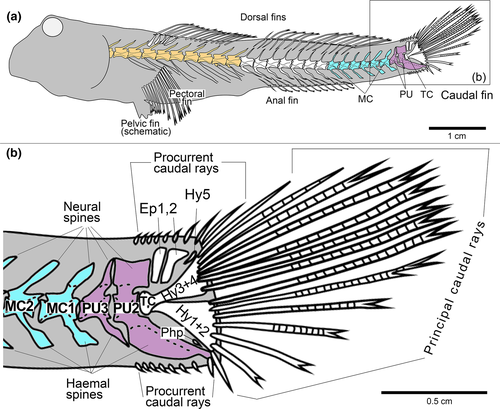
For the purpose of this study, we use as additional term 'modified caudal vertebra' (MC), which is numbered from the posteriormost to the anteriormost (like the preural vertebrae). This term is used for each vertebra that has, like a preural vertebra, a visible modification of its neural and/or haemal spine in comparison to a 'usual' caudal vertebra, but differs from a preural vertebra as it is not involved in caudal ray support (Figure 1b). The same terminology and definition are used in Charmpila et al. (in press).
3 RESULTS
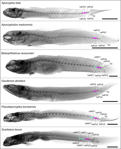
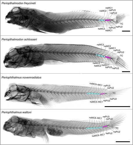
Representative X-ray images of each species, with depictions of their preural and modified caudal vertebrae, are provided in the Appendix Figures A1 and A2.
3.1 The posteriormost vertebral column
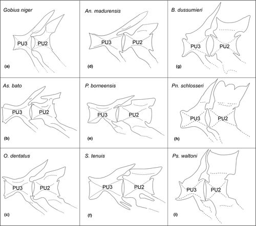
The presence of two preural vertebrae (PU2, PU3), as seen in Gobius niger (Figure 2a), and the absence of modified caudal vertebrae in the definition of the present study can be considered as the usual configuration in the Gobiidae; it also occurs in As. bato, O. dentatus, and An. madurensis (Figure 2b–d, Appendix: Figure A1).
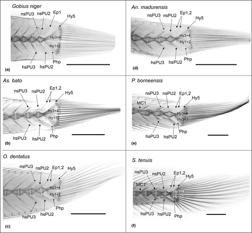
The addition of a single modified caudal vertebra (MC1) is seen in the posterior vertebral column of P. borneensis and B. dussumieri (Figures 2e, 3a, Appendix: Figure A1). Note that B. dussumieri exhibits a neural spine of MC1 that is not only slightly broadened (as in P. borneensis), but also relatively closer positioned to the neural spine of PU3 (Figure 3a). Two modified caudal vertebrae (MC1, MC2) occur in S. tenuis (Appendix: Figure A1) and Pn. freycineti (Appendix: Figure A2), and 3–6 modified caudal vertebrae are recorded in Pn. schlosseri (MC1–MC3, Appendix: Figure A2), Ps. novemradiatus (MC1–MC5, Appendix: Figure A2) and Ps. waltoni (MC1–MC6, Appendix: Figure A2).
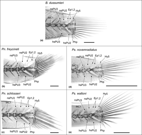
Furthermore, the shape, size and inclination of the neural and haemal spines of PU2 and PU3 revealed clear differences between the species (Figures 2-4), of which the details are specified in Table 2. Most noticeable is a particular close association between the neural spines of PU2 and PU3 in Ps. novemradiatus (Figure 3d) and Ps. waltoni (Figures 3e, 4i): the PU2 spine has a convex anterior margin that dovetails with the concave posterior margin of the PU3 spine.
| Characters |
G. ni. (for comparison) |
Studied mudkipper species | ||||||||||
|---|---|---|---|---|---|---|---|---|---|---|---|---|
| As. bato | O. de. | An. ma. | P. bo. | S. te. | B. du. | Pn. fr. | Pn. sch. | Ps. no. | Ps. wa. | |||
| Presence of preural vertebrae | PU2 + PU3 | x | x | x | x | x | x | x | x | x | x | x |
| Presence of modified caudal vertebrae | MC1 | (x) | x | x | x | x | x | x | ||||
| MC2 | (x) | (x) | x | x | x | |||||||
| MC3 | x | x | x | |||||||||
| MC4 + MC5 | x | x | ||||||||||
| MC6 | (x) | |||||||||||
| Shape neural spine PU2 (versus PU3) | Widened + pointed | (x) | (x) | (x) | ||||||||
| Widened + truncated | (x) | x | x | x | x | x | x | x | ||||
| Elevated | x | x | x | x | ||||||||
| Shape haemal spine PU2 (versus PU3) | Slightly elliptic | x | ||||||||||
| Plank-like | x | x | x | x | x | x | ||||||
| Fan-shaped | (x) | (x) | x | x | x | x | ||||||
| Proximity neural spines PU2 - PU3 | Distance in-between | x | x | x | x | x | x | |||||
| Very close | x | x | x | x | x | |||||||
| Dovetailing | x | x | ||||||||||
| Dip neural spine PU3 | Inclined | x | x | x | x | x | x | |||||
| Almost upright | x | x | x | x | x | |||||||
| Parhypural | Long | x | x | |||||||||
| Medium-sized or short | x | x | x | x | ||||||||
| tiny | x | x | x | x | x | x | ||||||
| Principal caudal rays | Relatively uniform | x | x | x | x | x | ||||||
| Ventralmost rays thickened + shortened | x | x | x | x | x | x | ||||||
- Abbreviations: As. bato, Apocryptes bato; An. ma., An. madurensis; B. du., B. dussumieri; G. ni., Gobius niger; MC, modified caudal vertebra; O. de., O. dentatus; P. bo., P. borneensis; Pn. fr., Pn. freycineti; Pn. sch., Pn. schlosseri; Ps. no., Ps. novemradiatus; Ps. wa., Ps. waltoni; PU, preural vertebra; S. te., S. tenuis; x, character well developed; (x), character weakly developed.
- [Correction added on 2 July 2020, after first online publication: the species names have been abbreviated in Table 2 to improve the layout in this version.]
3.2 Caudal skeleton
The configuration of the hypural plates is largely the same in all ten species (Figures 2, 3, Appendix: Figures A1 and A2). There are two large hypural plates (Hy1+2, Hy3+4), approximately equal in size and separated by a gap; HY3+4 is fused to the terminal centrum. Hypural plate 5 (Hy5) is a small, elongate or needle-shaped autogenous bone. Two relatively robust epural bones are present in all species. The parhypural is slender and not connected to the terminal centrum. In P. borneensis (Figure 2e), S. tenuis (Figure 2f), Pn. freycineti (Figure 3b), Pn. schlosseri (Figure 3c), Ps. novemradiatus (Figure 3d) and Ps. waltoni (Figure 3e), the parhypural is distinctly smaller than in the other species.
The principal caudal rays are relatively uniform in As. bato (Figure 2b), An. madurensis (Figure 2d), O. dentatus (Figure 2c) and P. borneensis (Figure 2e). In contrast, the principal caudal rays show an asymmetrical configuration in B. dussumieri (Figure 3a), Pn. freycineti (Figure 3b), Pn. schlosseri (Figure 3c), Ps. novemradiatus (Figure 3d), Ps. waltoni (Figure 3e) and S. tenuis (Figure 2f), with thickened and shortened ventralmost rays compared to their dorsal counterparts. This is most pronounced in Ps. waltoni, and least developed in S. tenuis.
4 DISCUSSION
Several of the studied mudskipper species displayed at least one modified caudal vertebra (versus not present in other gobiids), with highest numbers in Pn. schlosseri, Ps. novemradiatus, and Ps. waltoni (Table 2). Modified caudal vertebrae may provide extra robustness to the posterior vertebral column, and thus help support and stabilize the body when the fish moves on land. This would be in line with the occurrence of the highest counts of modified caudal vertebrae in Pn. schlosseri, Ps. novemradiatus, and Ps. waltoni, as these species are among the most amphibious of all oxudercine species (Murdy, 1989; Polgar et al., 2017; Polgar, Sacchetti, & Galli, 2010; Takita, Larson, & Ishimatsu, 2011).
The dovetailing configuration of the neural spines of PU2 and PU3 seen in the two studied species of Periophthalmus (Figure 4h, i) is also visible in the drawings of the caudal skeleton of Ps. kalolo by Fujita (1990: fig. 499) and Ps. barbarus by Harris (1960: fig. 4). Harris (1960) explained it as an adaptation that enables the fish to execute powerful terrestrial ‘jumps’ (‘skipping’ according to Harris, 1960, because the animal uses its tail to propel its body into the air, rather than the hind limbs, as in true jumping). Accordingly, the very close association between the neural spines of PU2 and PU3 represents a strengthening of these vertebrae, to withstand the strong forces that act upon them during skipping. That it is solely present in species of Periophthalmus correlates well with their high levels of adaptation to amphibious life (see Polgar et al., 2017).
A thickening and shortening of the ventralmost principal caudal rays was previously noted only for Periophthalmus (Harris, 1960; Murdy, 1989). It has been confirmed here for Ps. novemradiatus and Ps. waltoni and was also recorded for Pn. freycineti, Pn. schlosseri, B. dussumieri and S. tenuis (Figures 2f, 3a–e). The ventral portion of the caudal fin supports the body when the fish moves about on land or performs terrestrial jumps, e.g. during courtship or as an escape response (Pace, 2017; Zander, 2011), and the strengthening of the ventralmost principal caudal rays is an adaptation that supports these modes of locomotion (Harris, 1960). That the thickening and shortening of these rays is most pronounced in Ps. waltoni and least developed in S. tenuis correlates well with their relative levels of adaptation to amphibious life (see Polgar et al., 2017).
A further aspect of this study is that the newly recognized traits all relate to hard parts that can easily fossilize. At present, no fossil mudskippers are known, although they probably have a long evolutionary history (Polgar et al., 2014). The improved knowledge of their skeletal structures can also facilitate the identification of mudskipper fossils that may come to light in the future.
ACKNOWLEDGMENTS
For facilitating access to specimens we are grateful to Dr. Ulrich Schliewen and Dirk Neumann (SNSB-Bavarian State Collection of Zoology, Munich, Germany), Dr. Peter Bartsch and Edda Aßel (Museum für Naturkunde Berlin, Germany), Dr. Friedhelm Krupp and Dr. Moritz Sonnewald (Senckenberg Forschungsinstitut und Naturmuseum, Frankfurt, Germany), Dr. Stefan Merker and Dagmar Beermann (Staatliches Museum für Naturkunde Stuttgart, Germany). We thank Dr. Radek Šanda (National Museum Praha, Czech Republic) and Dr. Jasna Vukić (Charles University, Praha, Czech Republic) for personally delivering the specimen of Gobius niger. Dirk Neumann (ZSM) kindly conducted the X-rays of Pn. freycineti and Pn. schlosseri. The fieldworks in Iran were financially supported by Shiraz University and University of Sistan and Baluchestan. We acknowledge Mr. Abdulwahed Pehpouri (Mashad, Iran) for logistical help with field trip and accommodation. HRE thanks DAAD (German Academic Exchange Service) for financial support to visit Ludwig-Maximilians-Universität München to undertake the present study. The manuscript benefited greatly from the constructive comments of Dr. Gianluca Polgar (Western Kentucky University, USA) and two anonymous reviewers. Finally, we thank Dr. Paul Hardy (Düsseldorf, Germany) for critical reading of the manuscript. The authors have no conflict of interest to declare.
Appendix A
Open Research
DATA AVAILABILITY STATEMENT
X-ray images of all studied specimens are available upon request to the corresponding author.


