Assessment of liver function for safe hepatic resection
Abstract
The preoperative assessment of liver function is extremely important for preventing postoperative liver failure and mortality after hepatic resection. Liver function tests may be divided into three types; conventional liver function tests, general scores, and quantitative liver function tests. General scores are based on selected clinical symptoms and conventional test results. Child–Turcotte–Pugh score has been the gold standard for four decades, but the Child–Turcotte–Pugh score has difficulty discriminating a good risk from a poor risk in patients with mild to moderate liver dysfunction. The model for end-stage liver disease score has also been applied to predict short-term outcome after hepatectomy, but it is only useful in patients with advanced cirrhosis. Quantitative liver function tests overcome the drawbacks of general scores. The indocyanine green retention rate at 15 minutes (ICG R15) has been reported to be a significant predictor of postoperative liver failure and mortality. The safety limit of the hepatic parenchymal resection rate can be estimated using the ICG R15, and a decision tree (known as the Makuuchi criteria) for selecting patients and hepatectomy procedures has been proposed. Hepatic resection can be performed with a mortality rate of nearly zero using this decision tree. If the future remnant liver volume does not fulfill the Makuuchi criteria, preoperative portal vein embolization should be performed to prevent postoperative liver failure. Galactosyl human serum albumin-diethylenetriamine-pentaacetic acid scintigraphy also provides data that complement the ICG test. Other quantitative liver function tests, however, require further validation and simplification.
1. INTRODUCTION
There are many causes of liver dysfunction, including viral hepatitis, fatty liver disease, cholestasis, and chemotherapy-associated steatohepatitis. Liver cirrhosis arising from viral hepatitis is a major cause of liver dysfunction in patients with primary liver tumors.1–4 In patients with metastatic liver tumors, chemotherapy-associated liver dysfunction (including steatosis, steatohepatitis, and sinusoidal injury) has been reported as a risk factor for hepatectomy.5,6 Postoperative liver failure is a serious complication with a high mortality rate; therefore, the assessment of liver function, which can predict the risk of liver failure and mortality, is absolutely necessary for safe hepatic resection. However, the liver is a multi-functioned organ and a single comprehensive liver function test does not exist. General scores, including the Child-Turcotte-Pugh (CTP) score,7,8 the Model for End-Stage Liver Disease (MELD) score,9,10 and Makuuchi criteria,3 are useful for estimating integrated liver function. Although these general scores are useful, they do have some drawbacks. Several quantitative liver function tests have been introduced to compensate for these drawbacks of general scores.11 On the other hand, major hepatectomy is a risk in itself, even for patients with normal liver function, and the safety limit of the hepatic parenchymal resection rate must be considered according to the grade of liver dysfunction that is present.
The goals of hepatic functional assessment in liver surgery are proper patient selection and the prediction of the safety limit of the hepatic parenchymal resection rate. In this review article, we describe the present state of liver function assessments in liver surgery and provide guidance for safe hepatic resection.
2. LIVER FUNCTION TESTS
Hepatic function tests can be divided into three types (Table 1): conventional liver function tests, scoring systems that integrate clinical and laboratory variables, and quantitative liver function tests, the latter of which may theoretically be the most relevant to liver surgery. Conventional liver function tests include well-known and simple, but very important, laboratory parameters. They represent a variety of liver functions, as listed in Table 1; however, none of these parameters provide quantitative information regarding the hepatic functional reserve. Among these tests, the serum total bilirubin, prothrombin time, and serum albumin are integrated in the CTP criteria.
| 1. Conventional tests | |
|---|---|
| Test | Function/event measured |
| Serum bilirubin | Uptake, conjugation, excretion |
| Albumin | Synthesis |
| Alkaline phospatase | Cholestasis |
| Gamma-glutamyl transpeptidase | Cholestasis |
| Transaminases | Necrosis, enzyme induction, alcohol abuse |
| Coagulation factors: prothrombin time | Synthesis |
| Serum bile acids | Excretion, shunting |
| Pletelet count | Portal hypertension |
| 2. General score | |
|---|---|
| Score | Parameters |
| Child-Turcotte-Pugh score | serum bilirubin, albumin, PT%, ascites, encephalopathy |
| MELD score | serum bilirubin, creatinine, and INR |
| Makuuchi criteria (decision tree) | Ascites, serum bilirubin, ICG R15 |
| 3. Quantitative liver function tests | |
|---|---|
| Tests | Function tested |
| Aminopyrine breath test | Microsomal function |
| Antipyrine clearance | Microsomal function |
| Caffeine clearance | Microsomal function |
| Lidocaine clearance (MEGX) | Microsomal function |
| Methacetin breath test | Microsomal function |
| Galactose elimination capacity (GEC) | Cytosol function |
| Low-dose galactose clearance | Hepatic perfusion (liver blood flow) |
| Sorbitol clearance | Hepatic perfusion (liver blood flow) |
| ICG disappearance | Hepatic perfusion, anion excretion |
| Albumin synthesis | Synthetic function |
| Urea synthesis | Synthetic function |
| 99mTc-GSA | Functional hepatocyte mass |
3. GENERAL SCORE
3.1. Child-Turcotte-Pugh score
In 1964, Child and Turcotte proposed a grading system for liver function to predict the outcome of portal hypertensive patients undergoing a port-caval shut.7 This grading system was later modified by Pugh et al. in 1973, and the system has subsequently been known as the CTP score.8 Five simple parameters were selected based on their experiences: (i) the presence or absence of encephalopathy; (ii) the presence or absence of ascites; (iii) the serum total bilirubin level; (iv) the serum albumin level, (v) and the prothrombin time. The CTP score is a simple system for grading liver function based on these five easily measurable factors and has been considered a gold standard for more than four decades.
The CTP score has also been shown to predict the short-term and long-term outcomes of patients undergoing liver resection. Franco et al. showed that CTP A patients with HCC were good candidates for surgical resection.2 However, most hepatic dysfunction in surgical candidates is slight or moderate and might not be fully evaluated using the CTP score. A major drawback of the CTP scoring system is the so-called “floor and ceiling effect”. Most patients who undergo elective hepatic resection are classified as CTP A, but some risk postoperative liver failure and mortality exists even in this group of patients.2,4,12–15 The CTP scoring system is not able to discriminate the risk of liver failure in patients with low scores of 5–6 points in class A, that is, the “floor effect”. In fact, patients with CTP A are found to have a wide variety of liver functional reserves when assessed using quantitative tests (Fig. 1).16,17 In other words, we must be able to identify “good-risk” and “poor-risk” CTP A patients to determine the safety margin for liver resection. Quantitative liver function tests are useful for this purpose. On the other hand, patients with CTP C exhibit a wide range of cirrhosis severity, that is, the “ceiling effect”. Huo et al. proposed a modified CTP scoring system containing a class D to discriminate the severity of cirrhosis.18
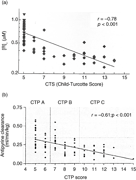
Relationship between Child–Turcotte–Pugh (CTP) score and receptor amount of (a) Galactosyl human serum albumin-diethylenetriamine-pentaacetic acid scintigrams TcGSA and (b) antipyrine clearance. (From Pimstone et al. and Mahmoud et al., with permission.)
3.2. The MELD score
Malinchoc et al. performed a multivariate analysis to predict the short-term results after transjugular intrahepatic portosystemic shunt (TIPS) procedure performed at the Mayo Clinic and reported that the serum concentrations of bilirubin and creatinine, the international normalized ratio for prothrombin time (INR), and the cause of the underlying liver disease were significant predictors of survival and, furthermore, that the Mayo model was superior to CTP classification.9 This model was slightly modified and is now widely used as the Model for END-Stage Liver Disease (MELD) score; the formula for the MLED score is 3.8*loge(bilirubin [mg/dL]) + 11.2* loge(INR) + 9.6*loge(creatinine [mg/dL]) + 6.4*(etiology: 0 if cholestatic or alcoholic, 1 otherwise).10 The MELD score can be used to predict the mortality risk of patients with end-stage liver disease and is applied for the allocation of donor livers.10,19 Considering its origin, the MELD score may overcome the drawback of the CTP score's ceiling effect.
The MELD score has also been applied to predict the postoperative mortality risk of patients undergoing hepatic resection. In a study of 82 patients with cirrhosis, 13 of whom died postoperatively, Teh et al. reported that a MELD score ≧ 9, the clinical tumor symptoms, and the ASA (American Society of Anesthesiologists) score were independent predictors of mortality after the hepatic resection of hepatocellular carcinoma.20 Similarly, Cucchetti and colleagues showed that cirrhotic patients with a MELD score equal to or above 11 had a high risk of postoperative liver failure in their report, 11 of 154 cirrhotic patients who underwent a hepatectomy experienced liver failure.21 Of note, however, the MELD score was not useful for predicting postoperative mortality in patients without cirrhosis.14,15 Schroeder and coworkers reviewed the records of 587 patients who had undergone elective hepatic resection for primary or metastatic liver tumors or benign diseases and showed that their mean MELD score, which was 6.5 ± 4.5 (SD) was not significantly related to mortality.14 Instead, they insisted that the CTP and ASA scores were superior for predicting the short-term outcomes. Teh et al. performed a case-controlled study of consecutive patients with or without cirrhosis undergoing hepatic resection for hepatocellular carcinoma and reported that the MELD score failed to predict postoperative outcomes in patients without cirrhosis.15 Presumably, the majority of patients undergoing elective hepatectomy have a low MELD score compared to that of patients undergoing TIPS or liver transplantation. The MELD score may be useful only in patients with advanced cirrhosis. An on-line worksheet for assessing postoperative mortality risk in patients with cirrhosis using age, the ASA score, the serum bilirubin level, the serum creatinine level, the INR, and the etiology of the cirrhosis is available at http://www.mayoclinic.org/meld/mayomodel9.html.
3.3. Makuuchi criteria
A decision tree for hepatectomy procedure has been proposed by Makuuchi et al. (Fig. 2).3 This surgical algorithm is the most popular in Japan and has had a great impact on the improvement of operative mortality and morbidity in hepatoma patients. The decision tree involves only three parameters: the presence or absence of uncontrollable ascites, the serum bilirubin level, and the indocyanine green retention rate at 15 minutes (ICG R15). Patients with uncontrollable ascites and a high bilirubin level are not candidates for hepatic resection. The extent of the hepatectomy procedure is selected according to the ICG R15 value, which appears at the bottom of the decision tree. Because the ICG R15 is not a linear quantitative parameter, only the surgical procedure, and not the exact numbers for the hepatic parenchymal resection rate, is presented for each ICG category. For example, a cirrhotic patient with an ICG value of 20% does not have twice the hepatic functional reserve of a patient with an ICG value of 40%. Using this decision tree, hepatic resection can be performed with almost zero mortality.3,13,22 These results suggest that hepatic resection can be safely performed in patients meeting the Makuuchi criteria, with the extent of the resection based on the ICG R15 value. Thus, the ICG test might be useful for discriminating good and poor risk CTP A patients.
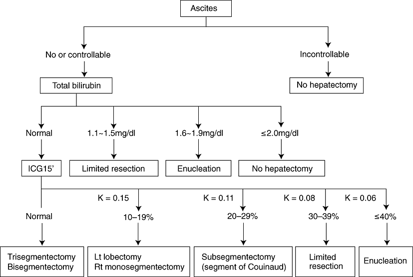
Makuuchi criteria for safe hepatic resection. (From Makuuchi et al., with permission.)
4. QUANTITATIVE LIVER FUNCTION TESTS
A number of quantitative liver function tests have been reported to meet the needs of hepatic surgeons. Quantitative liver function tests assess a specific aspect of hepatic function, that is, the liver functions of the microsome, cytosol, anion excretion, urea synthesis, or receptors on the hepatocyte membrane (Table 1).11 The clearance of intravenously administered exogenous substances that are eliminated or metabolized only by the liver are used for quantitative liver function tests, and the intrinsic clearance and hepatic flow are regards as determinants. The clearance of low extractable substances represents microsomal function (aminopyrine, antipyrine, caffeine, lidocaine, and metatherin) or cytosol function (galactose). On the other, the clearance of high extractable substances, such as sorbitol, low dose galactose, and ICG, represents hepatic flow.23 Galactosyl human serum albumin-diethylenetriamine-pentaacetic acid scintigrams (TcGSA) measure receptors on the hepatocyte membrane and indicate the functional hepatic mass. Breath tests using an inactive isotope carbon 13, such as 13C-Phenylalanine21 or 13C-methacetin, have been increasingly used for research purposes.24,25
Most of these quantitative function tests, however, have not been shown to be superior to conventional general scores, such as the CTP score, for predicting the surgical outcome of hepatectomy.26 Furthermore, the methods of quantitative liver functional tests are generally more complex than the ICG clearance test; consequently, these tests tends to be used for clinical research, but not for routine practice. At present, only the ICG test and GSA scintigraphy are widely accepted for clinical use in Japan.
4.1. ICG clearance test
After a bolus injection of indocyanine green, the dye binds to plasma proteins, and is removed exclusively by the liver through a carrier-mediated mechanism; the dye is ultimately excreted unchanged into the bile. It is not metabolized and does not undergo enterohepatic circulation. The disappearance curve of ICG has two components, a distribution and an elimination phase, and the turning point of these two phases is 20–30 min. ICG has a relatively high intrinsic clearance; therefore ICG retention at 15 minutes (ICG R15) represents hepatic perfusion. ICG R15 most likely increases in patients with cirrhosis because of intrahepatic shunt and sinusoidal capillarization. Intrahepatic shunts decrease the actual hepatic perfusion. Sinusoidal capillarization prevents the free diffusion of protein with high molecular weight such as albumin, and the uptake of ICG-bound protein also decreases. As a result, the clearance of ICG is delayed in patients with cirrhosis, and the extent of the increases in ICG R15 reflects the degree of liver dysfunction. The transportation of ICG competes with that of bilirubin; therefore, the ICG test is not suitable for patients with jaundice.
The ICG test provides additional information to the CTP score, and may overcome the drawbacks of the latter system. The ICG R15 was recommended as grade B for the assessment of liver function before surgery in recently developed evidence-based clinical guidelines for the treatment of hepatocellular carcinoma in Japan.27 The ICG test is particularly useful as a predictor of postoperative death. Hemming et al. reported that ICG clearance was the only test that predicted the risk of liver failure and mortality after hepatic resection and that no other liver function test was useful for this purpose.12 In a study of 315 patients, Nonami et al. found 24 patients with liver failure and showed that the ICG clearance and intraoperative blood loss were significant predictors of postoperative liver failure.28 Fan et al. reported that 101 patients underwent major hepatic resection, with a mortality rate of 13.8%; an ICG R15 value of 14% was the cutoff point for patient short-term survival according to a discriminant analysis.4 Lau et al. reported that 127 patients underwent hepatic resection and 14 patients (11%) died postoperatively; in their study, ICG R15 was the only test that could discriminate between survivors and non-survivors.26 They presumed that the safety limit was an ICG R15 value of 14% for major hepatectomy and 23% for minor hepatectomy. However, their decision algorism did not include the resection rate of the hepatic parenchymal volume.
The hepatic parenchymal resection rate calculated by computed tomography, that is, the remnant liver volume rate, is another important aspect of preventing postoperative liver failure.1,29,30 Okamoto et al. reported a close relationship between the ICG R15 and the parenchymal hepatic resection rate relative to postoperative liver failure in a study of 38 patients, 10 of whom died (Fig. 3).1 A two-pronged approach using the ICG R15 and the liver resection rate (remnant rate) is extremely important for predicting the surgical risk of liver failure and mortality and to decide on the surgical procedure that should be used to perform a hepatectomy in patients with impaired livers. If the future remnant liver (FRL) volume does not fulfill the Makuuchi criteria, preoperative portal vein embolization (PVE) should be performed to prevent postoperative liver failure as mentioned later.29,31,32
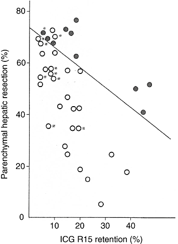
Relationship between indocyanine green retention rate at 15 minutes (ICG R15) and parenchymal hepatic resection rate relative to patient outcome. ○, survivors; ●, non-survivors;  , patients who did not have cirrhosis (n = 10). (From Okamoto et al., with permission.)
4.2. TcGSA scintigram
The asialoglycoprotein (ASGP) receptor is a hepatic cell surface receptor observed only in mammals. Intravenously administered TcGSA (galactosyl human serum albumin –diethylenetriamine- pentaacetic acid) rapidly binds to the asialoglycoprotein receptor and is taken up by the hepatocytes.33 A typical time activity curve after the injection of TcGSA in healthy subjects is shown in Figure 4.34 The characteristics of TcGSA are as follows: (i) GSA is taken up only by the liver; (ii) accumulation in the liver is rapid; (iii) not only blood clearance, but also hepatic uptake of GSA can be estimated; (iv) continuous time activity data can be obtained; and (v) the ligand-receptor binding is a second-order process. In other words, we can safely inject an amount of GSA that will occupy a significant fraction of the receptor.
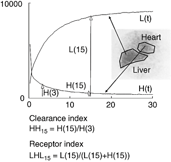
HH15 and LHL15. The clearance index HH15 and the receptor index LHL15 are calculated from three points of data in the time activity curves. (From Torizuka et al., with permission.)
The clearance index HH15 (heart 15/heart 3) and the receptor index LHL15 (liver 15/heart 15 + liver 15) are simple indicators calculated from three points of data in the time activity curves (Fig. 4).34 The LHL15 has been reported to be associated with ICG R15 and postoperative complications.35 To be precise, HH15 and LHL15 are not indices of total functional hepatocyte mass; therefore, R0 (receptor amount),36 Rmax (maximal removal rate of ASGP),37 Rtotal (total ASGP receptor amount),38 R0-remnant (total amount of the remnant liver),39 and GSA-RL (Rmax in the future remnant liver)40 are calculated using a kinetic model and are used as quantitative parameters. TcGSA scintigrams have a history of being used for 17 years but have become popular in only the last 10 years, and the modality is now known in Japan as a useful parameter, in addition to ICG R15, for predicting the outcome of hepatic resection.27,41 As the R0-remnant and GSA-RL represent the functional reserve after hepatectomy, these parameters may be superior to ICG R15 for predicting postoperative liver failure.40,42 Kokudo et al. reported a close relationship between the amount of receptors in the remnant liver and postoperative liver failure (Fig. 5).42
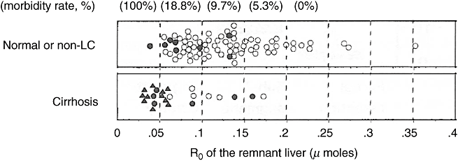
Relation between receptor amount in the remnant liver and postoperative liver failure. The close and open circles indicate patients with and those without complications indicating liver failure, respectively. The morbidity rate in patients with a remnant receptor amount of less than 0.05 µmoles was 100% and the rate decreased in inverse proportion to the remnant receptor amount. (From Kwon et al., with permission.)
Because of the above-mentioned excellent short-term outcomes after hepatic resection when performed according to the ICG R15 value, new liver function tests, including TcGSA scintigrams, are now used only for patients whose liver function cannot be fully estimated using an ICG based algorithm, such as patients with jaundice or ICG intolerance.
4.3. Galactose elimination capacity
Galactose is a high extractable substance after low dose administration, and its clearance represents hepatic flow. However, galactose is a harmless substance and is unique because it can be administered as a saturating dose. Galactose elimination capacity (GEC) is determined by serial measurement of serum concentration after administration of saturating dose of galactose (0.5mg/kg), and represents cytosolic metabolic capacity of the liver.11 GEC has been used to determine functional reserve of the severe liver disease, such as fulminant liver failure, primary biliary cirrhosis, or chronic active hepatitis. Recently, Redaelli et al. reported that preoperative GEC predicted the complications and mortality after hepatic resection.43 In the study of 258 patients with liver resection, 78 for HCC and 180 for non-HCC, GEC less than 4 mg/min/kg and 6 mg/min/kg was significantly associated with postoperative complications and death in patients with HCC and non HCC, respectively. GEC has been used to measure liver cytosolic liver function after in donors and recipients of living donor liver transplantation.44 The drawback of GEC is that it is of no value in detecting minor functional impairment. GEC has possibility to complement the CTP score and ICG R15, but a strategy of safe hepatic resection based on GEC has not yet been described. To determine the safety limit for hepatic resection rate in relation to the GEC is a future issue.
5. SELECTION OF HEPATECTOMY PROCEDURE
The mortality rate after hepatic resection has been improved thanks to advances in liver surgery and is now reported to be less than 5% in high-volume centers.45–47 Patient selection is usually made based on the CTP score. In principle, patients with CTP A are candidates for hepatic resection in western countries. Furthermore, a Barcelona group insisted that portal pressure evaluated using the hepatic venous pressure gradient (HVPG) was the only significant predictive factor of postoperative hepatic decompensation and that hepatectomies should be restricted to patients with portal hypertension.48 Portal hypertension has been adopted by the Barcelona Clinic Liver Cancer (BCLC) staging classification, in which the treatment schedule is determined according to both tumor progression and liver function.49
In Japan and other Eastern countries, the ICG test is routinely performed in addition to the CTP score, although the ICG test is not as popular in western countries. Patients with CTP A and some patients with CTP B are considered candidates for hepatectomy. The safety limit of the hepatectomy procedure is determined using ICG-based criteria, such as the Makuuchi decision tree, and this method has become a gold standard, in addition to the CTP score, in Japan. As a result, the average mortality rate after hepatectomy was 0.9% in Japan according to a nation-wide study,50 and zero mortality among 1056 consecutive patients over an 8-years period has been reported by our institution.13 Evidence-based clinical guidelines have proposed an algorithm for the treatment of HCC, which is determined according to the degree of liver damage and the tumor status.27 The degree of liver damage is classified as class A, B, and C using the presence of ascites, the serum total bilirubin, the serum albumin, the ICG R15, and the prothrombin time,51 and patients with class A or B are recommended to undergo resection if the number of tumors is less than three.27
ICG R15 was shown to be a significant predictor of liver failure and mortality after hepatectomy in the 1980s and early 1990s when the mortality rate was over 10%. The safety limit for the hepatic resection rate in relation to the ICG R15 had been estimated. Recently, 3D virtual hepatectomy simulation based on liver circulation has been developed, and the liver resection volume of planned hepatectomy can be more precisely predicted.52,53 The safety limit for hepatic resection rate may be reevaluated using the new approach in the future. In the era of nearly zero mortality, prospective studies of new assessments of hepatic function are difficult to plan using mortality after hepatectomy as an endpoint. New liver function tests must instead be more convenient and precise than the CTP score, the ICG test, or the TcGSA scintigrams.
The risk of liver dysfunction as a result of chemotherapy, that is, steatosis, steatohepatitis, and sinusoidal obstructive syndrome, has been described in patients who have received chemotherapy for colorectal cancer and liver metastasis, and microscopic findings in background liver tissue from resected specimens are related to surgical outcome after hepatic resection.5,6,54 However, the preoperative assessment of liver function after neo-adjuvant chemotherapy has not yet been established.
6. INDICATION FOR PORTAL VEIN EMBOLIZATION
In addition to liver function tests, the remnant liver volume is another important aspect of safe hepatic resection.30,55 If FRL volume is not enough to perform hepatectomy safely, indication of PVE should be considered.31,32 Originally, PVE was indicated if the rate of FRL volume was fewer than 40% in the patients with normal liver, and fewer than 50% in those with injured liver, i.e. with an ICG R15 value between 10–20% or with jaundice.29,31 These extensions of indication of major hepatic resection have been proved useful for both patients with hilar cholangiocarcinoma and with hepatocellular carcinoma.56,57 On the other hand, Vauthy et al. described the indication of PVE as followings: PVE was indicated when the FRL volume was ≤ 20% of the estimated total liver volume in patients with normal liver, ≤ 30% in patients with fibrosis or sever liver injury, and ≤ 40% in patients with cirrhosis.55,58 Furthermore, Clavien et al. proposed the decision tree for major hepatectomy using CTP score, presence of portal hypertension, and ICG R15 together with future remnant liver volume.59 In the review article, the authors proposed that the cutoff points of FRL for major hepatectomy was proposed 30% and 50% for patients with normal and cirrhotic liver, respectively, and that ICG R15 less than 14% is safety limit for major hepatectomy without PVE in patients with cirrhosis. As mentioned above, Makuuchi criteria have been expanded in both aspects of ICGR R15 and the rate of FRL volume in relation to the indication of PVE because the criteria is so strict that patients can underwent safe hepatic resection. However, extension of Makuuchi criteria should be carefully tried and validated in a large number of patients.
7. CONCLUSION
In summary, the CTP system is still useful for selecting surgical candidates for liver resection. The MELD score is helpful for predicting surgical outcome only in patients with advanced liver cirrhosis. The safety limit for the parenchymal resection rate can be determined using “criteria” based on the ICG-R15 score. GSA scintigrams provide data that complements the ICG test. Other quantitative liver function tests require further validation and simplification.


