Reading ability and stereoacuity with combined implantation of refractive and diffractive multifocal intraocular lenses
Abstract.
Purpose: To evaluate reading ability and stereoscopic vision with combined implantation of refractive and diffractive multifocal intraocular lenses (IOLs).
Methods: Thirty-one cataract patients (62 eyes) were assigned to receive either a ReZoom NXG1 IOL in the dominant eye and a Tecnis ZM900 IOL in the fellow eye (MIOL group), or Sensar AR40e IOLs bilaterally (SIOL group). The uncorrected visual acuity (UCVA) at 500 cm, best spectacle-corrected visual acuity (BSCVA) at 500 cm, reading acuity, reading speed, near stereoacuity and questionnaire were assessed 3 months postoperatively.
Results: Three months postoperatively, monocular and binocular UCVA and BSCVA at 500 cm showed no significant differences in both groups. The uncorrected reading acuity and reading speed in the MIOL group were significantly better than those in the SIOL group and were similar to that with correction in the SIOL group. The uncorrected mean near stereoacuity in the MIOL group was significantly better than that in the SIOL group (69 ± 50 seconds of arc in the MIOL group versus 180 ± 160 seconds of arc in the SIOL group). Patients in the MIOL group had a high level of satisfaction and more than 80% of them had an increased independence from spectacles for brief reading.
Conclusion: The combined implantation of refractive and diffractive multifocal IOLs was effective in improving reading ability and near stereoacuity with a good visual quality.
Introduction
With great advances in phacoemulsification technology, as well as intraocular lens (IOL) designs, much attention has been paid to improving postoperative optical quality. Although monofocal IOLs [or single-focus IOLs (SIOLs)] can provide good distance vision, the ability to focus at varying distances is lost with monofocal IOL implantation. Therefore, the multifocal IOL (MIOL), whether it is a refractive MIOL (RMIOL) or a diffractive MIOL (DMIOL), is designed to provide patients with an expanded range of vision. Unfortunately, because of the limitation of the design, none of the available MIOLs can provide satisfactory vision in postoperative patients, like the accommodative IOL (Mastropasqua et al. 2007). It may be beneficial to combine different types of lenses to enhance visual quality and reduce unwanted side-effects.
Most recent studies involving a mix or match of two MIOLs are focused on evaluating the visual acuity outcomes. These results showed that a combination of MIOLs can improve near, intermediate and distance vision (Blaylock et al. 2006; Chiam et al. 2007; Mester et al. 2007; Pepos et al. 2007; Toto et al. 2007; Goes 2008). However, little is known about functional vision with combined refractive and diffractive MIOLs. In this prospective study, a mixed combination of RMIOLs and DMIOLs were implanted. Subjective and objective measures of functional vision, including reading acuity, reading speed, near stereoacuity and questionnaire, were evaluated and compared.
Materials and Methods
Thirty-one age-related cataract patients (62 eyes) were recruited from the Zhongshan Ophthalmic Centre of Sun Yat-Sen University (Guangzhou, China) from September 2007 to April 2008. The MIOL group and the SIOL group were divided according to the type of IOL implanted. Patients in the MIOL group who wished to reduce dependence on spectacles after surgery were implanted with a combination of a ReZoom NXG1 RMIOL (Advanced Medical Optics, Santa Ana, California, USA) in the dominant eye and a Tecnis ZM900 DMIOL (Advanced Medical Optics) in the non-dominant eye. Patients in the SIOL group were implanted bilaterally with Sensar AR40e monofocal IOLs (Advanced Medical Optics).
The inclusion criteria were age between 50 and 80 years, corneal preoperative astigmatism < 1.5 dioptre (D) and availability for postoperative examinations. Exclusion criteria included severe systemic diseases, ocular diseases other than cataract (e.g. amblyopia, corneal disease, uveitis, retinopathy or glaucoma), previous ocular surgery, or intraoperative or postoperative complications.
All patients provided informed consent before surgery in accordance with the Declaration of Helsinki, and institutional review board approval was obtained from the hospital ethics committee. Bilateral implantation was performed within 1 week of the first IOL implantation. The target refraction was 0 to + 0.25 D for the MIOL and 0 to −0.25 D for the monofocal IOL. The SRK-T formula was used for IOL power calculations (Olsen 2007).
MIOL
The ReZoom NXG1 refractive multifocal lens is a three-piece hydrophobic acrylic with five refractive zones: zones 1, 3 and 5 are distance-dominant; zones 2 and 4 are near-dominant; and an aspheric transition between the zones provides balanced intermediate vision. This design, called Balanced View Optics Technology, provides good visual function across a range of distances in varying light conditions. It uses a near power of 3.5 D at the lens plane, which corresponds to approximately C2.8D at the spectacle plane.
The Tecnis ZM900, made with a silicone material, is a three-piece DMIOL. The diffractive design on the posterior surface creates two focal points and light is distributed evenly between near and distance vision with independent pupil size. A modified prolate anterior surface is designed to compensate the spherical aberration of the cornea. Its added power is 4.0 D at the lens plane, which corresponds to C3.2D at the spectacle plane.
Surgical technique
All surgeries were performed by the same surgeon (W.C.). After topical anaesthesia with Alcaine 0.05% and a temporal 3.2 mm clear corneal incision, a continuous curvilinear capsulorhexis was created. Phacoemulsification was followed by irrigation and aspiration of the cortex, and IOL implantation in the capsular bag with an injector. Postoperative therapy consisted of tobramycin 0.3% and dexamethasone 0.2% eyedrops administered six times daily for 4 weeks.
Postoperative clinical evaluations were set at 1 day, 1 week, 1 month and 3 months. Patients who had a visual function assessment by a ‘blinded’ examiner at the 3-month follow-up were included in this study.
Visual acuity
Monocular and binocular uncorrected visual acuities (UCVA) and best spectacle-corrected visual acuities (BSCVA) were recorded in decimal units with a standard logarithm chart at 500 cm. Under low-light and bright-light condition, the mean light densities in the perimeter hemisphere were 6 and 100 cd/m2 respectively (TES-1332A digital luminometer; TES, Taibei, Taiwan).
Reading acuity and reading speed
Reading acuity was tested using the Chinese Reading Charts (Jia Qu). In accordance with the Radner Reading Charts and Hütz et al. (2006), reading speed was tested using a third-grade elementary school text with a 12-point print size and a 1.5 line spacing. Patients were asked to read a sentence binocularly as quickly and accurately as possible while the other sentences were covered with a piece of paper. The number of words read per minute was recorded. All measurements of reading ability were performed at a standard reading distance (25 cm) and an optimal reading distance under bright-light (100 cd/m2) and low-light (6 cd/m2) conditions. Two refractive conditions were used to test reading ability: either without correction or with best near correction.
Near stereoacuity
Near stereoacuity was measured at 40 cm using the Randot stereotests (Stereo Optical Company, Chicago, Illinois, USA) using the accompanying polarizing spectacles under a luminance of 100 cd/m2. The Randot stereotests consist of three subsets of vectographs. The first component consists of six shapes used for testing gross stereopsis (500 and 250 seconds of arc disparity). The second component consists of three rows of five cartoon animals per row, with the figures in each row having the same disparity (400, 200 and 100 seconds of arc, respectively). The last component consists of 10 circular disparate areas, the disparities of which range from 400 to 20 seconds of arc. The level of stereopsis of the last correctly chosen target was recorded.
Questionnaire
Subjective vision-related quality of life was evaluated using the National Institute of Health Refractive Error Quality of Life Instrument-42 (NEI-RQL-42) scale, which was translated into Chinese for this study.
Statistical analysis
All visual acuity values were converted to the logarithm of the minimum angle of resolution (logMAR) for statistical analysis. Reading acuity was recorded by noting point size and converted to the logarithm for statistical analysis. Postoperative 3-month data were analysed using the spss 12.0 statistical software system (SPSS, Inc. Chicago, Illinois, USA) by a two-tail t-test. Results were expressed as mean ± standard deviation (SD). A p-value < 0.05 was considered statistically significant.
Results
The MIOL group comprised 30 eyes from 15 patients (10 male, five female) with an average age of 71 ± 7 years, while the SIOL group consisted of 32 eyes from 16 patients (11 male, five female) with an average age of 69 ± 9 years. Age and sex showed no significant differences between groups.
Monocular and binocular distance acuity
Under bright-light (100 cd/m2) and low-light (6 cd/m2) conditions, the monocular UCVA and BSCVA were not statistically different in the ReZoom NXG1 eyes, Tecnis ZM900 eyes and Sensar AR40e eyes at 500 cm. Similarly, the binocular UCVA and BSCVA between the MIOL group and the SIOL group were not statistically significant (Table 1).
| Group | 100 cd/m2 | 6 cd/m2 | ||
|---|---|---|---|---|
| UCVA | BSCVA | UCVA | BSCVA | |
| NXG1 | 0.02 ± 0.12 | 0.01 ± 0.11 | 0.14 ± 0.14 | 0.13 ± 0.13 |
| ZM900 | 0.07 ± 0.13 | 0.02 ± 0.08 | 0.24 ± 0.19 | 0.17 ± 0.13 |
| AR40e | 0.04 ± 0.15 | −0.00 ± 0.13 | 0.17 ± 0.17 | 0.11 ± 0.13 |
| p-value | 0.554 | 0.790 | 0.194 | 0.388 |
| MIOL-ou | −0.02 ± 0.10 | −0.04 ± 0.09 | 0.08 ± 0.14 | 0.05 ± 0.13 |
| SIOL-ou | −0.02 ± 0.12 | −0.03 ± 0.11 | 0.09 ± 0.16 | 0.07 ± 0.16 |
| p-value | 0.903 | 0.813 | 0.746 | 0.725 |
- UCVA, uncorrected visual acuity; BSCVA, best spectacle-corrected visual acuity; NXG1, ReZoom NXG1 intraocular lens; ZM900, Tecnis ZM900 intraocular lens; AR40e, Sensar AR40e intraocular lenses; MIOL, multifocal intraocular lens; SIOL, single-focus intraocular lens; ou, both eyes.
Reading acuity
As shown in Table 2, under a luminance of 100 cd/m2, the reading acuities without correction were significantly better in the MIOL group at either standard reading distance (25 cm) or optimal reading distance compared to the SIOL group. The reading acuity with best near correction was improved, but did not reach a significant difference compared to uncorrected reading acuity. Although the reading acuity with best near correction improved significantly in the SIOL group, the result was similar to that in the MIOL group with or without correction. The optimal reading distance for reading acuity was not different between the two groups. Similar results were obtained under a luminance of 6 cd/m2.
| Group | 100 cd/m2 | 6 cd/m2 | ||||
|---|---|---|---|---|---|---|
| Reading acuity at standard reading distance | Reading acuity at optimal reading distance | Optimal reading distance (cm) | Reading acuity at standard reading distance | Reading acuity at optimal reading distance | Optimal reading distance (cm) | |
| MIOL group | ||||||
| Uncorrected | 0.6 ± 0.2 | 0.6 ± 0.1 | 29.0 ± 5.7 | 0.7 ± 0.1 | 0.7 ± 0.1 | 28.8 ± 5.6 |
| Corrected | 0.6 ± 0.1 | 0.6 ± 0.1 | 28.4 ± 5.2 | 0.6 ± 0.1 | 0.6 ± 0.1 | 27.6 ± 4.8 |
| SIOL group | ||||||
| Uncorrected | 0.8 ± 0.2 | 0.8 ± 0.2 | 30.3 ± 6.4 | 0.9 ± 0.2 | 0.9 ± 0.1 | 30.6 ± 7.2 |
| Corrected | 0.6 ± 0.1 | 0.5 ± 0.1 | 28.4 ± 4.1 | 0.7 ± 0.1 | 0.7 ± 0.2 | 28.5 ± 4.3 |
| p-value* | 0.005 | 0.004 | 0.628 | 0.000 | 0.000 | 0.535 |
| p-value† | 0.068 | 0.054 | – | 0.546 | 0.546 | – |
| p-value‡ | 0.083 | 0.100 | – | 0.270 | 0.270 | – |
| p-value§ | 0.000 | 0.000 | – | 0.000 | 0.000 | – |
- MIOL, multifocal intraocular lens; SIOL, single-focus intraocular lens.
- * MIOL group without correction versus SIOL group without correction.
- † MIOL group without correction versus SIOL group with best near correction.
- ‡ MIOL group without correction versus MIOL group with best near correction.
- § SIOL group without correction versus SIOL group with best near correction
Reading speed
As shown in Table 3, under a luminance of 100 and 6 cd/m2, the mean reading speed without correction at optimal reading distance was significantly faster in the MIOL group compared to the SIOL group. The uncorrected and corrected mean reading speeds were similar in the MIOL group. The corrected mean reading speed improved significantly compared to the uncorrected mean reading speed in the SIOL group. There was no significant difference between the MIOL group with or without correction and the SIOL group with best near correction. Without correction, the optimal reading distance for reading speed was significantly higher in the SIOL group compared to the MIOL group.
| Group | 100 cd/m2 | 6 cd/m2 | ||
|---|---|---|---|---|
| Optimal reading distance (cm) | Reading speed (words/min) | Optimal reading distance (cm) | Reading speed (words/min) | |
| MIOL group | ||||
| Uncorrected | 33.6 ± 5.0 | 197.7 ± 37.8 | 29.7 ± 5.3 | 179.8 ± 30.6 |
| Corrected | 33.1 ± 5.1 | 201.3 ± 62.0 | 29.7 ± 5.1 | 191.9 ± 39.2 |
| SIOL group | ||||
| Uncorrected | 38.1 ± 7.2 | 136.2 ± 71.0 | 39.5 ± 8.7 | 79.2 ± 75.4 |
| Corrected | 32.6 ± 4.3 | 206.5 ± 34.9 | 31.3 ± 3.9 | 181.2 ± 36.4 |
| p-value* | 0.047 | 0.015 | 0.001 | 0.001 |
| p-value† | – | 0.336 | – | 0.868 |
| p-value‡ | – | 0.849 | – | 0.353 |
| p-value§ | – | 0.004 | – | 0.000 |
- MIOL, multifocal intraocular lens; SIOL, single-focus intraocular lens.
- * MIOL group without correction versus SIOL group without correction.
- † MIOL group without correction versus SIOL group with best near correction.
- ‡ MIOL group without correction versus MIOL group with best near correction.
- §SIOL group without correction versus SIOL group with best near correction.
The mean spherical equivalent of best near correction was 1.0 ± 0.7 D in the MIOL group, which was significantly lower than the 2.5 ± 0.6 D achieved in the SIOL group (p < 0.0001).
Near stereoacuity outcome
In the MIOL group, the uncorrected near stereoacuity was 69 ± 50 seconds of arc and corrected near stereoacuity was 62 ± 34 seconds of arc. In the SIOL group, the near stereoacuity without and with correction were 180 ± 160 seconds of arc and 136 ± 117 seconds of arc, respectively. The near stereoacuity without correction in the MIOL group was significantly better than that with or without correction in the SIOL group (p = 0.021 and p = 0.023, respectively).
The number of patients who achieved each level of near stereoacuity is shown in Table 4. In the MIOL group, all patients had uncorrected stereoacuity of 200 seconds of arc or better, and 40% of them (six of 15 patients) achieved a stereoacuity better than 60 seconds of arc. However, in the SIOL group, only 75% of patients had uncorrected stereoacuity of 200 seconds of arc or better, and only one of 16 patients (6.25%) achieved a stereoacuity better than 60 seconds of arc. The stereoacuity with best near correction in the two groups did not improve significantly.
| Group | Near stereoacuity without correction (seconds of arc) | Near stereoacuity with best near correction (seconds of arc) | ||||
|---|---|---|---|---|---|---|
| ≤ 60 | 70–200 | > 200 | ≤ 60 | 70–200 | > 200 | |
| MIOL group | 6 (40%) | 9 (60%) | 0 | 6 (40%) | 9 (60%) | 0 |
| SIOL group | 1 (6.25%) | 11 (68.75%) | 4 (25%) | 1 (6.25%) | 12 (75%) | 3 (18.75%) |
- MIOL, multifocal intraocular lens; SIOL, single-focus intraocular lens.
Subjective outcomes
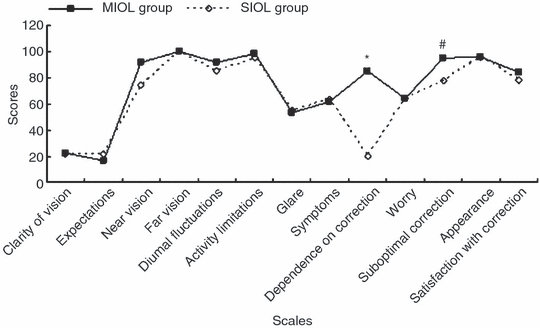
Figure 1 shows the subjective outcomes in the two groups using the NEI-RQL-42 scores. There were no significant differences between the MIOL and SIOL groups in scores for 11 of 13 scales. Although the score for near vision was higher in the MIOL group than in the SIOL group, the difference was not significant (p = 0.423). However, the scores for dependence on correction and suboptimal correction were significantly improved in the MIOL group compared to the SIOL group (p < 0.0001, p < 0.0001).

Comparison of mean scores of the Refractive Error Quality of Life Instrument-42 (NEI-RQL-42) scale for vision-related quality of life between the multifocal intraocular lens (MIOL) group and the single-focus intraocular lens (SIOL) group. *Score for dependence on correction, p < 0.0001. #Score for suboptimal correction, p < 0.0001.
In the MIOL group, the rate of independence from spectacles was 60% when long words in books or newspapers were read, and 86.7% when brief words (such as those in directions or menus) were read. In contrast, in the SIOL group only one of 16 patients did not need reading glasses when he read something brief.
Discussion
It has been demonstrated that as a standard procedure, bilateral implantation of monofocal IOLs after cataract extraction can fully improve distance vision and near vision with spectacle correction. Therefore, in this study, patients with bilateral monofocal IOLs were used as controls. Our results showed that compared to bilateral monofocal IOL implantation, combined refractive and diffractive MIOL implantation could provide satisfactory reading acuity, reading speed and near stereoacuity without obvious photic phenomena. More than 80% of patients who had combined refractive and diffractive MIOLs implanted were not dependent on using their spectacles for brief reading after surgery.
Reading is an essential skill to functioning effectively in society. Return of good reading ability is the principal motivation in a patient’s decision to have cataract surgery. Because reading involves a larger retinal area and provides topographic information, measurement of reading abilities provides much more information about functional vision than visual acuity testing. Therefore, the present study evaluated reading performance. Our results showed that a combined implantation of refractive and diffractive MIOLs can improve distance vision without correction. More importantly, our results indicated that patients with mixed MIOLs can achieve satisfactory reading acuity and reading speed under bright- and low-light conditions compared to bilateral implantation of monofocal IOLs. In this study, a Chinese reading chart was designed to test reading speed. This reading chart’s print size and contents are adapted to Chinese patients and closely simulate everyday life situations such as reading newspapers, books and magazines. Furthermore, patients were asked to comprehend the contents while they were reading – transmitting information to the brain is an essential task of visual processing. Additionally, we found that the mean spherical equivalent of best near correction was about 1.0 D in the MIOL group and 2.5 D in the SIOL group. This result indicated that the added powers of these MIOLs were not enough and patients with them preferred using reading glasses when long or smaller words were read.
The ability to perceive depth or relative distance on the basis of retinal disparity clues is termed stereopsis. The smallest amount of horizontal retinal image disparity that gives rise to a sensation of depth is called stereoacuity. Because all optical, neural and motor components in both eyes must be working together for a normal stereoacuity to be achieved, stereoacuity is of major importance in the clinical assessment of the presence and status of binocular vision. Our results showed that patients with combined MIOLs had a better near stereoacuity than those with bilateral monofocal IOLs, which is consistent with other studies (Katsumi et al. 1992; Häring et al. 1999; Jacobi et al. 2002). Häring et al. used the Stiles–Crawford effect to explain this phenomenon: the MIOL is designed to simultaneously create multiple images on the retina, which ‘wrap’ the retinal receptor cells within ‘scattered’ light and consequently improve visual quality (Lakshminarayanan et al. 1993; Häring et al. 1999). Therefore, one could hypothesize that the MIOL comes closer to the optical performance of the natural human lens than the unnaturally clear monofocal IOL, with its physically perfect image formation at one focal point. However, the precise theory is still unknown. Moreover, although refractive errors causing reduced visual acuity have the potential to increase the stereoacuity threshold (Larson & Lachance 1983), our results suggest that corrected near visual acuity does not markedly improve near stereoacuity of patients with monofocal IOL. These optical mechanisms regarding the influence of IOL on stereopsis will be investigated further.
It is known that standard performance-based clinical measures of vision such as visual acuity often fail to evaluate all the aspects of visual function and vision-related quality of life. Many studies have demonstrated that the NEI-RQL instrument is efficient and valuable in detecting changes in health-related quality of life in response to different methods of refractive error correction, such as keratorefractive surgery, MIOLs, contact lenses and phakic IOLs (McDonnell et al. 2003; Blaylock et al. 2006; Richdale et al. 2006). Using the NEI-RQL-42, the present study found a high level of satisfaction among patients who underwent combined diffractive and refractive IOL implantation, especially in their dependence on correction and suboptimal correction scales. In our follow-up, we found that the patients in our SIOL group paid more attention to their distance vision than their near vision in terms of satisfaction. Therefore, it was not difficult to explain why no differences were found between the two groups in the near vision and satisfaction with correction scales. Our questionnaire showed that the frequency of glare was not increased in the MIOL group. It is possible to hypothesize that the ReZoom NXG1 RMIOL has aspheric smooth transitions in the five concentric refractive zones, the Tecnis ZM900 DMIOL has a modified prolate anterior surface design, and both of these characteristics possibly improve contrast sensitivity and decrease halos and glare. Further studies on the wavefront aberration and contrast sensitivity should be performed to clarify these phenomena. It is worth mentioning that because all of our patients did not drive, the questions about driving in the NEI-RQL-42 were not valid for this study and our questionnaire’s results are inapplicable to evaluating the beneficial and adverse impact on driving.
In conclusion, our study showed that the combined implantation of ReZoom NXG1 RMIOL and Tecnis ZM900 DMIOL provided satisfactory reading ability and near stereoacuity with reduced spectacle wear. However, some limitations still exist, and the results cannot be generalized to all populations and viewing conditions. Further studies should be performed using different controls – such as bilateral refractive or diffractive MIOLs – and methods of analysis, as well as larger groups of patients, in order to draw definite conclusions.


