Acute visual loss after spinal surgery
Abstract.
Purpose: To report visual loss after prone spinal surgery.
Methods: Computed tomography scan, fundus photography, optical coherence tomography (OCT).
Results: A 56-year-old man demonstrated loss of vision in the left eye after cervical spinal surgery. Clinical examination revealed loss of vision to finger counting, severe visual field defect and blurred neural rim area around the optic disc in the left eye. Six weeks later, visual acuity in the left eye was 6/9 and there was inferior visual field defect. Six months after the surgery, significant reduction of retinal nerve fibre layer thickness around the optic nerve head was measured with OCT, consistent with the visual field defect.
Conclusion: Ischemic optic neuropathy is the most common cause of visual loss after spine surgery and special emphasis should be given to protect the eye against possible pressure during the surgery.
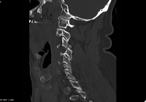
A 56-year-old man was admitted to the Department of Neurosurgery at Haukeland University Hospital, Bergen, after a neck trauma. His medical history included an aortic aneurysm and six myocardial infarctions. Examinations and computed tomography (CT) scan of the cervical spine revealed a dislocated fracture at the level C3/C4 (Fig. 1). There were no neurological deficits. Routine surgical reposition and osteosynthesis at the level of the fracture was performed 2 days later with the patient in prone position. The surgery lasted for 2 hr and blood loss was estimated to be 800 ml. The average blood pressure during surgery was 110/60, but he had a few dips down to 80/50.

Preoperative computed tomography scan: at the level of C3, a fracture of processus articularis inferior is present, with prominent gliding and subluxation C3/C4.
When the patient woke up after the operation, he complained of loss of visual field in the left eye. He described it as a huge black spot, but also claimed to see coloured stripes and circles related to this spot.
He was examined by an ophthalmologist the same day. She found a relative afferent pupillary defect in the affected eye with a visual acuity of counting fingers. Only the upper nasal part of the visual field was preserved. The neural rim around the optic disc, especially on the temporal side, was blurred. The diameter of the arterial blood vessels in the proximity of the disc seemed attenuated, but no embolus could be seen. Intraocular pressure was normal. Differential diagnosis included a perioperative partial occlusion of the central retinal artery or ischemic optic neuropathy. Doppler ultrasound of the carotid arteries revealed no significant stenosis on either side. CT of the brain did not show any signs of infarction or haemorrhage.
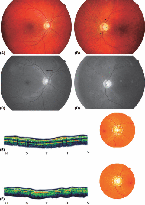
When the patient was reviewed 6 weeks later, visual acuity in the left eye had improved to 6/9, although he still had a significant defect in the lower parts of the visual field. The left optic disc was slightly pale with blurred nerve fibres on its temporal side (Fig. 2A,B). Six months after the spinal surgery, retinal nerve fibre layer (RNFL) photography and optical coherence tomography (OCT) measurement of RNFL thickness was performed, revealing a significant reduction in the left eye compared to the right eye (Fig. 2C–F). This finding supported the diagnosis of ischemic optic neuropathy, related to the spinal surgery.

Fundus picture of the right (A) and left (B) eyes showing healthy reddish colour of the neural rim (asterisk) and normal calibre of the vessels (A) and atrophy of the optic nerve head, with a sharply delineated margin and a pale neural rim (asterisk) (B). Atrophy of the retinal nerve fibre layer (RNFL) causes an increased transparency of the retina, leaving the mottled appearance of the choroid more visible than in the right eye. Note the attenuation of retinal arterioles (short arrows). Blue-filtered photograph of the fundus showing light striation along the major vessel arcades (long arrows), representing the normal RNFL in the right eye (C) and the absence of striation in the left eye (D), corresponding to atrophy of the RNFL. (E, F) Optical coherence tomography (OCT): circumferential scan of the optic nerve head. Image on the right: a wheel-shaped grid superimposed on the fundus image, numbers indicating thickness (in μm) of the RNFL in each sector. Image on the left: the actual 360° OCT scan of the retina, aligned horizontally for convenience. The layer between the white lines is the RNFL. The letters below the figure indicate the position in the circular arrangement: N, nasal; S, superior; T, temporal; I, inferior. (E) Right eye. The overall thickness of the RNFL is within normal limits. The uneven distribution of nerve fibres is also according to the normal pattern, with the inferior and superior parts thicker than the nasal and temporal parts. (F) Left eye. The RNFL thickness is outside normal limits. In the inferior and superior parts, the thickness is only half of that in the corresponding areas in the right eye.
Postoperative visual loss after prone spine surgery is a relatively uncommon but devastating complication most often associated with cardiac, spine, head and neck surgery (Lee et al. 2006). During the 1990s, concerns were voiced that the occurrence of postoperative visual loss was increasing and thus, the American Society of Anesthesiologists Committee on Professional Liability established a registry to collect detailed information on cases occurring after non-ocular surgery (Lee et al. 2006). Ischaemic optic neuropathy was the most common cause of visual loss after spine surgery.
The aetiology of postoperative ischaemic optic neuropathy is still not clear. It has been suggested that the condition is a ‘compartment syndrome of the eye’, created by increased venous pressure and interstitial fluid accumulation either within lamina cribrosa at the optic nerve head or within the bony optic canal (Lee et al. 2006). Anaemia and hypotension have been described as particularly potent risk factors (Patil et al. 2008). However, because they also occur during many surgical procedures that do not have an increased risk for postoperative ischaemic optic neuropathy, one must look for additional risk factors: high intraocular pressure (IOP) decreases the perfusion pressure of the optic nerve head; in awake volunteers, the prone position increases IOP compared to the spine position (Lam & Douthwaite 1997). This increase in IOP may explain why patients undergoing prone surgery are more likely to develop visual loss, especially if they have experienced anaemia and hypotension during surgery. Systemic hypertension, diabetes mellitus, arteriosclerosis and smoking have been identified as additional risk factors (Lee et al. 2006; Pane et al. 2007).
Postoperative visual loss is a serious but rare complication to spine surgery. The incidence reported varies, but lies between 0.2% (Stevens et al. 1997) and 0.1% (Patil et al. 2008). Although uncommon, it has serious consequences for the patient. Thus, in patients with predisposing factors, special emphasis should be given to protect the eye against pressure during spinal surgery, avoiding hypotension and hypovolaemia (Stevens et al. 1997; Patil et al. 2008). Vision should be monitored in the recovery room, and it is important to take postoperative complaints about loss of vision seriously. Postoperative hypotension and anaemia should be identified and treated rapidly (Stevens et al. 1997). Our patient experienced a blood loss of 800 ml during the procedure and, although not severe, this may have contributed to his postoperative ischaemic neuropathy, together with his pre-existing hypertension and arteriosclerosis.
Acknowledgements
The authors thank Dr Gesche Neckelmann of the Department of Radiology at Haukeland University Hospital, Bergen, Norway.


